4404
The value of radiomics models based on DTI and DWI sequences in identifying cervical adenocarcinoma and squamous cell carcinoma1the First Affiliated Hospital of Dalian Medical University, Dalian, China
Synopsis
Keywords: fMRI Analysis, Uterus
Motivation: To explore the feasibility of differentiating pathological types of cervical adenocarcinoma and squamous cell carcinoma by radiomics models based on DTI and DWI sequences.
Goal(s): To explore the value of radiomics models based on DTI and DWI sequences in differentiating pathological types of cervical adenocarcinoma and squamous cell carcinoma.
Approach: Based on the DTI and DWI sequence images of cervical cancer patients , this study quantitatively analyzed the differences in the radiomics feature parameters of adenocarcinoma and squamous cell carcinoma.
Results: The radiomics models based on DTI and DWI sequences are valuable in differentiating the pathological types of cervical adenocarcinoma and squamous cell carcinoma.
Impact: Radiomics can transform images into data that can be mining, providing a non-invasive and effective auxiliary method for determining tumor heterogeneity before clinical treatment of cervical cancer, guiding clinical practice, and enabling patients to achieve personalized diagnosis and treatment.
Objective
To explore the feasibility of differentiating pathological types of cervical adenocarcinoma and squamous cell carcinoma by radiomics models based on DTI and DWI sequences, so as to improve preoperative evaluation of cervical cancer and guide clinical treatment strategy.Materials and Methods
This retrospective study included 147 cervical cancer patients with MRI in our hospital, including 41 adenocarcinoma and 106 squamous carcinoma. All enrolled patients underwent 1.5T MRI (GE 1.5T Signa HDXT MR) scanning two weeks before surgery, and the scanning sequences included T1WI, T2WI, DWI and DTI sequences.The original DWI images were transferred to the ADW 4.6 workstation, and the Functool function was applied to generate the Apparent diffusion coefficient (ADC) images.The original DTI images were transferred to the ADW 4.6 workstation, and the Functool function was applied to generate the flip angle (FA) images and the Apparent Diffusion Coefficient time (ADCT) images.By using ITK-SNAP software (version 3.6, open source software, http://www.itksnap.org), the primary lesions of cervical cancer were mapped manually on ADC images, FA images and ADCT images in full layer as Region of interest (ROI).On the uAI Research Portal platform, 464 radiomics features were extracted from ADC, FA and ADCT images respectively, including 14 shape features, 18 first-order features, 21 texture features and 411 Laplacian coefficient features of Gaussian transform.Ultimately, a total of 1392 features were extracted from the three sequences.The image data were randomly divided into the training set and the test set according to the ratio of 7:3. Through minimum Redundancy Maximum Relevance (mRMR), Recursive Feature Eliminationusing Cross Validation (RFECV) and Least absolute shrinkage and selection operator (LASSO) algorithms reduce the dimension of features. ADC , FA and ADCT images finally screened out 10, 10 and 5 radiomics features, respectively. Based on the final selected features, partial least squares discriminant analysis( PLS-DA ) was used to establish the radiomics models: including ADC, FA, ADCT and combined radiomics models, and the models were verified by the 5 fold cross verification(5-FCV)method.The area under the receiver operating characteristics curve (AUC), sensitivity, specificity and accuracy of each radiomics model were recorded.According to the AUC values of the training set and the test set, the effectiveness of each radiomics model in differentiating pathological types of cervical adenocarcinoma and squamous cell carcinoma was evaluated.Results
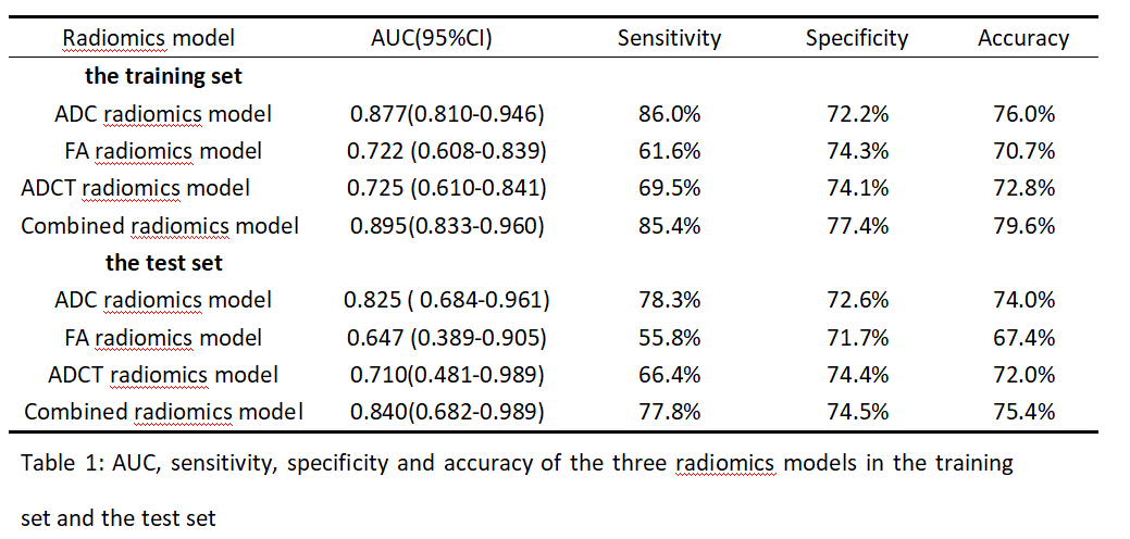
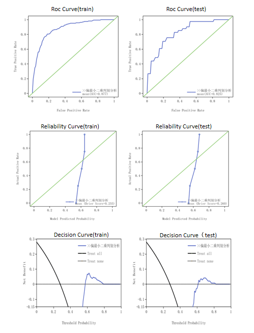
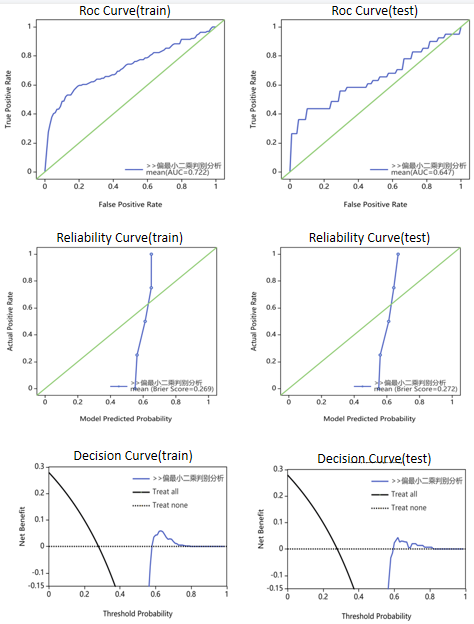
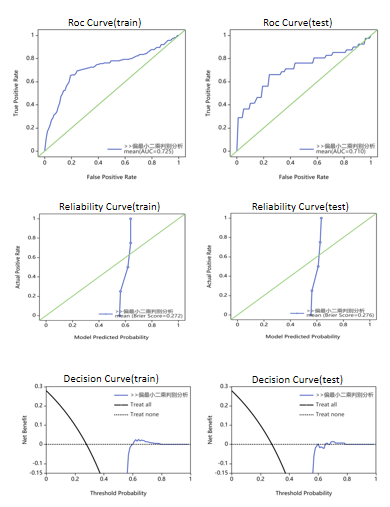
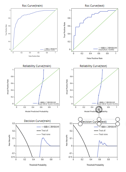
The AUC of ADC radiomics model in the training set and test set were 0.877 (95%CI 0.810-0.946) and 0.825 (95%CI 0.684-0.961), the sensitivity was 86.0% and 78.3%, the specificity was 72.2% and 72.6%, and the accuracy was 76.0% and 74.0%, respectively.The AUC of FA radiomics model in the training set and test set were 0.722 (95%CI 0.608-0.839) and 0.647 (95%CI 0.389-0.905), the sensitivity was 61.6% and 55.8%, the specificity was 74.3% and 71.1%, and the accuracy was 70.7% and 67.4%, respectively.The AUC of ADCT radiomics model in the training set and test set were 0.725 (95%CI 0.610-0.841) and 0.71 (95%CI 0.481-0.934), the sensitivity was 69.5% and 66.4%, the specificity was 74.1% and 74.4%, and the accuracy was 72.8% and 72.0%, respectively. The AUC of combined radiomics model in the training set and test set were 0.895 (95%CI 0.833-0.960) and 0.840 (95%CI 0.682-0.989), the sensitivity was 85.4% and 77.8%, the specificity was 77.4% and 74.5%, and the accuracy was 79.6% and 75.4%, respectively.Conclusions
The radiomics models based on DTI and DWI sequences are valuable in differentiating the pathological types of cervical adenocarcinoma and squamous cell carcinoma, and can provide a non-invasive and effective auxiliary method for judging tumor heterogeneity before clinical treatment of cervical cancer.Acknowledgements
No acknowledgement found.References
[1]LAMBIN P,RIOS—VELAZQUEZ E,LEIJENAAR R,et a1.Radiomics:extracting more information from medical images USing advanced feature analysiS[J].Eur J Cancer,2012,48(4):441—6.
[2]PINKER K,SHITANO F,SALA E,et a1.Background,current role,and potential appl ications of radiogenomics [J]. Journal of magnetic resonance imaging:JMRI,2018,47(3):604—20.
[3]KANG D,PARK J E,KIM Y H,et a1.Diffusion radiomics as a diagnostic model for atypical manifestation of primary central nervous system lymphoma:development and multicenter external validation[J].Neuro Oncol,2018,20(9):1 251—61.
Figures