4403
Functional Brain Alterations in Children with Autism Spectrum Disorder after Fecal Microbiota Transplantation: a preliminary study.1Department of Radiology, Shanghai Tenth People’s Hospital, Tongji University School of Medicine, Shanghai, China, 2Philips Healthcare, Shanghai, China
Synopsis
Keywords: fMRI Analysis, Pediatric, Autism
Motivation: The combination of fecal microbiota transplantation (FMT) and resting-state fMRI (rs-fMRI) can provide a new imaging perspective for understanding the neural mechanism of gut microbiota in ASD.
Goal(s): This study is to evaluate the functional alterations and investigate the potential mechanisms of FMT treatment in ASD.
Approach: Amplitude of low-frequency fluctuation (ALFF) and Regional Homogeneity (ReHo) based on rs-fMRI are useful methods to characterize brain function and activity.
Results: After FMT treatments, the higher ALFF and ReHo values in the left angular gyrus and bilateral lingual gyrus, which could become the underlying mechanism of improvements.
Impact: FMT can improve the symptoms of ASD and influence the brain functional activity. Combining FMT and MRI can provide a new imaging perspective for understanding the neural mechanism and assist clinical follow-up of ASD.
Introduction
Autism spectrum disorder (ASD) has been widely recognized as a complex neurodevelopmental disorder characterized by repetitive behaviors and social-communication deficits. Currently, there is still no specific treatment for ASD. Gut microbiota is known to play the main role in regulating health and disease and modulating brain function and social behavior1. Gastrointestinal dysfunction including constipation, diarrhea, and abdominal pain becomes one of the most prevalent physiological symptoms of ASD2. The most representative method is fecal microbiota transplantation (FMT). FMT is a therapy that transplants the gut microbiota from healthy people into the intestines of patients to reconstruct the gut microbiota. The previous study suggested that FMT can help improve the symptoms of ASD3. Neuroimaging provides an objective basis for clinical functional changes and is helpful in the follow-up of changes in ASD. Amplitude of low-frequency punctuation (ALFF) and regional homogeneity (ReHo) based on resting-state fMRI (rs-fMRI) are useful data-driven methods to characterize brain function and activity. This study is to evaluate the functional alterations and investigate the potential mechanisms of FMT treatment in ASD.Methods
Eighty children with ASD completed rs-fMRI follow-up 6 months after FMT treatment in this study from June 2021 to September 2023. This study is a double-blind experiment. After enrollment, subjects were divided into an experimental group and a control group in a ratio of 2:1. The experimental group received the FMT capsules and the control group received the placebo. Resting-state fMRI images were acquired on an Ingenia CX 3.0-T MRI scanner (Philips Healthcare, Best, the Netherlands) with a 32-channel head coil. All children were examined under sedation using chloral hydrate and a caregiver and a doctor for each participant were present throughout the duration of the scan. The preprocessing of resting-state fMRI data was performed in MATLAB R2013b (Mathworks, Natick, MA) with RESTplus software4. The First ten time points of fMRI data were removed. Because of using multiband scanning, slice timing was not performed5. After realigning, nobody had excessive head motion (≥±3 mm in any axis). The processed images were then normalized by the DARTEL toolbox using the T1 image new segment. A Gaussian filter (a 6mm FWHM) was used to smooth the data (for ReHo analysis, this step was performed during ReHo and DC calculation). The Friston 24 motion parameters, white matter, and the cerebrospinal fluid were regressed as covariates. Finally, band-pass filtering (0.01–0.08 Hz) was performed to remove the effects of high-frequency noise (for ALFF analysis, the ALFF calculation replaced this step). To explore the functional alterations before and after treatment, paired t-tests were performed on the ALFF and ReHo maps.Results
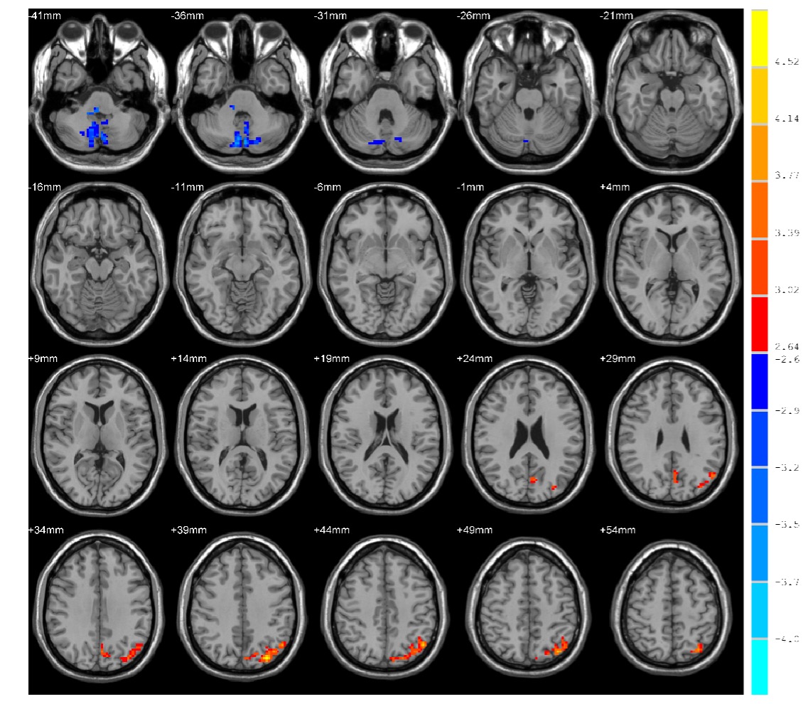
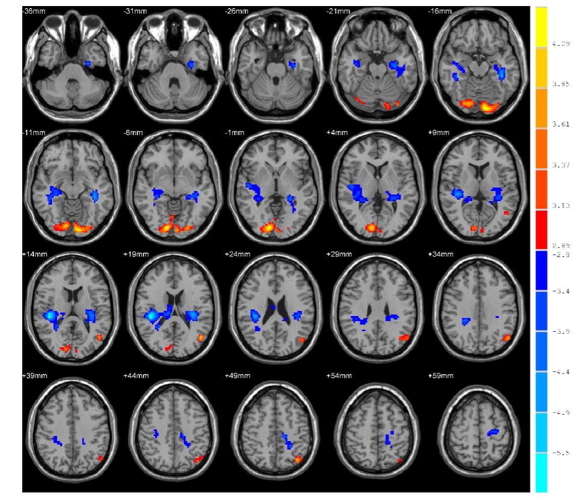
After FMT treatment, ASD children showed increased ALFF in the left angular gyrus, while showed decreased ALFF in the bilateral cerebellum crus2 (p < 0.01, cluster size > 100 clusters) (Fig 1). The ReHo was higher in the bilateral lingual gyrus and left angular gyrus, and lower in the bilateral hippocampus and left precentral gyrus (p < 0.005, cluster size > 100 clusters) (Fig 2).Discussion
ALFF and ReHo are useful methods to characterize local spontaneous brain activity in fMRI studies6. ALFF measures the amplitude of time series fluctuations at each voxel. ReHo measures the local synchronization of the time series of neighboring voxels. The angular gyrus is engaged in transferring visual information and involved in a number of processes related to language, number processing and spatial cognition7. The lingual gyrus, part of the occipital lobe, is primarily responsible for the processing of visual memory and logical processing. After FMT treatments, the higher ALFF and ReHo values in the left angular gyrus and bilateral lingual gyrus meant neuronal activities increased and could become the underlying mechanism of improvements. We also found lower ALFF and ReHo values in several brain regions after FMT treatment. The experiment is still collecting follow-up cases and the grouping situation has not been revealed yet. It is unknown which autistic children are taking the real capsule. There is currently no research on the functional alterations in brain before and after FMT treatment for ASD. This study is a preliminary exploration of the results of the current follow-up cases. The analysis will be further improved based on the revealed results.Conclusion
FMT can improve the symptoms of ASD and influence the brain functional activity. Combining FMT and MRI can provide a new imaging perspective for understanding the neural mechanism and assisting in clinical follow-up of ASD.Acknowledgements
We would like to thank Shanghai Tenth People’s Hospital and Philips Healthcare for supporting this research. We also thank all participants and their families for their cooperation in this study.References
1. Martínez-González AE, Andreo-Martínez P. Prebiotics, probiotics and fecal microbiota transplantation in autism: A systematic review. Prebióticos, probióticos y trasplante de microbiota fecal en el autismo: una revisión sistemática. Rev Psiquiatr Salud Ment (Engl Ed). 2020;13(3):150-164.
2. Leader G, Abberton C, Cunningham S, et al. Gastrointestinal Symptoms in Autism Spectrum Disorder: A Systematic Review. Nutrients. 2022, 14(7):1471.
3. Bibbo S, Ianiro G, Gasbarrini A, Cammarota G. Fecal microbiota transplantation: past, present and future perspectives. Minerva Gastroenterol Dietol, 2017, 63: 420-430.
4. Jia X, Wang J, Sun H, Zhang H, Liao W, Wang Z, et al. RESTplus: an improved toolkit for resting-state functional magnetic resonance imaging data processing. Science Bulletin, 2019, 64: 953-954.
5. Smitha K A, Arun K M, Rajesh P G, Joel S E, Venkatesan R, Thomas B, et al. Multiband fMRI as a plausible, time-saving technique for resting-state data acquisition: Study on functional connectivity mapping using graph theoretical measures. Magn Reson Imaging, 2018, 53: 1-6.
6. Lv H, Wang Z, Tong E, Williams L M, Zaharchuk G, Zeineh M, et al. Resting-State Functional MRI: Everything That Nonexperts Have Always Wanted to Know. American Journal of Neuroradiology, 2018, 39: 1390-1399.
7. Rockland K S , Graves W W .The angular gyrus: a special issue on its complex anatomy and function[J].Brain structure & function, 2023, 228(1):1-5.
Figures