4401
Resting-State Dynamic Functional Gradient Stability Disturbance by Task fMRI in Lifelong Premature Ejaculation1Nanjing Drum Tower Hospital, Affiliated Hospital of Medical School, Nanjing University, Nanjing, China
Synopsis
Keywords: fMRI Analysis, fMRI (resting state)
Motivation: Premature ejaculation (PE) is a major component of male sexual disfunction. The brain's recovery process following erotic stimulation in PE patients remains unclear.
Goal(s): In this study, we aimed to explore the brain network hierarchies by brain gradient and the dynamic gradient stability modified by erotic stimulation.
Approach: We explored the brain network hierarchies by brain gradient in PE and NC group across pre- and post-task resting-state and brain dynamic BOLD phase-locking functional gradient stability.
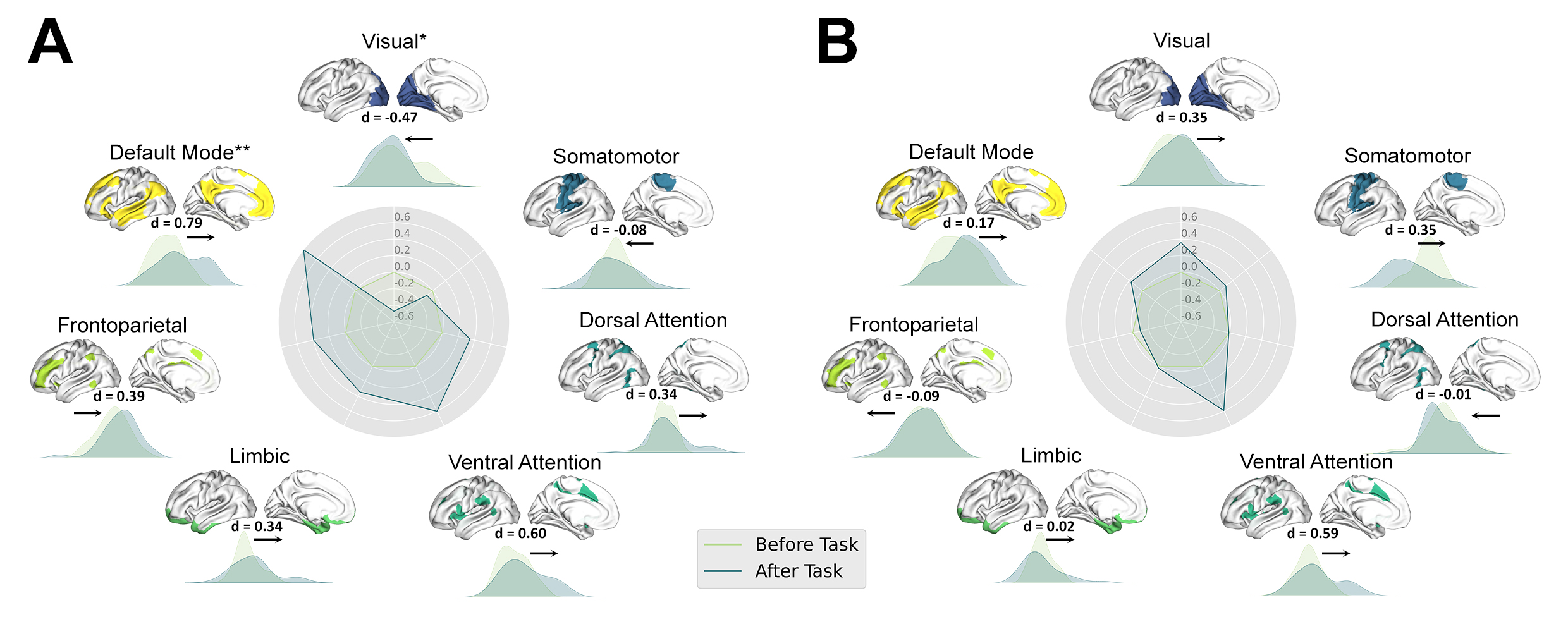
Results: The PE patients have decreased dynamic functional gradient stability of visual network and increased stability of the default mode network.
Impact: The dynamic functional gradient stability of visual network and the default mode network were altered in lifelong PE compared to normal controls. These findings revealed that brain spontaneous activity in PE patients can hardly recovery as soon as normal control.
INTRODUCTION
Premature ejaculation (PE) is a major component of male sexual disfunction[1]. The brain's recovery process following erotic stimulation in PE patients remains unclear. In this study, we aimed to explore the brain network hierarchies by brain gradient in PE and NC group across pre- and post-task resting-state and to explored how brain gradient stability was modified by erotic stimulation.METHODS
Twenty-eight right-hand patients with lifelong PE and seventeen right-hand normal controls (NCs) were recruited. All MRI data were collected on the Phillps Ingenia 3.0T CX MR scanner. Every subject in this study first went through a resting state fMRI scan (rs-fMRI) before fMRI task (task free rs-fMRI dataset). Followed by two tasks fMRI, another rs-fMRI scan was scanned (task modulated rs-fMRI dataset). After the rs-fMRI preprocess, ROI-wise gradient maps were calculated using the BrainSpace Toolbox (https://github.com/MICA-MNI/BrainSpace)[2]. Further dynamic BOLD phase-locking functional gradient and stability of dynamic functional gradient (DFG) mapping were also calculated. Group difference was examined between patients with LPE and NC across the two timepoint using linear mixed effects (LME) model.RESULTS
Both LPE patients and NC in two rs-fMRI datasets, showed a clear transition from a unimodal system to a transmodal system in the principal gradient, and separated primary networks in the secondary gradient, with visual network suited on one end, and the somatomotor on the other end. Compared to task free resting state, there was an apparent increase stability in the DMN of patients with LPE (Cohen’s d = 0.79, K-S stat =0.36, P = 1.03×10⁻⁵, Bonferroni corrected) and decrease stability in the visual network (Cohen’s d = -0.47, K-S stat = 0.25, P = 0.031, uncorrected) in the principal gradient in the task modulated rs-fMRI dataset. There were increase stability in right prefrontal dorsal medial (PFCdPFCm_5 and PFCdPFCm_8, P < 0.05, Bonferroni corrected) and decreased stability in left visual cortex (Vis_10, P < 0.05, Bonferroni corrected) in the principal functional gradient in PLE patients between the task free and task modulated rs-fMRI dataset. In second functional gradient, there were increased stability in left prefrontal cortex (PFC_9, P < 0.05, Bonferroni corrected) and right temporal occipital parietal cortex (TempOccPar_6, P < 0.05, Bonferroni corrected) in PLE patients. The IELT score was significantly negatively correlated with the delta gradient stability in right PFCdPFCm_5 cortex (r = -0.74, P < 0.001) in the principal functional gradient in NC group.DISCUSSION and CONCLUSION
In this study, we explored the brain network hierarchies by brain gradient in PE and NC group across pre- and post-task resting-state and brain dynamic BOLD phase-locking functional gradient stability. The dynamic functional gradient stability of visual network was significantly decreased and the default mode network was significantly increased in LPE patients after the fMRI task modulation, while no significantly alterations were observed in NC group. ROI level analysis found the dynamic functional gradient stability was decreased in the left visual cortex and increased in the left PFC and right PFCdPFCm and TempOccPar cortex. These findings revealed that brain spontaneous activity in PE patients can hardly recovery as soon as normal control.Acknowledgements
No acknowledgement found.References
1. Serefoglu, E.C., et al., An evidence‐based unified definition of lifelong and acquired premature ejaculation: Report of the second International Society for Sexual Medicine Ad Hoc Committee for the Definition of Premature Ejaculation. The journal of sexual medicine, 2014. 11(6): p. 1423-1441.
2. Vos de Wael, R., et al., BrainSpace: a toolbox for the analysis of macroscale gradients in neuroimaging and connectomics datasets. Communications Biology, 2020. 3(1): p. 103.
Figures