4399
The application value of multimodal functional magnetic resonance imaging in differentiating type I and type II endometrial cancer1the First Affiliated Hospital of Dalian Medical University, Dalian, China, 2the First Affiliated Hospital of Dalian Medical University, DaLian, China
Synopsis
Keywords: fMRI Analysis, Uterus
Motivation: Preoperative assessment of the histological type of EC based on multimodal functional MRI is expected to provide personalized treatment for patients and improve their prognosis.
Goal(s): To explore the application value of APT, IVIM and DCE techniques in distinguishing between Type I and Type II endometrial cancer.
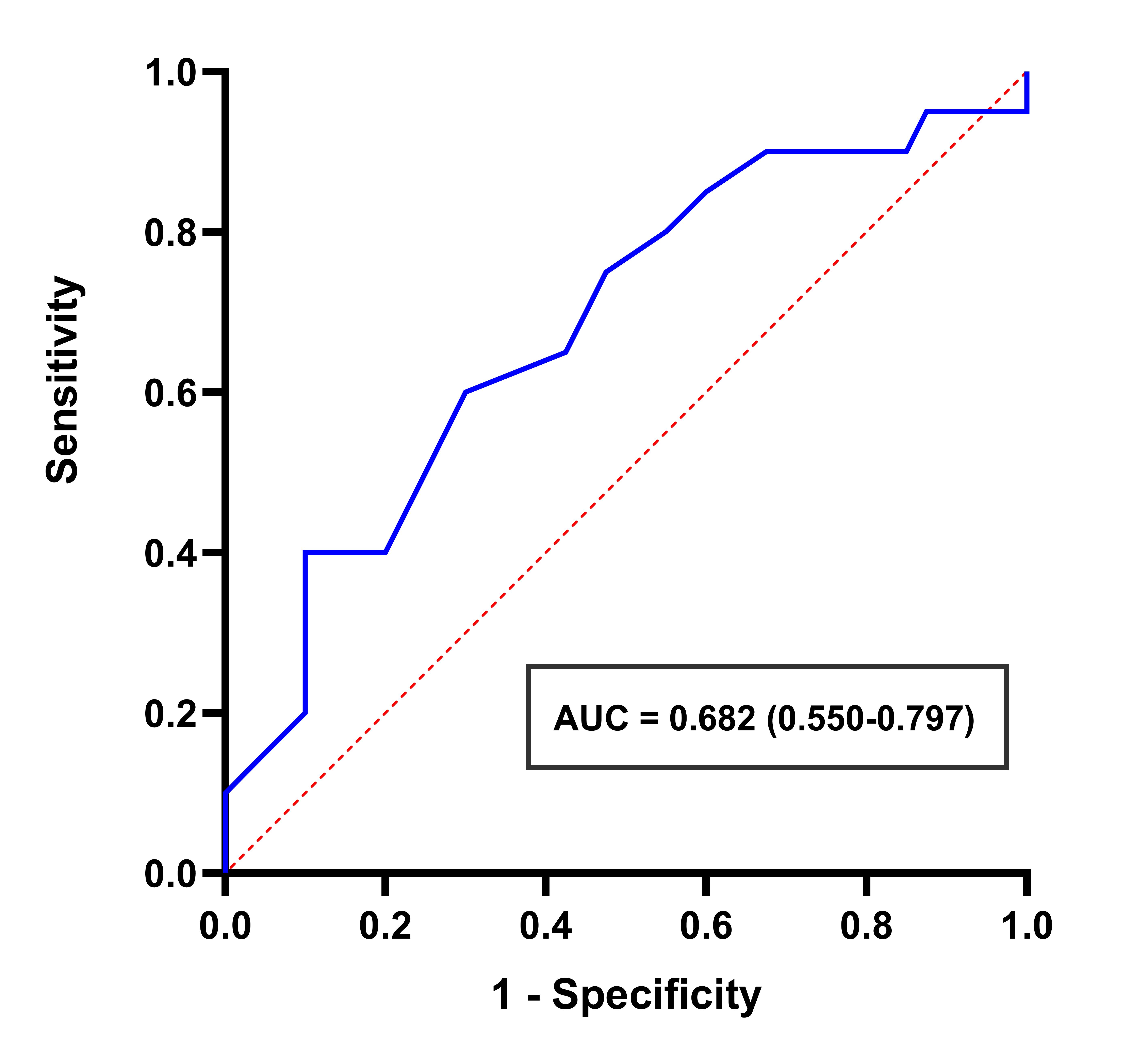
Approach: The ROC curve were used to analyze and evaluate the predictive ability of the independent risk factor for the histological type of EC.
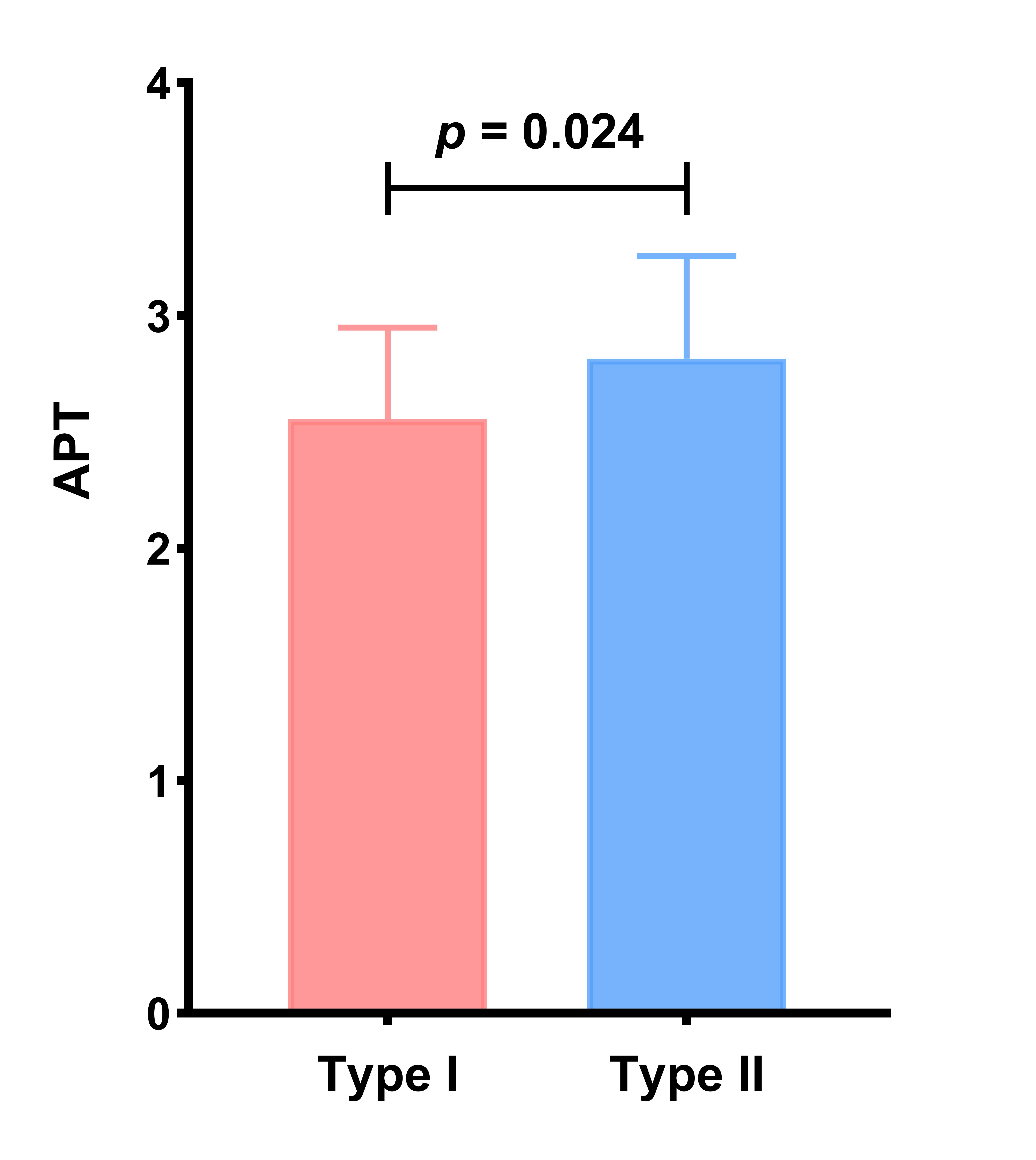
Results: The APT value of the type II group were higher than the the type I group and the AUC value of APT was 0.682 for identifying the histological types of EC.
Impact: APT technique can be utilized for quantitative identification of type I and type II EC, and has broader clinical application prospects.
Introduction
Endometrial cancer (EC) is the sixth most common female cancer worldwide[1]. According to the 2023 FIGO staging system[2], EC is divided into two main histological types: invasive (Type I) and non-invasive (Type II). This staging system, for the first time, integrates histological types and emphasizes that different histological types of EC may have different biological characteristics and prognosis. Preoperative assessment of the histological type of EC is crucial for providing the personalized treatment for patients and improving their prognosis. Therefore, This study aimed to explore the application value of intravoxel incoherent motion (IVIM), amide proton transfer (APT) and dynamic contrast-enhanced (DCE) magnetic resonance imaging (MRI) in distinguishing between Type I and Type II endometrial cancer.Methods
A total of 60 EC patients who underwent 1.5 Tor 3.0 T MRI examination of upper abdomen in our hospital were included in our study, including 40 patients in type I group and 20 patients in type II group. The MRI images of all patients included IVIM, APT and DCE sequence. The APT, diffusion coefficient (D), pseudo diffusion coefficient (D*), perfusion fraction (f), volume transfer constant (Ktrans), rate transfer constant (Kep) and volume of extravascular extracellular space per unit volume of tissue (Ve) values of the two groups of lesions were measured using the Philips post-processing workstation. The receiver operating characteristic (ROC) curve were used to analyze and evaluate the predictive ability of the independent risk factor for the histological type of EC.Results
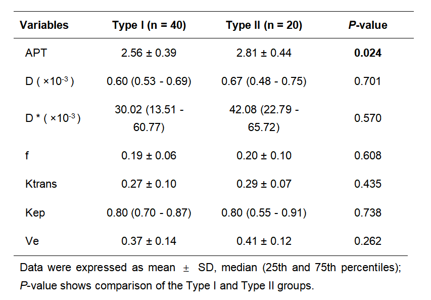
The APT value of the type II group were higher than the type I group, The difference was statistically significant (P < 0.05). However there was no statistically significant difference in the value of D, D*, f, Ktrans, Kep, Ve between two groups (P > 0.05) (Table 1) (Fig 1). The ROC curve shown that area under the curve (AUC) (95%CI) value of APT was 0.682 (0.550-0.797), and the sensitivity and specificity were 40%, 90% respectively (Fig 2).Discussion
In this study, our findings indicate that APT is an independent risk factor for identifying the histological types of EC. However, IVIM and DCE did not show significant differences in distinguishing between Type I and Type II EC. We speculate that this may be due to the propensity of Type II EC to undergo necrosis, leading to a reduction in tumor parenchymal cell death and microvascular regeneration, which results in the parameter values of IVIM and DCE reflecting diffusion and perfusion not being statistically significant. Considering the relatively small sample size, we believe that it is necessary to further expand the sample size to conduct more in-depth research.Conclusion
APT technique can be utilized for quantitative identification of type I and type II EC. This technology provides a new method for preoperative quantitative assessment of the histological type of EC, which is expected to provide patients with more accurate and faster diagnostic results, assist clinicians in formulating more effective treatment strategies, thereby improving treatment efficacy and survival rates.Acknowledgements
No acknowledgement found.References
[1]. Berek JS, Matias-Guiu X, Creutzberg C, et al. FIGO staging of endometrial cancer: 2023 [published correction appears in Int J Gynaecol Obstet. 2023 Oct 6;:]. Int J Gynaecol Obstet. 2023;162(2):383-394. doi:10.1002/ijgo.14923
[2]. Sung H, Ferlay J, Siegel RL, et al. Global Cancer Statistics 2020: GLOBOCAN Estimates of Incidence and Mortality Worldwide for 36 Cancers in 185 Countries. CA Cancer J Clin. 2021;71(3):209-249. doi:10.3322/caac.21660
Figures