4397
The altered brain structure and function in patients with chronic prostatitis/chronic pelvic pain syndrome: a neuroimaging study1Department of Medical Imaging, The Affiliated Hospital of Northwest University·Xi’an No.3 Hospital, Xi'an, China, 2Department of Medical Imaging, Xi'an Jiaotong University Medical College First Affiliated Hospital, Xi'an, China
Synopsis
Keywords: Gray Matter, fMRI (resting state), chronic prostatitis/chronic pelvic pain syndrome
Motivation: Chronic prostatitis/chronic pelvic pain syndrome (CP/CPPS) is mainly characterized by persistent pain and discomfort in the pelvic region, which seriously affects the young and middle-aged men in China.
Goal(s): Further exploration of the central mechanisms is essential to gain a deeper understanding of this chronic pain condition.
Approach: This study, based on multimodal MRI and combined with clinical questionnaires and scales, adopts a cross-sectional design to systematically analyze the changes in gray matter and resting-state functional connectivity abnormalities.
Results: The structure and function of the striatum are altered in CP/CPPS patients.
Impact: The study results provide objective evidence of cerebral abnormalities to further understand the central pathophysiological mechanisms of CP/CPPS. They also offer valuable neuroimaging-based insights for future clinical advancements in precise diagnosis and treatment of CP/CPPS.
Introduction
Chronic prostatitis/chronic pelvic pain syndrome (CP/CPPS) is a common chronic pain syndrome that affects males and is characterized by recurrent or persistent pelvic pain/discomfort and/or lower urinary tract symptoms[1,2,3]. Based on the hypothesis of end-organ pathologies, the current general clinical diagnosis and treatment was not fully satisfy both physicians and patients[4,5]. Neuroimaging studies in heterogeneous patients with CP/CPPS have demonstrated the significant role of supraspinal mechanisms [6]. Consequently, we conducted a cross-sectional study to investigate structural and functional brain changes in young and middle-aged CP/CPPS patients.Method
This study was reviewed and approved by the Medical Ethics Review Board of Xi’an No.3 Hospital. Additionally, written informed consent was obtained from all participants and in accordance with the Declaration of Helsinki.A total of 29 CP/CPPS patients and 29 healthy controls (HCs) were enrolled in the study, and data were collected accordingly. MR examinations were conducted using a 3.0T MR scanner (Philips Ingenia, Philips Healthcare, Best, the Netherlands) with a 32-channel head-neck coil. High-resolution brain structural images were obtained from each participant [repetition time (TR) = 8.2 ms, echo time (TE) = 3.8 ms, flip angle = 8°, slice thickness = 1 mm, field of view (FOV) = 24 × 24 cm2, matrix size = 240 × 240, voxel size = 1 × 1 × 1 mm3]. The resting state functional MRI images were obtained from gradient-echo-planar imaging sequence (TR = 2,500 ms, TE = 30 ms, flip angle = 90°, voxel size = 3 × 3 × 3 mm3, slice thickness = 3 mm with no gap, slices = 50, matrix size = 80 × 80, FOV = 24 × 24 cm2, volumes = 180).
All neuroimaging data underwent preprocessing using fmriprep [7]. Subsequently, the volume of subcortical nuclei was extracted, and analysis of variance was conducted to compare intergroup differences in different nuclei while controlling for intracranial volume and age using R (R version 4.0.2, R Foundation for Statistical Computing, Vienna, Austria). The preprocessed data was input into CONN[8] to select brain regions with structural changes as seeds and analyze their resting state functional connectivity (rs-FC) changes with the entire brain. Additionally, we investigated the association between alterations in brain structure and function and clinical variables in patients using correlation analysis.
Results
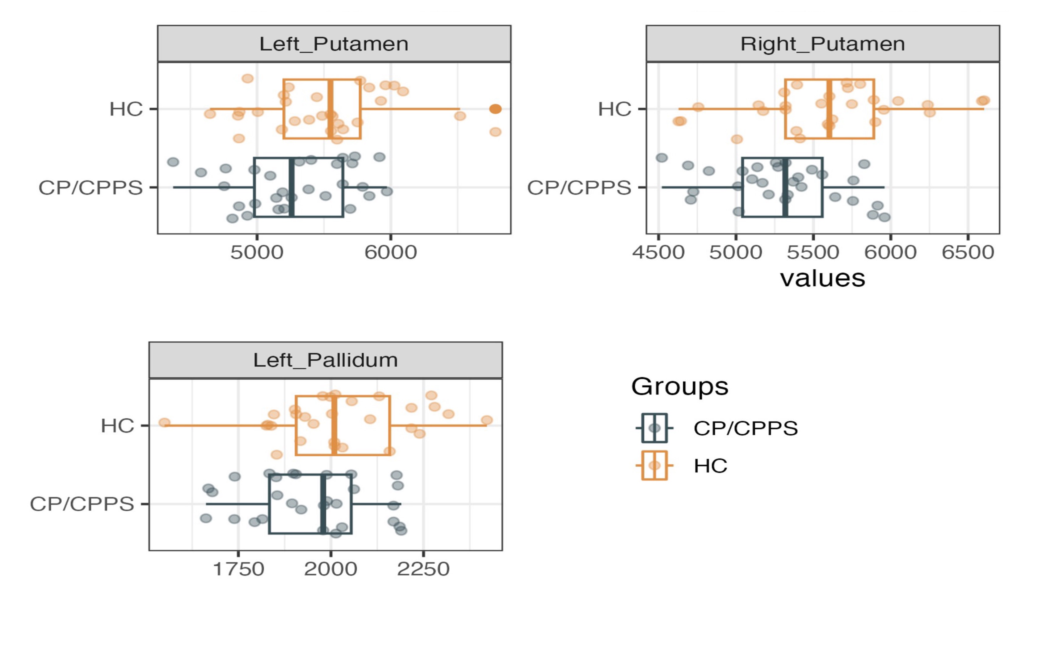
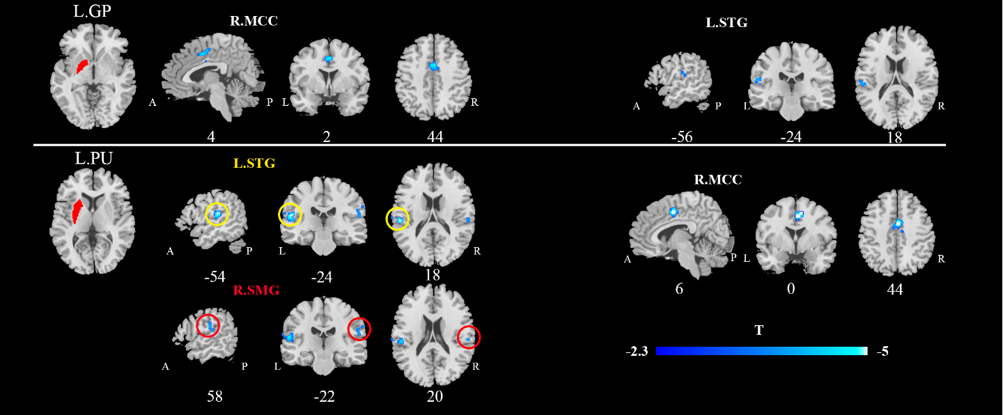
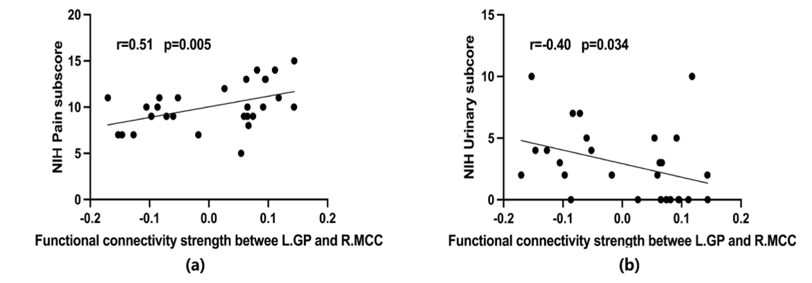
Compared to the healthy controls, morphological analysis indicated a significant reduction in the volume of both the bilateral putamen and the left globus pallidus (Figure 1). The seed-based functional analysis revealed a decrease in rs-FC between the left putamen and the right middle cingulate gyrus, superior temporal gyrus, and left superior frontal gyrus. Furthermore, it showed a decrease in rs-FC between the left globus pallidus and the right middle cingulate gyrus and left superior frontal gyrus (Figure 2). The altered volume and rs-FC of the left putamen and left globus pallidus were found to be associated with clinical symptoms. Partial correlation analysis demonstrated that pain scores were positively correlated with the volume of the left putamen (r = 0.53, P = 0.015) and the left globus pallidus (r = 0.50, P = 0.035). Additionally, the rs-FC between the left globus pallidus and the right middle cingulate gyrus was positively correlated with NIH-CPSI pain scores (r = 0.51, P = 0.005) and negatively correlated with urinary symptom scores (r = -0.4, P = 0.034), as illustrated in Figure 3.Discussion
The structural and functional alterations in the left caudate nucleus and globus pallidus of young and middle-aged CP/CPPS patients are closely related to the patient's clinical characteristics. This finding suggests that there is a disruption in the cortico-basal ganglia circuitry in CP/CPPS [9], which may be involved in abnormalities in somatic sensation and motor function, affecting the patients' pain perception and regulation, ultimately leading to recurrent or persistent pelvic pain/discomfort.Conclusion
Patients with CP/CPPS exhibited altered brain morphology and function, which influenced their clinical symptoms. This study not only further highlights the significant role of supraspinal mechanisms in the brain's somatosensory and motor functions in CP/CPPS but also provides valuable neuroimaging evidence for future clinical diagnostic and therapeutic assessments.Acknowledgements
We would like to thank all patients and healthy volunteers who participated in the study.References
- Habermacher, G. M., Chason, J. T., & Schaeffer, A. J. (2006). Prostatitis/chronic pelvic pain syndrome. Annual review of medicine, 57, 195–206. https://doi.org/10.1146/annurev.med.57.011205.1356541356542.
- Krieger, J. N., Nyberg, L., Jr, & Nickel, J. C. (1999). NIH consensus definition and classification of prostatitis. JAMA, 282(3), 236–237. https://doi.org/10.1001/jama.282.3.2363.
- Walz et al., 20073. Walz, J., Perrotte, P., Hutterer, G., Suardi, N., Jeldres, C., Bénard, F., Valiquette, L., & Karakiewicz, P. I. (2007). Impact of chronic prostatitis-like symptoms on the quality of life in a large group of men. BJU international, 100(6), 1307–1311. https://doi.org/10.1111/j.1464-410X.2007.07250.x4.
- Franco, J. V., Turk, T., Jung, J. H., Xiao, Y. T., Iakhno, S., Garrote, V., & Vietto, V. (2018). Non-pharmacological interventions for treating chronic prostatitis/chronic pelvic pain syndrome. The Cochrane database of systematic reviews, 5(5), CD012551. https://doi.org/10.1002/14651858.CD012551.pub35
- Qin, Z., Zhang, C., Guo, J., Kwong, J. S. W., Li, X., Pang, R., Doiron, R. C., Nickel, J. C., & Wu, J. (2022). Oral pharmacological treatments for chronic prostatitis/chronic pelvic pain syndrome: A systematic review and network meta-analysis of randomised controlled trials. EClinicalMedicine, 48, 101457. https://doi.org/10.1016/j.eclinm.2022.1014576.
- Clemens, J. Q., Mullins, C., Ackerman, A. L., Bavendam, T., van Bokhoven, A., Ellingson, B. M., Harte, S. E., Kutch, J. J., Lai, H. H., Martucci, K. T., Moldwin, R., Naliboff, B. D., Pontari, M. A., Sutcliffe, S., Landis, J. R., & MAPP Research Network Study Group (2019). Urologic chronic pelvic pain syndrome: insights from the MAPP Research Network. Nature reviews. Urology, 16(3), 187–200. https://doi.org/10.1038/s41585-018-0135-57.
- Esteban, O., Markiewicz, C. J., Blair, R. W., Moodie, C. A., Isik, A. I., Erramuzpe, A., Kent, J. D., Goncalves, M., DuPre, E., Snyder, M., Oya, H., Ghosh, S. S., Wright, J., Durnez, J., Poldrack, R. A., & Gorgolewski, K. J. (2019). fMRIPrep: a robust preprocessing pipeline for functional MRI. Nature methods, 16(1), 111–116. https://doi.org/10.1038/s41592-018-0235-48.
- Whitfield-Gabrieli, S., & Nieto-Castanon, A. (2012). Conn: a functional connectivity toolbox for correlated and anticorrelated brain networks. Brain connectivity, 2(3), 125–141. https://doi.org/10.1089/brain.2012.00739.
- Borsook, D., Upadhyay, J., Chudler, E. H., & Becerra, L. (2010). A key role of the basal ganglia in pain and analgesia--insights gained through human functional imaging. Molecular pain, 6, 27. https://doi.org/10.1186/1744-8069-6-27
Figures