4396
Altered topological organization and functional brain connectome in breast cancer patients with fear of cancer recurrence1Radiology, Peking University Cancer Hospital & Institute, Beijing, China, 2Beijing University of Posts and Telecommunications, Beijing, China, 3Peking University Cancer Hospital & Institute, Beijing, China
Synopsis
Keywords: Functional Connectivity, fMRI (resting state)
Motivation: Fear of cancer recurrence (FCR) is a common psychological condition in breast cancer patients.
Goal(s): To reveal the FCR neural mechanism on the whole-brain connectome network level.
Approach: We collected resting-state functional MRI from 43 breast cancer patients and 40 healthy controls (HCs).
Results: Graph theory analyses revealed a decreased clustering coefficient in breast cancer patients compared to healthy control subjects. Breast cancer patients showed lower FCS in hub regions of DMN and higher FCS in frontoparietal, DMN, dorsal attention and primary somatomotor networks. Among the hyperconnected regions, the left inferior frontal operculum (IFO) showed a significant positive correlation with FCRI-severity.
Impact: Our study indicated that breast cancer patients have a more random-like functional network. FCS in some specific brain regions of breast cancer patients such as left IFO was associated with FCR severity.
Background
Breast cancer surpassed lung cancer to become the most commonly diagnosed cancer worldwide in 2020[1]. The survival rate and life span of breast cancer patients also extended. Consequently, in the management of breast cancer, the aim is to preserve quality of life with prolonged life expectancy. Fear of cancer recurrence (FCR), defined as ‘the fear or worry that the cancer will return or progress in the same organ or in another part of the body’ [2, 3], is a common emotional disturbance in breast cancer patients and has negative effects on patients’ quality of life[4]. Using rs-fMRI, many studies have shown altered brain network topology[5] and network functional connectivity[6] in chemotherapy-treated breast cancer patients. To date, only two studies using rs-fMRI assessed the association of FCR and neural disruption in patients with cancer[7] [8]. They focused on regional spontaneous brain activity(i.e. amplitude of low-frequency fluctuations (ALFF), regional homogeneity(ReHo) and voxel-mirrored homotopic connectivity (VMHC)). However, we still lack evidence of FCR neural mechanism on the whole-brain connectome network level. In this study, we employed rs-fMRI seed-based voxel-wise functional connectivity analysis and graph theory analysis to compare the brain connectome and topological organization in non-metastatic breast cancer patients and healthy control subjects. Furthermore, we also explored the relationships between the brain network indicators and FCR in breast cancer patients.Methods
We collected resting-state functional MRI on a 3.0T uMRI 790 scanner using a 32 channels head coil from 43 breast cancer patients and 40 healthy controls (HCs). The rs-fMRI data were obtained by a gradient-echo planar imaging sequence, repetition time (TR)= 700 ms, echo time (TE) = 30 ms, flip Angle (FA) = 52°, field of view (FOV) = 210x210 mm, voxel size = 2.5x2.5x2.5mm, matrix = 84x84, multi-layer excitation acceleration: 7 times, scan time: 8min22s. FCR was evaluated for all patients using a self-report scale FCRI and a shorter form subscale FCRI-severity[9, 10]. A higher score indicates higher levels of FCR. We calculated global network metrics include small world property, gamma, lambda, clustering coefficient and characteristic path length and their relationship with FCR. We conducted whole-brain voxel-wise functional connectivity strength (FCS) analysis followed by seed-based functional connectivity (FC) analysis. Non-parametric permutation statistical tests were utilized to identify brain regions with altered connectivity in breast cancer patients. We also calculated the correlation between brain functions (i.e., FCS and FC) and FCR scores (i.e., FCRI and FCRI-severity).Results
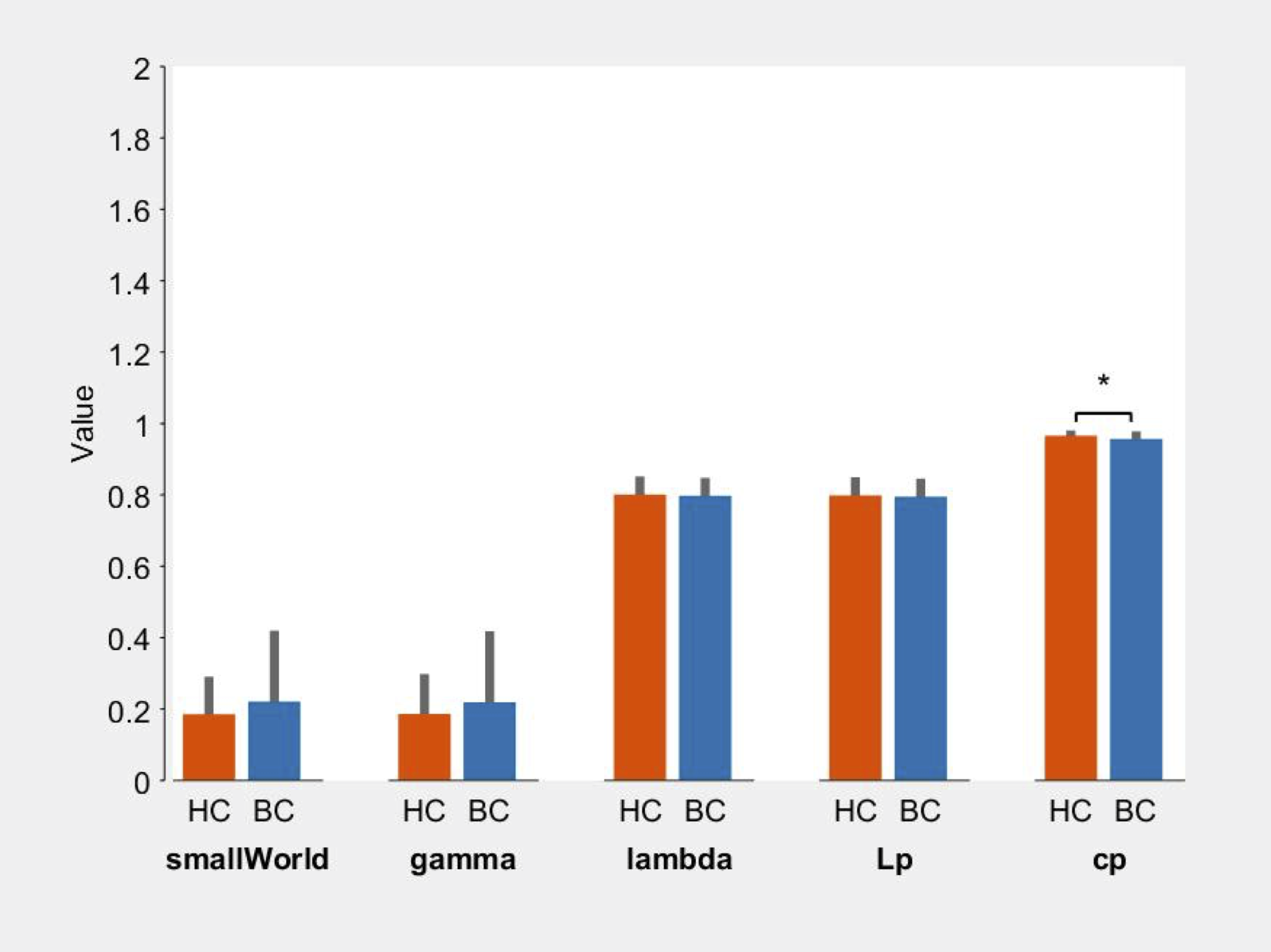
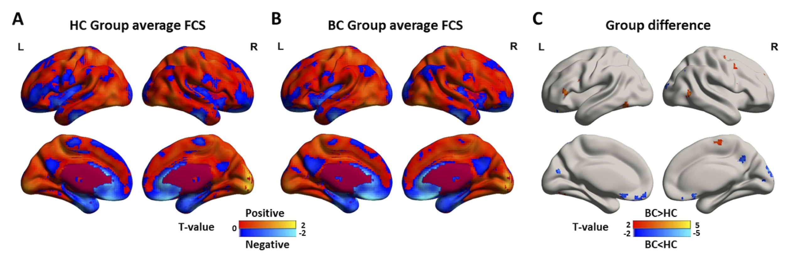
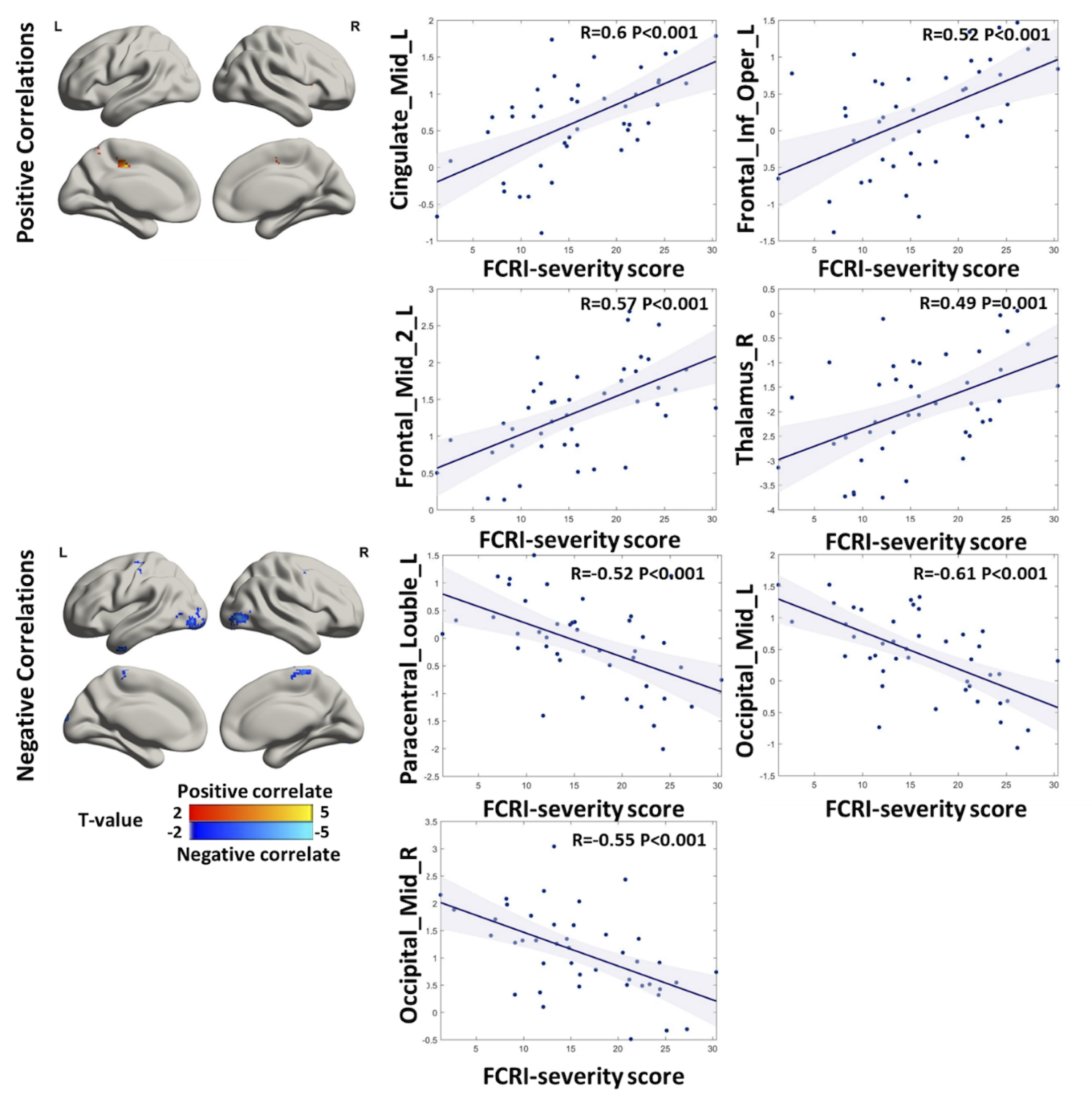
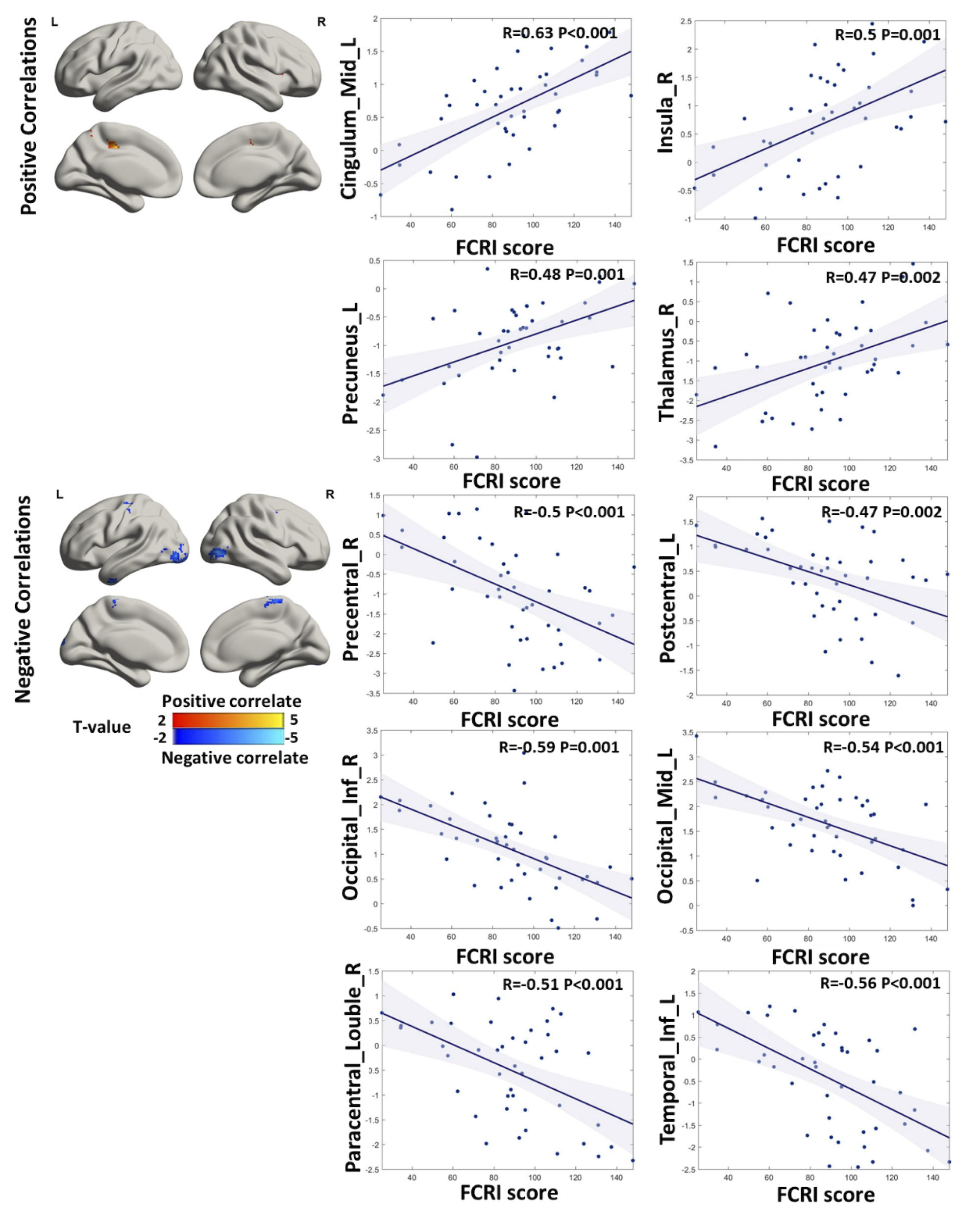
Graph theory analyses revealed a decreased clustering coefficient in breast cancer patients compared to healthy control subjects (Figure 1, P=0.04), but no global network metrics were associated with FCR. Breast cancer patients showed lower FCS regions in right inferior frontal orbital, right superior frontal gyrus, right cuneus and precuneus, right angular, left inferior parietal, left rectus and left calcarine, many of these are hub regions of DMN (Figure 2). Breast cancer patients showed higher FCS in both high-order function (frontoparietal, default mode and dorsal attention systems) and primary somatomotor networks (Figure 2). The FCRI and FCRI-severity related FCS clusters in breast cancer patients had similar spatial distributions (Figure 3, Figure 4). Among the hyperconnected regions in breast cancer, the left inferior frontal operculum (IFO) showed a significant positive correlation with FCRI-severity.Conclusions and discussion
Our study indicated that breast cancer patients have a more random-like functional network. FCS in some specific brain regions of breast cancer patients such as left IFO was associated with FCR severity. The IFO has been implicated in various aspects of emotion processing. A meta-analysis study showed that the IFO and the anterior insula were activated by both pain-related and non-pain related fear [11]. Moreover, panic disorder patients showed prolonged fear activation in the left IFO and right insula during fear extinction recall compared to healthy controls [12]. These findings provided insights into neural mechanism of FCR in breast cancer patients on brain connectome level.Acknowledgements
No acknowledgement found.References
[1] Sung H, Ferlay J, Siegel RL, Laversanne M, Soerjomataram I, Jemal A, et al. Global Cancer Statistics 2020: GLOBOCAN Estimates of Incidence and Mortality Worldwide for 36 Cancers in 185 Countries. CA Cancer J Clin. 2021;71:209-49.
[2] Lebel S, Ozakinci G, Humphris G, Thewes B, Prins J, Dinkel A, et al. Current state and future prospects of research on fear of cancer recurrence. Psychooncology. 2017;26:424-7.
[3] Vickberg SM. The Concerns About Recurrence Scale (CARS): a systematic measure of women's fears about the possibility of breast cancer recurrence. Ann Behav Med. 2003;25:16-24.
[4] Simard S, Thewes B, Humphris G, Dixon M, Hayden C, Mireskandari S, et al. Fear of cancer recurrence in adult cancer survivors: a systematic review of quantitative studies. J Cancer Surviv. 2013;7:300-22.
[5] Bruno J, Hosseini SM, Kesler S. Altered resting state functional brain network topology in chemotherapy-treated breast cancer survivors. Neurobiol Dis. 2012;48:329-38.
[6] Feng Y, Wang YF, Zheng LJ, Shi Z, Huang W, Zhang LJ. Network-level functional connectivity alterations in chemotherapy treated breast cancer patients: a longitudinal resting state functional MRI study. Cancer Imaging. 2020;20:73.
[7] Zhou J, Feng P, Lu X, Han X, Yang Y, Song J, et al. Do Future Limitation Perspective in Cancer Patients Predict Fear of Cancer Recurrence, Mental Distress, and the Ventromedial Prefrontal Cortex Activity? Front Psychol. 2018;9:420.
[8] Peng L, Hu X, Xu C, Xu Y, Lai H, Yang Y, et al. Altered regional homogeneity and homotopic connectivity in Chinese breast cancer survivors with fear of cancer recurrence: A resting-state fMRI study. J Psychosom Res. 2023;173:111454.
[9] Simard S, Savard J. Fear of Cancer Recurrence Inventory: development and initial validation of a multidimensional measure of fear of cancer recurrence. Support Care Cancer. 2009;17:241-51.
[10] Simard S, Savard J. Screening and comorbidity of clinical levels of fear of cancer recurrence. J Cancer Surviv. 2015;9:481-91.
[11] Biggs EE, Timmers I, Meulders A, Vlaeyen JWS, Goebel R, Kaas AL. The neural correlates of pain-related fear: A meta-analysis comparing fear conditioning studies using painful and non-painful stimuli. Neurosci Biobehav Rev. 2020;119:52-65.
[12] Schwarzmeier H, Kleint NI, Wittchen HU, Strohle A, Hamm AO, Lueken U. Characterizing the nature of emotional-associative learning deficits in panic disorder: An fMRI study on fear conditioning, extinction training and recall. Eur Neuropsychopharmacol. 2019;29:306-18
Figures