4394
Cerebral neurovascular coupling dysfunction is associated with cognitive decline in non-dialysis patients with chronic kidney disease1Department of Radiology, Beijing Friendship Hospital, Capital Medical University, Beijing, China, 2GE HealthCare, MR Research, Beijing, China, Beijing, China
Synopsis
Keywords: fMRI Analysis, Quantitative Susceptibility mapping, Non-dialysis patients with chronic kidney disease, cognitive decline, neurovascular coupling, arterial spin labeling, functional magnetic resonance imaging
Motivation: The relationship between neurovascular coupling (NVC) dysfunction and cognitive decline in non-dialyzed patients with chronic kidney disease is unclear.
Goal(s): Our goal was to explore the pattern of NVC changes in non-dialysis patients with chronic kidney disease and its relationship with cognitive decline.
Approach: Analyzing neurovascular coupling patterns at the whole-brain and regional levels based on multimodal coupled neuroimaging.
Results: Neurovascular coupling dysfunction associated with cognitive decline in non-dialysis patients with chronic kidney disease.
Impact: Neurovascular coupling may be a potential mechanism for cognitive decline in non-dialyzed patients with chronic kidney disease.
1. Introduction and Purpose
Chronic kidney disease (CKD), defined as the presence of impaired renal function (estimated glomerular filtration rate(eGFR) <60 mL/min/1.73m2) or proteinuria (urine albumin-to-creatinine ratio < 30 mg/g), has become a globally recognized public health problem1. CKD patients experience cognitive decline early in life, which is particularly severe in patients with CKD stage 5. Previous studies have demonstrated that non-dialyzed patients with CKD have dysfunction in neural activity, cerebral blood flow (CBF) and sensitivity associated with cognitive decline2, 3. However, the presence of NVC dysfunction in non-dialyzed patients with CKD and its relationship with cognitive function remains unclear. This study aims to investigate the NVC changes in non-dialyzed patients with CKD. This study also aims at correlating NVC parameters with montreal cognitive assessment (MoCA) scores with a view to finding potential biological markers.2. Materials and methods
Patients: The study was approved by the Ethics Committee of Beijing Friendship Hospital of Capital Medical University and implemented in accordance with the Declaration of Helsinki. A total of 43 patients with stage 1–4 chronic kidney disease (CKD1-4) and 69 patients with non-dialysis-dependent stage 5 chronic kidney disease (CKD5 ND) were recruited. In addition, 44 sex-, age-, and education-matched healthy controls (HCs) were aslo enrolled in the study. Imaging: Resting-state functional magnetic resonance imaging (fMRI) data, high-resolution T1-weighted imaging, perfusion imaging and quantitative susceptibility mapping (QSM) data were obtained sequentially using a 3.0T magnetic resonance scanner (Discovery MR750w, General Electric, USA) with an eight-channel phased array coil. Data processing: The rs-fMRI, arterial spin labeling (ASL) and QSM data were processed on MATLAB 2018b (https://www.mathworks.com/) platform using DPABI V6.0 (http://rfmri.org/dpabi) software package, spm12, and STISuite V3.0, respectively. The amplitude of low-frequency fluctuation (ALFF), regional homogeneity (ReHo), cerebral blood flow (CBF) values and sensibility values were obtained based on the above methods and voxel-based comparisons were made between the three groups. Finally the evaluation of four NVC coefficients (ALFF-CBF, ReHO-CBF, susceptibility -ALFF value, and susceptibility- ReHo value) was carried out using the written code, and inter-group comparisons were performed using spss 25.0. Multiple comparisons were corrected using Bonferroni's method (P < 0.05/116 = 0.0004) .3. Results
ALLF and ReHo in the CKD5 ND and CKD1-4 were dominated by decreases in the default mode network region (DMN) compared with HCs (Figure 3a-b);CBF in the putamen(PUT) was elevated and CBF in the middle temporal gyrus was decreased in the CKD5 ND and CKD1-4 groups compared with HC; CBF in the left hippocampus (HIP.L) and the left superior occipital gyrus (SOG.L) was elevated in the patients in the CKD5 ND group compared with the CKD1-4 group (Figure 3c); and CKD5ND and CKD1-4 groups had higher sensitivity values in the bilateral caudate nucleus (CAU), bilateral thalamus (THA) and bilateral PUT, and lower sensitivity values in the bilateral HIP (Figure 3d). At the whole-brain level, the coupling coefficients (CBF-ALFF, CBF-ReHo, and sensitivity-ALFF) were lower in the CKD1-4 and CKD5 ND groups than in the HCs, which were positively correlated with the MoCA scores. At the ROI level, the CBF-ALFF coupling and CBF-ReHo coupling in the HIP and basal ganglia regions were lower in the CKD1-4 and CKD5 ND groups than in the HCs (Figure 4). Most importantly, sensitivity-ALFF coupling of the THA.L were positively correlated with MoCA scores (r = 0.317, P = 0.045) in the CKD1-4 group (Figure5).4. Discussion
The CKD1-4 and CKD5 ND groups exhibited reduced NVC at the whole-brain level, and a significant positive correlation between NVC and MoCA scores was observed in CKD1-4 group. This suggests that a mismatch between oxygen demand and blood supply and extensive neurovascular unit dysfunction is prevalent in non-dialysis patients with CKD4, which may be a potential mechanism for the development of cognitive deficits in non-dialysis patients with CKD. At the whole-brain level, the susceptibility-ALFF of the CKD1-4 group showed a significant positive correlation with MoCA, indicating that the higher the susceptibility-ALFF , the better the cognitive function. This suggests that QSM may be as important as CBF in assessing cerebrovascular status in CKD patients.5. Conclusion
Non-dialysis CKD patients exhibit abnormal NVC and these changes are associated with cognitive decline. This suggests that NVC may be a potential mechanism for cognitive decline in non-dialyzed patients with CKD, in which the susceptibility-ALFF coupling may serve as a potential new biomarker for early assessment of cognitive decline in non-dialyzed patients with CKD.Acknowledgements
This work was supported by the National Natural Science Foundation of China (No. 82202099), the Beijing Municipal Administration of Hospitals Clinical Medicine Development of Special Funding Support (No. ZYLX201824 and ZYLX202101), the Beijing Municipal Administration of Hospital’s Mission Plan (No. SML20150101), the Beijing Scholars Program (No. [2015] 160), the Beijing Friendship Hospital, Capital Medical University (seed project No. YYZZ202129), and the Training Fund for Open Projects at Clinical Institutes and Departments of Capital Medical University (No. CCMU2022ZKYXY011).
References
1. Wang, L.; Xu, X.; Zhang, M.; Hu, C.; Zhang, X.; Li, C.; Nie, S.; Huang, Z.; Zhao, Z.; Hou, F. F.; Zhou, M., Prevalence of Chronic Kidney Disease in China: Results From the Sixth China Chronic Disease and Risk Factor Surveillance. JAMA internal medicine 2023, 183 (4), 298-310.
2. Wang, H.; Liu, X.; Song, L.; Yang, W.; Li, M.; Chen, Q.; Lv, H.; Zhao, P.; Yang, Z.; Liu, W.; Wang, Z. C., Dysfunctional Coupling of Cerebral Blood Flow and Susceptibility Value in the Bilateral Hippocampus is Associated with Cognitive Decline in Nondialysis Patients with CKD. Journal of the American Society of Nephrology : JASN 2023.
3. Chen, H. J.; Qi, R.; Kong, X.; Wen, J.; Liang, X.; Zhang, Z.; Li, X.; Lu, G. M.; Zhang, L. J., The impact of hemodialysis on cognitive dysfunction in patients with end-stage renal disease: a resting-state functional MRI study. Metabolic brain disease 2015, 30 (5), 1247-56.
4. Hu, B.; Yan, L. F.; Sun, Q.; Yu, Y.; Zhang, J.; Dai, Y. J.; Yang, Y.; Hu, Y. C.; Nan, H. Y.; Zhang, X.; Heng, C. N.; Hou, J. F.; Liu, Q. Q.; Shao, C. H.; Li, F.; Zhou, K. X.; Guo, H.; Cui, G. B.; Wang, W., Disturbed neurovascular coupling in type 2 diabetes mellitus patients: Evidence from a comprehensive fMRI analysis. NeuroImage. Clinical 2019, 22, 101802.Figures
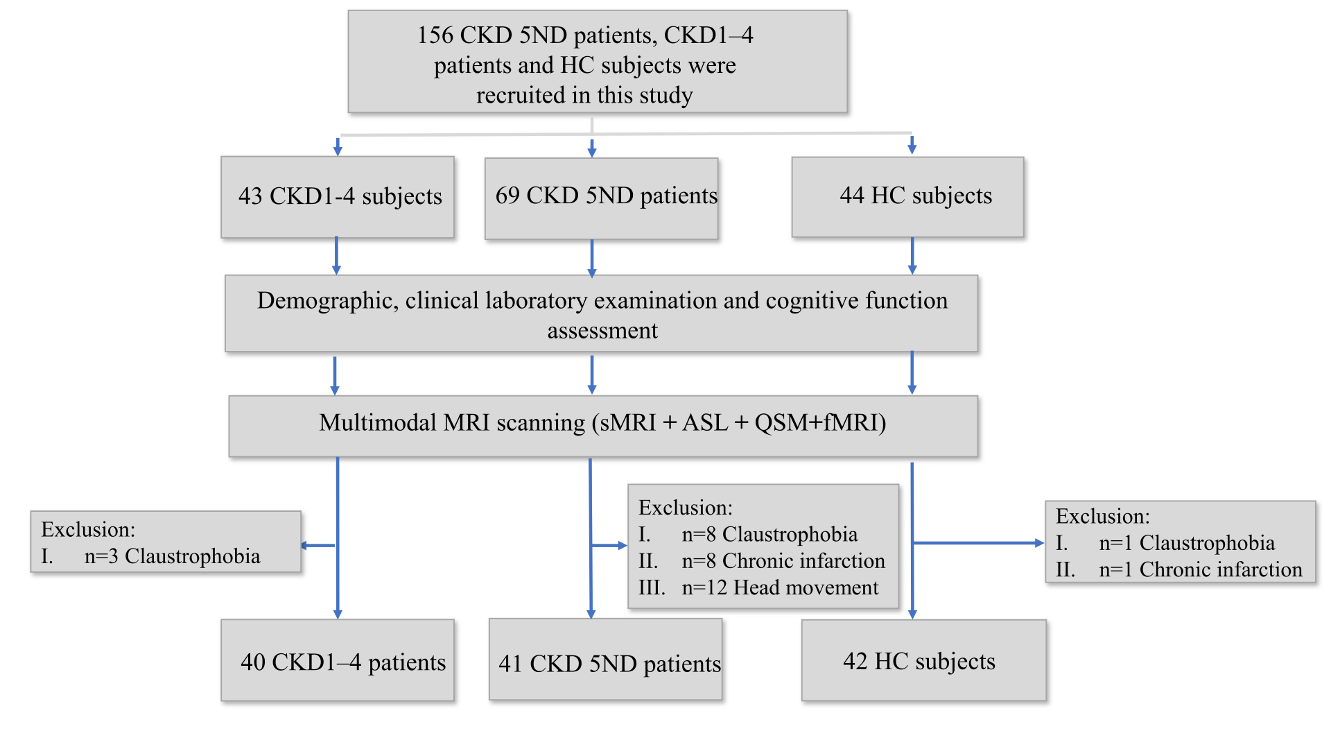
Fig. 1. Summary of patient recruitment and exclusions. CKD, chronic kidney disease; CKD 5ND, patients with non-dialysis-dependent stage 5 chronic kidney disease; CKD1–4, patients with stage 1–4 chronic kidney disease; HC, healthy control; sMRI, structural MRI; ASL, arterial spin labeling; QSM, quantitative susceptibility mapping; fMRI, functional magnetic resonance imaging.
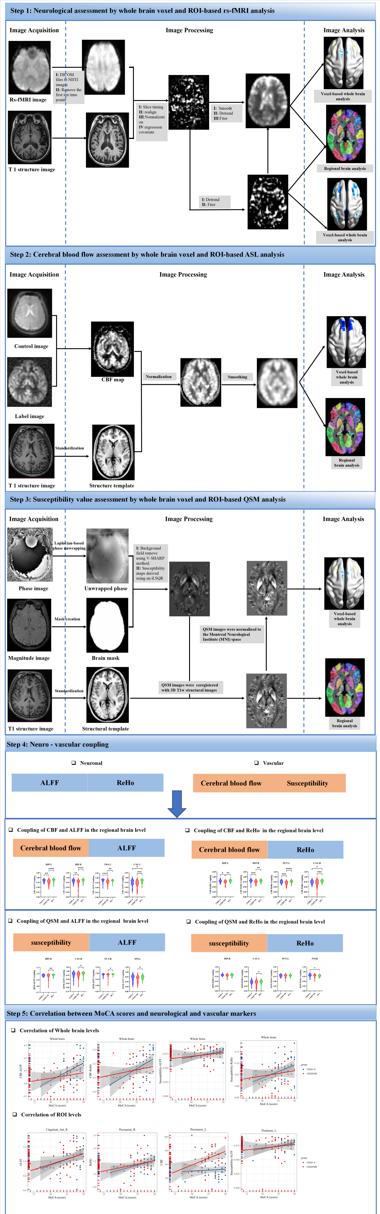
Figure 2. Flow chart of the experiment. CBF, cerebral blood flow; QSM, quantitative susceptibility mapping; ROI, region of interest; ALFF, amplitude of low frequency fluctuation; ReHo, regional homogeneity; rs-fMRI, resting-state functional magnetic resonance imaging.
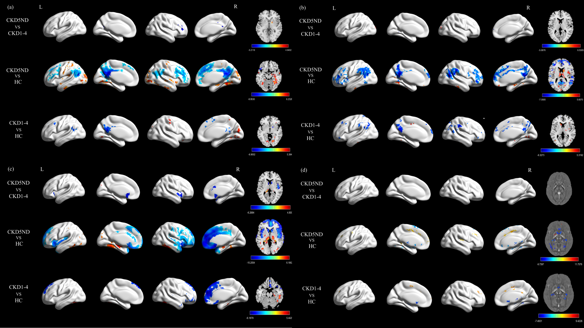
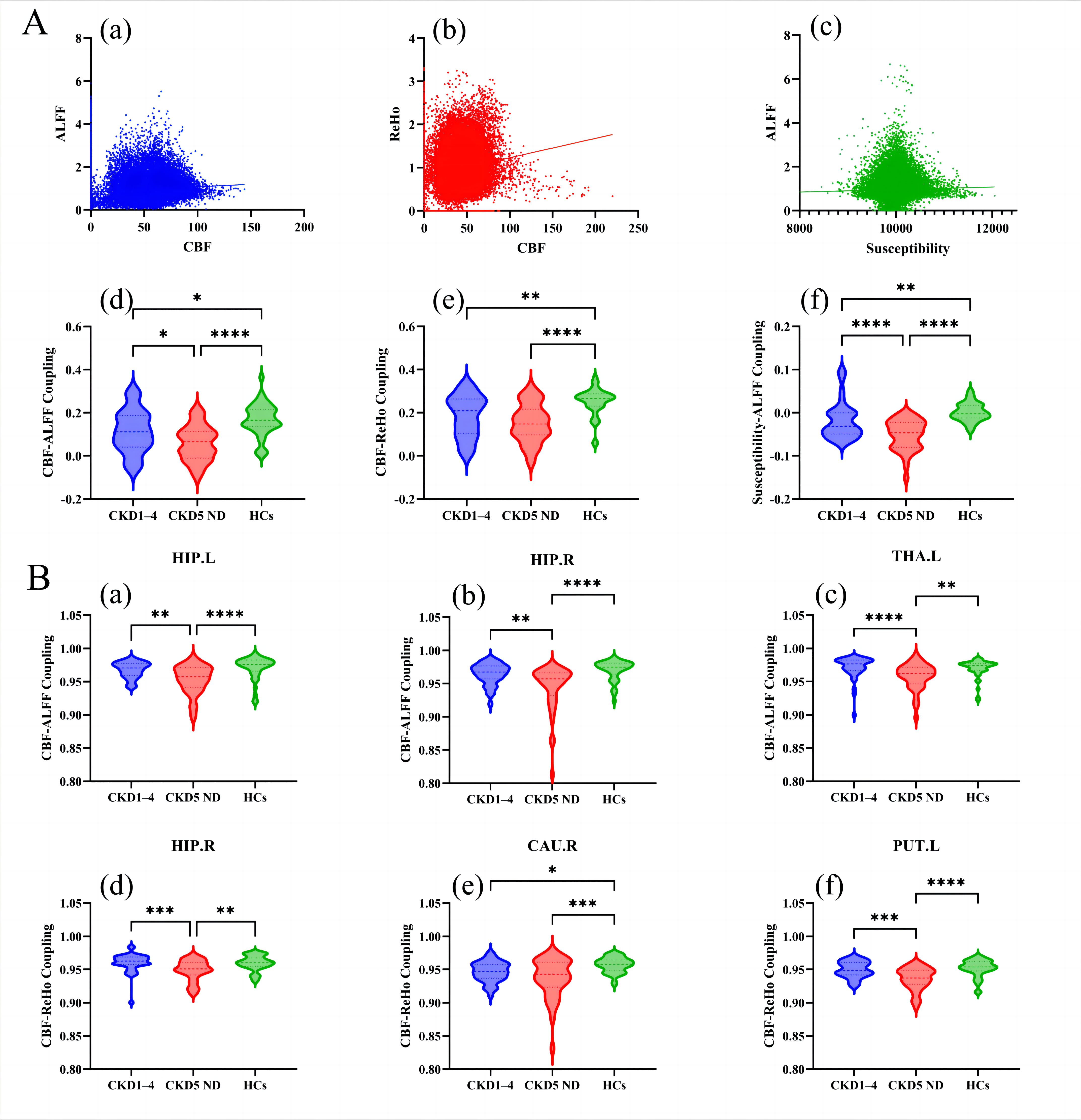
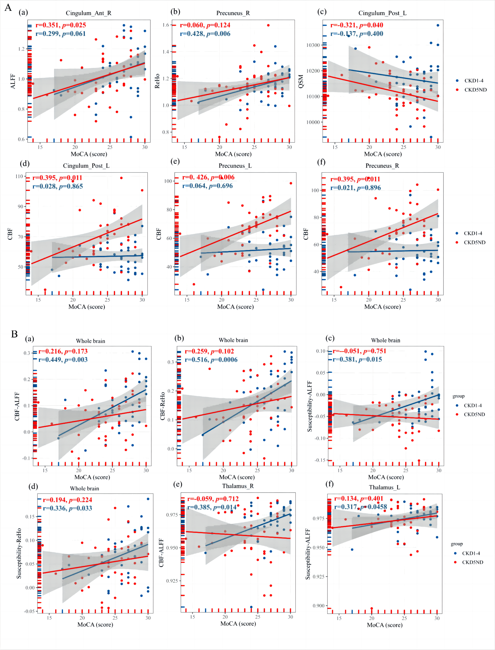
Fig. 5. A Scatter plot showing the correlation between roi-based neurovascular indicators and MoCA scores in CKD 1-4 patients, CKD 5ND patients. B Scatter plots of whole-brain and ROI-based correlations of neurovascular coupling metrics with MoCA scores in patients with CKD 1 - 4 and CKD 5ND. ROI, region of interest; Cingulum_Ant_R, right anterior cingulate and paracingulate gyri; Cingulum_Post_L, left posterior cingulate gyrus; R, right; L, left.