4393
Abnormal Resting-state Functional Connectivity and cerebral blood flow in Overactive Bladder Syndrome1Computer Science, State University of New York at Binghamton, Binghamton, NY, United States, 2New York State Psychiatric Institute, New York, NY, United States, 3Urology, State University of New York at Stony Brook, Stony Brook, NY, United States, 4Radiology, State University of New York at Stony Brook, Stony Brook, NY, United States
Synopsis
Keywords: Functional Connectivity, Perfusion, fMRI (resting state)
Motivation: Overactive Bladder (OAB) significantly impacts patients' quality of life. However, the brain-bladder control mechanism during the bladder filling was not known.
Goal(s): To investigate abnormality of cerebral blood flow (CBF) and functional connectivity (FC) during the bladder filling in OAB patients.
Approach: Arterial spin labeling (ASL) images were acquired with bladder filling volumes of 0, 50, 100, 200, 350 and 500mL.
Results: We observed medio-prefrontal cortex (mPFC) CBF and mPFC-PCC (posterior cingulate cortex) FC are compromised in OAB patients during bladder filling.
Impact: We offered a new perspective for the role of ASL perfusion and functional connectivity in understanding brain mechanism that controls urinary continence. ASL may be used to monitor treatment effects of OAB patients.
Introduction
Overactive bladder (OAB) is highly prevalent syndrome which affects the life quality in the social and functional domain1. Functional neuroimaging studies, such as cerebral blood flow (CBF) and task-based functional magnetic resonance imaging (fMRI) studies, have demonstrated regional abnormality of neural activities in OAB patients2, 3. However, little is known on how the brain-bladder neural control breaks down during the bladder filling and micturition process in OAB patients. In this study, we investigate the brain CBF and functional connectivity (FC) responses to different levels of bladder filling in OAB patients compared to controls.Methods
Ten healthy female participants (28±11 years old) and twelve female OAB patients (52±21 years old) were imaged on a 3T Siemens Prisma MRI with a 64-channel head/neck coil. During bladder filling, we acquired up to six sets of fMRI images with filling volumes of 0, 50, 100, 200, 350, and 500 mL using the Double echo-2D-EPI (DE-2D-EPI) sequence, which can capture ASL and BOLD images simultaneously4. We also acquired high-resolution T1 structural MPRAGE images for image registration.All DE-2D-EPI ASL image time series (BOLD images were not analyzed yet) were realigned to correct for motion and registered to the standard MNI space using MPRAGE images as intermediates with SPM12. The CBF map was quantified from the average ASL image and the reference image5. Motion parameters from realignment, white matter signals, and the linear trend were regressed out from the ASL time series. The seed region of interest (ROI)6 was chosen as the hub of the default mode network, the posterior cingulate cortex (PCC). We calculated individual PCC FC maps from denoised ASL time series by computing the Pearson correlation coefficient between the PCC seed ROI time series and individual voxels throughout the entire brain. These maps were then transformed into z-score maps using Fisher's z transformation to improve normality for group-level t-tests. Individual PCC FC maps and CBF maps were smoothed with an 8mm FWHM Gaussian kernel.
To test the potential differential responses of CBF/PCC FC to the levels of bladder filling between controls and OAB groups, we adopted a 2x2 factorial design of SPM12. The two independent variables are group (control, OAB) and bladder filling (small, large), each with two levels, while age serves as a covariate due to the significant difference (p=0.0039) between the control and OAB groups. Bladder filling volumes of 0, 50, and 100 mL were categorized as 'small filling,' while volumes of 200, 350, and 500 mL were considered 'large filling. We tested the main effects (control vs. OAB, small filling vs. large filling) and interaction effects, i.e., whether brain responses from small filling to large filling are significantly different between controls and OAB patients. The significance maps (positive and negative effects) were thresholded using a voxel-level p value of 0.005. A cluster-level p value of 0.05 was used to guard against false positives from multiple comparisons.
Results & Discussion
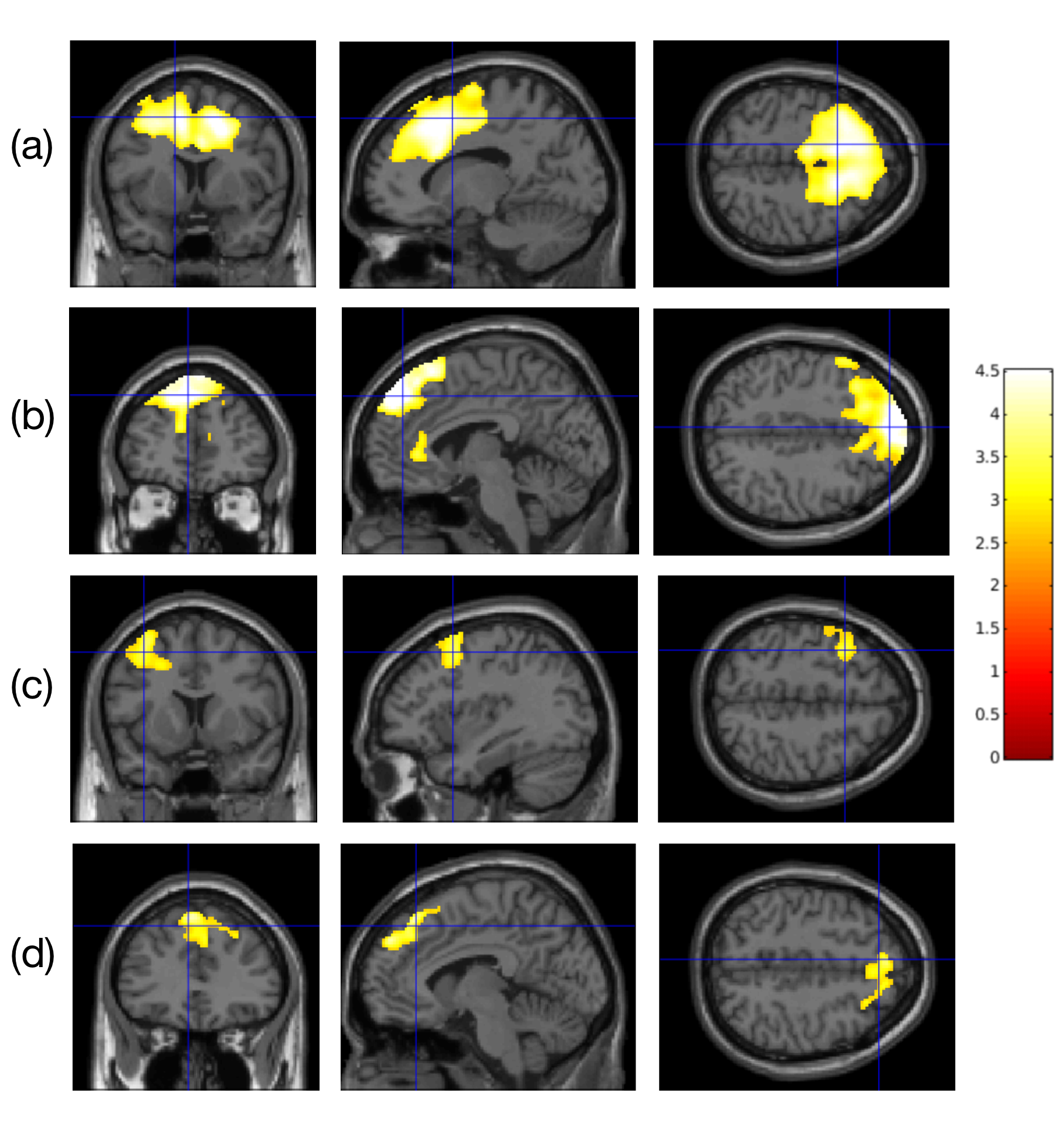
The mPFC CBF (Fig. 1a) and PCC FC with mPFC (Fig. 1b) increased from small filling to large filling in the control group. Our results agree with and extend increased regional homogeneity7 and increased FC8, 9 within the default mode network (DMN) region with a full bladder/strong desire to void/bladder distension compared to empty bladder. We observed reduced PCC FC in the angular, inferior parietal, thalamus, cerebellum, and dorsolateral prefrontal (a key hub of frontoparietal control network, FPN) regions of the OAB group compared to controls. Previous works reported the decreased FC within DMN2 and FPN10 at the resting state in OAB patients. Our results extend the decreased FC from the resting state to the bladder filling state of OAB patients.For the interaction effects, we observed that the increased mPFC CBF (Fig. 1c) and increased PCC-mPFC FC (Fig. 1d) during bladder filling in the OAB group were much weaker than that in the control group. These results are consistent with the abnormal brain-bladder control in the mPFC using fNIRS11 but with deactivation. This deactivation may be caused by different image processing methods. The reduced activation of mPFC and decreased mPFC-PCC FC (in general, reduced FC within DMN) during bladder filling in OAB patients may result in reduced inhibition of the voiding reflex, leading to typical OAB symptoms, such as urgency urinary incontinence.
Conclusion
mPFC CBF and mPFC-PCC FC are compromised in OAB patients during bladder filling. We offered a new perspective for the role of ASL perfusion and FC in understanding brain mechanism that controls urinary continence.Acknowledgements
References
1. Reynolds, W.S., J. Fowke, and R. Dmochowski, The Burden of Overactive Bladder on US Public Health. Curr Bladder Dysfunct Rep, 2016. 11(1): p. 8-13.
2. Zuo, L., et al., Abnormal Brain Functional Connectivity Strength in the Overactive Bladder Syndrome: A Resting-State fMRI Study. Urology, 2019. 131: p. 64-70.
3. Athwal, B.S., et al., Brain responses to changes in bladder volume and urge to void in healthy men.Brain, 2001. 124(Pt 2): p. 369-77.
4. Wengler, K., et al., Bladder Filling Induced Changes to Cerebral Blood Flow and BOLD Response.Proceedings of the 26th ISMRM Annual Scientific Meeting & Exhibition, 2018, Paris, France 2018. 2018.
5. Buxton, R.B., et al., A general kinetic model for quantitative perfusion imaging with arterial spin labeling. Magn Reson Med, 1998. 40: p. 383-96.
6. Vincent, J.L., et al., Evidence for a frontoparietal control system revealed by intrinsic functional connectivity. J Neurophysiol, 2008. 100(6): p. 3328-42.
7. Pang, D., Y. Gao, and L. Liao, Responses of functional brain networks to bladder control in healthy adults: a study using regional homogeneity combined with independent component analysis methods.Int Urol Nephrol, 2021. 53(5): p. 883-891.
8. Jarrahi, B., et al., Differential functional brain network connectivity during visceral interoception as revealed by independent component analysis of fMRI TIME-series. Hum Brain Mapp, 2015. 36(11): p. 4438-68.
9. Pang, D., et al., Brain functional network alterations caused by a strong desire to void in healthy adults: a graph theory analysis study. Neurourol Urodyn, 2020. 39(7): p. 1966-1976.
10. Zuo, L., et al., Intra- and inter-resting-state networks abnormalities in overactive bladder syndrome patients: an independent component analysis of resting-state fMRI. World J Urol, 2020. 38(4): p. 1027-1034.
11. Pang, D., et al., Sacral Neuromodulation Improves Abnormal Prefrontal Brain Activity in Patients with Overactive Bladder: A Possible Central Mechanism. J Urol, 2022. 207(6): p. 1256-1267.
Figures