4391
Reduced functional connectivity induced by longitudinal changes of structure and perfusion in hemodialysis patients with cognitive impairment1Department of Radiology, Beijing Friendship Hospital, Capital Medical University, Beijing, China, 2MR Research, GE Healthcare, Beijing, China
Synopsis
Keywords: Gray Matter, Neuro, cognitive impairment
Motivation: Hemodialysis (HD) leads to cognitive impairment; however, the pathophysiology of maintenance HD remains unclear.
Goal(s): This study aimed to investigate the longitudinal alterations in gray matter volume (GMV), cerebral blood flow (CBF) and functional connectivity (FC) in patients on follow-up HD compared with baseline HD.
Approach: All participants underwent high-resolution T1-weighted imaging, arterial spin labeling, and functional MR imaging to measure GMV, CBF, and FC.
Results: The right and left medial superior frontal gyrus (SFGmed.L) exhibited significantly lower GMV and CBF in patients on follow-up HD compared with baseline HD and lower FC between the SFGmed.L and left middle temporal gyrus.
Impact: Our findings provide unique insights into the pathological mechanisms of patients on maintenance HD with cognitive impairment.
Introduction
Hemodialysis (HD) is the most common kidney replacement therapy for patients with end-stage renal disease,1 and cognitive impairment is extremely common in patients on HD, with a prevalence of 80%.2 However, the pathophysiology of cognitive impairment in patients on HD remains unclear. Previous cross-sectional studies have demonstrated an association between cognitive impairment and GMV, CBF, and FC in patients on HD.3,4,5But the influence of individual factors on their results cannot be ignored in cross-sectional studies. Therefore, further longitudinal studies are required to determine the long-term consequences of HD on brain changes and whether these changes correlate with cognitive impairment. In the current study, we aimed to clarify the longitudinal variations of GMV and CBF in patients on HD, define co-changed brain regions as seed points and illustrate abnormal seed-based FC in patients on HD, and investigate the correlation between the co-changed brain regions and neuropsychological test scores.Methods
Twenty-seven patients with HD underwent brain MR imaging assessment (T1-weighted imaging, arterial spin labeling, and functional MR imaging), laboratory examinations, and Montreal Cognitive Assessment (MoCA) evaluation at baseline and after 3 years. Thirty heathy controls (HCs) were recruited in the study and underwent brain MR imaging assessment. A 3.0 Tesla magnetic resonance system (Discovery MR750W, General Electric, Milwaukee, Wisconsin, USA) with an eight-channel phased array coil was used to capture images. VBM data was processed using the MATLAB-based (R2018b; MathWorks) Statistical Parametric Mapping software (SPM12). We normalized, segmented, and smoothed T1-weighted images. SPM12 normalized, standardized, and smoothed CBF images derived from ASL difference images. To preprocess functional imaging data, we used DPARSF Advanced Edition software, which includes slice timing correction, motion correction, nuisance regression, co-registration, normalization, and filtering. We selected the left medial superior frontal gyrus (SFGmed.L) and the right medial superior frontal gyrus (SFGmed.R) as seed points. FC maps were generated using Fisher's Z transformation and smoothing.Results
No significant differences in age, TIV, or sex were observed between the two groups (P > 0.05).The follow-up HD group showed decreased GMV in both the bilateral medial superior frontal gyrus (SFGmed) and the bilateral dorsolateral superior frontal gyrus (SFGdor) compared to the baseline HD group (Fig. 1). In comparison to the baseline HD group, the follow-up HD group displayed reduced CBF in the bilateral SFGmed, bilateral SFGdor, bilateral middle temporal gyrus (MTG), and the right MFG (Fig. 2). Based on seed point SFGmed.L, the follow-up HD group had decreased FC with the left MTG (MTG.L), left parahippocampal gyrus (PHG), and left posterior cingulate gyrus (PCC) compared to the baseline HD group (Fig. 3 A). Based on seed point SFGmed.R, compared with the baseline HD group, the follow-up HD group exhibited lower FC with the MTG.L and right PHG (P < 0.05, cluster-level GRF-corrected) (Fig. 3 B).The decrease in GMV in the SFGmed.L was positively correlated with the MoCA scores in the baseline HD group (r = 0.411, P = 0.033). The reduction in FC between SFGmed.L and MTG.L was positively correlated with the MoCA scores in the follow-up HD group (r = 0.4468, P = 0.014) (Fig. 4). No significant results were found in other correlation analyses.Discussion
Alterations in CBF and GMV may have contributed to abnormal brain activity in patients on HD. In our study, compared with the baseline HD group, the follow-up HD group exhibited significantly lower FC between the SFGmed.L with the left PCG, left MTG, and left PHG, exhibiting significantly decreased FC between the SFGmed.R, bilateral MTG, and right PHG. Interestingly, these regions are mostly located within the default model network. Besides, at baseline, the decreased GMV in the SFGmed.L was positively associated with MoCA scores, indicating that cognitive impairment is likely to be the predominant structural impairment in patients on baseline HD. After 3 years, a reduction in FC between SFGmed.L and MTG.L was found to have a positive correlation with MoCA scores, suggesting a potential relationship between decreased functional connectivity and cognitive impairment.Conclusion
HD may cause changes in the GMV and CBF of patients on maintenance HD. Reduced GMV and CBF may result in decreased FC between the SFGmed.L and MTG.L, which may be associated with cognitive impairment in patients on maintenance HD. Our findings provide unique insights into the pathological mechanisms of patients on maintenance HD with cognitive impairment.Acknowledgements
This work was supported by the Beijing Municipal Administration of Hospitals Clinical Medicine Development of Special Funding Support (contract grant numbers: ZYLX201824 and ZYLX202101), Beijing Municipal Administration of Hospital’s Mission Plan (contract grant number: SML20150101), Beijing Scholars Program (contract grant number:[2015] 160), Beijing Friendship Hospital, Capital Medical University (contract grant number: seed project YYZZ202129), Training Fund for Open Projects at Clinical Institutes and Departments of Capital Medical University (CCMU2022ZKYXY011), and Natural Science Foundation of China (82202099).References
1. Romagnani P, Remuzzi G, Glassock R, et al. Chronic kidney disease. Nature Reviews Disease Primers. 2017;3(1):17088.
2. van Zwieten A, Wong G, Ruospo M, et al. Prevalence and patterns of cognitive impairment in adult hemodialysis patients: the COGNITIVE-HD study. Nephrol Dial Transplant. 2018;33(7):1197-1206.
3. Jin M, Wang L, Wang H, et al. Structural and Functional Alterations in Hemodialysis Patients: A Voxel- Based Morphometry and Functional Connectivity Study. Front Hum Neurosci. 2020;14:80.
4. Jiang XL, Wen JQ, Zhang LJ, et al. Cerebral blood flow changes in hemodialysis and peritoneal dialysis patients: an arterial-spin labeling MR imaging. Metab Brain Dis. 2016;31(4):929-936.
5. Zheng J, Sun Q, Wu X, et al. Brain Micro-Structural and Functional Alterations for Cognitive Function Prediction in the End-Stage Renal Disease Patients Undergoing Maintenance Hemodialysis. Acad Radiol. 2023;30(6):1047-1055.
Figures
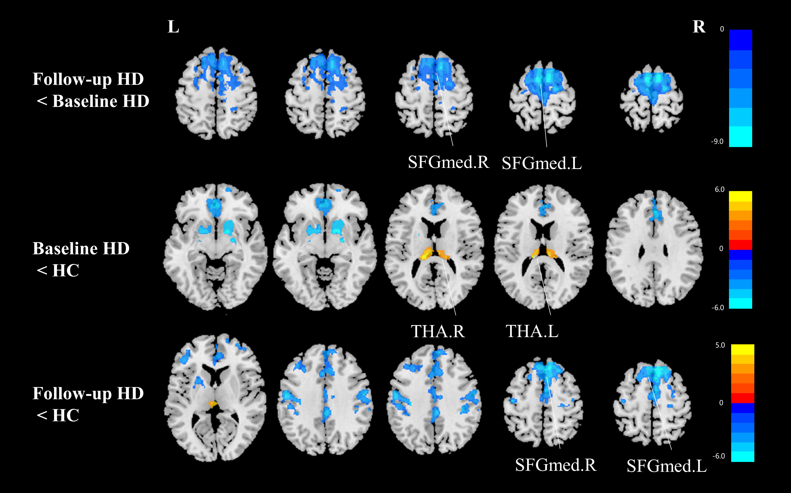
SFGmed.L, left superior frontal gyrus, medial; SFGmed.R, right superior frontal gyrus, medial; THA.L, left thalamus; THA.R, right thalamus; HD, hemodialysis; HC, healthy control; GMV, gray matter volume; FDR, false discovery rate.
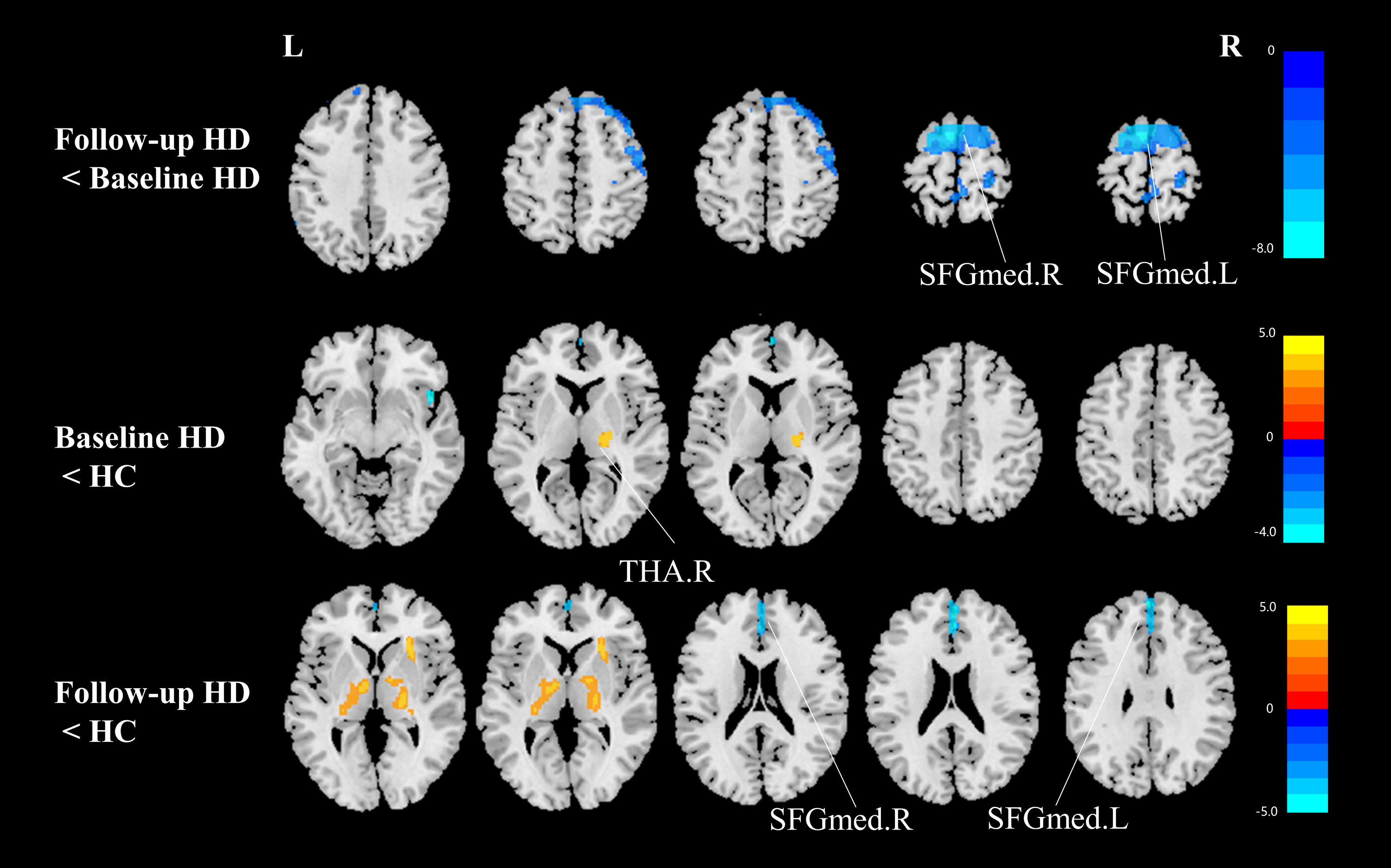
SFGmed.L, left superior frontal gyrus, medial; SFGmed.R, right superior frontal gyrus, medial; THA.R, right thalamus; HD, hemodialysis; HC, healthy control; CBF, cerebral blood flow; FDR, false discovery rate.
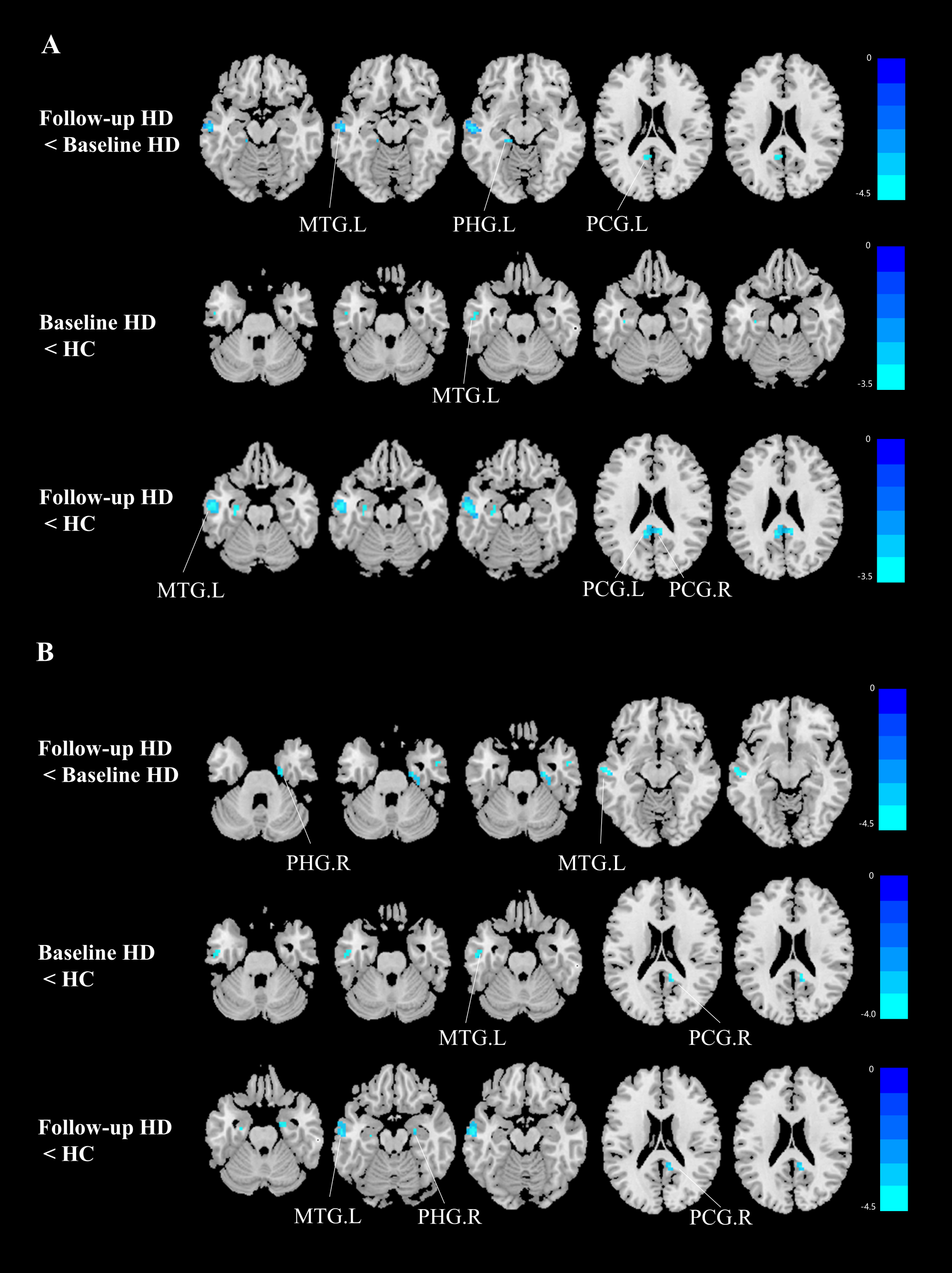
Fig 3 A. Difference among groups in FC between SFGmed.L and the rest of the whole brain. B. Difference among groups in FC between SFGmed.R and the rest of the whole brain.
MTG.L, left middle temporal gyrus; PHG.L, left parahippocampal gyrus; PHG.R, right parahippocampal gyrus; PCG.L, left posterior cingulate gyrus; PCG.R, right posterior cingulate gyrus; SFGmed.L, left superior frontal gyrus, medial; SFGmed.R, right superior frontal gyrus, medial; HD, hemodialysis; HC, healthy control; FC, functional connectivity; GRF, Gaussian random field.
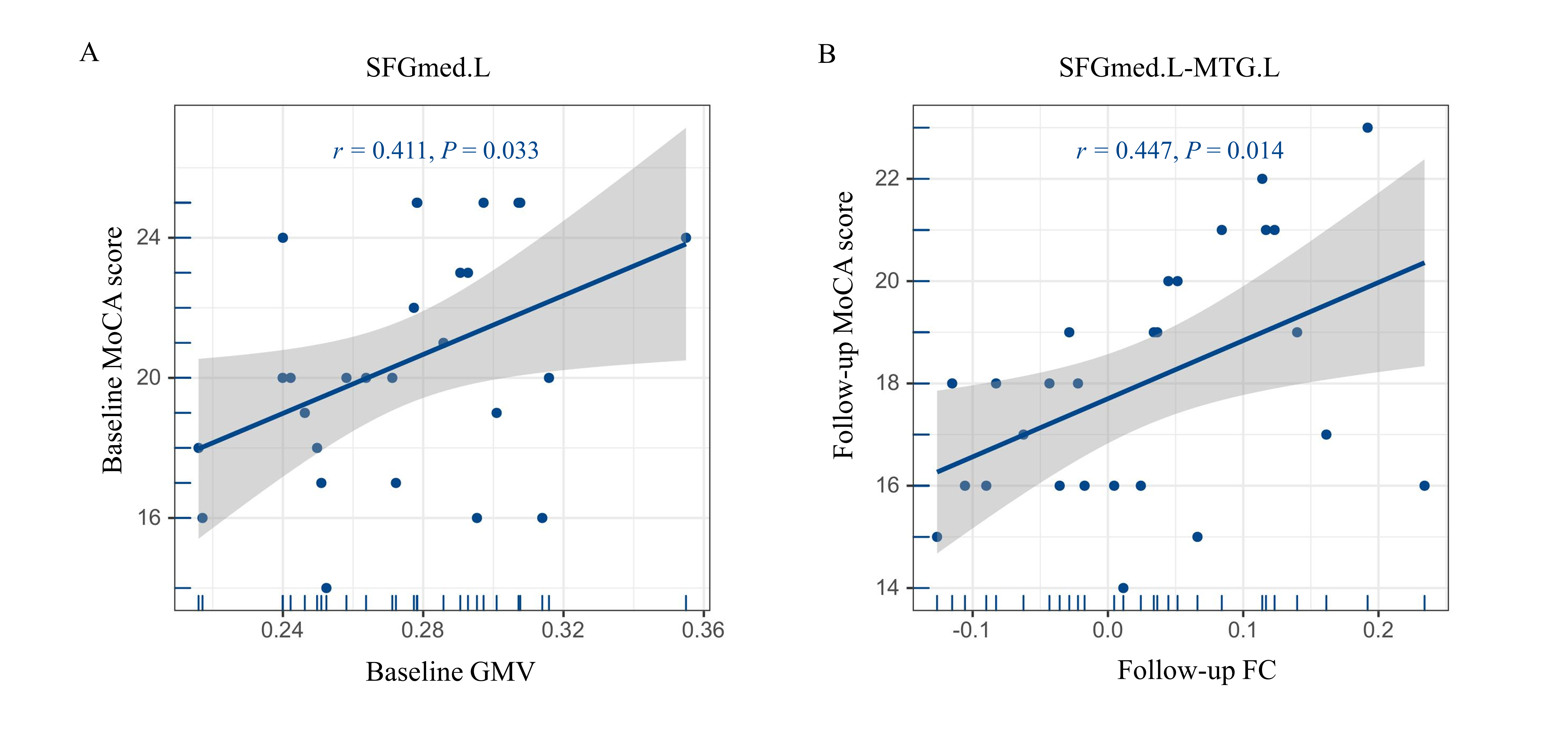
Figure 4 A Correlation between MoCA scores and decreased GMV in the SFGmed.L in the baseline HD group (r = 0.411, P = 0.033). B Correlation between MoCA scores and decreased FC in the SFGmed.L-MTG.L in the follow-up HD group (r = 0.447, P = 0.014).
GMV, gray matter volume; FC, functional connectivity; MoCA, Montreal Cognitive Assessment; HD, hemodialysis; SFGmed.L, left superior frontal gyrus, medial; MTG.L, left middle temporal gyrus.