4390
Abnormal dynamics function network during pain-free period: a resting-state co-activation pattern analysis in primary dysmenorrhea1School of Future Technology, Xi'an Jiaotong University, Xi'an, China, 2Xi'an Jiaotong University Bachelor of Dental Medicine, Xi'an, China, 3The Key Laboratory of Biomedical Information Engineering of Ministry of Education, Institute of Health and Rehabilitation Science, School of Life Science and Technology, Xi'an Jiaotong University, Xi'an, China, 4Xi'an Jiaotong University Health Science Center, Xi'an, China, 5Rehabilitation Medicine Department, The First Affiliated Hospital of Xi'an Jiaotong University, Xi'an, China, 6Department of Medical Imaging, The First Affiliated Hospital of Xi’an Jiaotong University, Xi'an, China
Synopsis
Keywords: Head & Neck/ENT, Brain, Co-activation pattern
Motivation: Brain mechanisms for the pain of primary dysmenorrhea (PDM) patients pain remain unclear.
Goal(s): To investigate dynamic brain functional networks in women with PDM during pain-free periods and explore the relationship between brain functional networks and the psycho-emotional states.
Approach: Applied the CAP method to investigate the dynamic network connectivity characteristics of the brain in 59 PDM patients, as compared with 57 healthy controls.
Results: The dynamic interaction of rs-fMRI brain networks in PDM patients were abnormal. The pain of PDM patients may related to the abnormal brain dynamic interaction of brain networks.
Impact: This study provided new insights into the neural mechanisms underlying recurrent chronic pain. PDM patients exhibited atypical dynamic interactions within their brain networks during pain-free ovulation cycles, and these alterations corresponded to emotions related to pain.
Introduction
Primary dysmenorrhea (PDM) is a chronic visceral pain that affects a significant number of women, with prevalence rates of up to 90%1,2. Previous studies have explored the impact of chronic pain on brain function using different approaches. Nevertheless, it remains unclear whether changes in brain dynamics occur in PDM patients during pain-free intervals. The use of Co-activation pattern (CAP) analysis, a method for examining brain dynamics that identifies inter-regional co-activation states across time series and labels each time point as one of these states3,4, may provide a more comprehensive understanding of this investigation. Incorporating the CAP methodology can contribute to further insights into the neural mechanisms underlying PDM. The purpose of this study is to investigated the dynamic network connectivity characteristics of the brain in women with PDM during pain-free periods and their relationship with pain-related psychological emotions.Methods
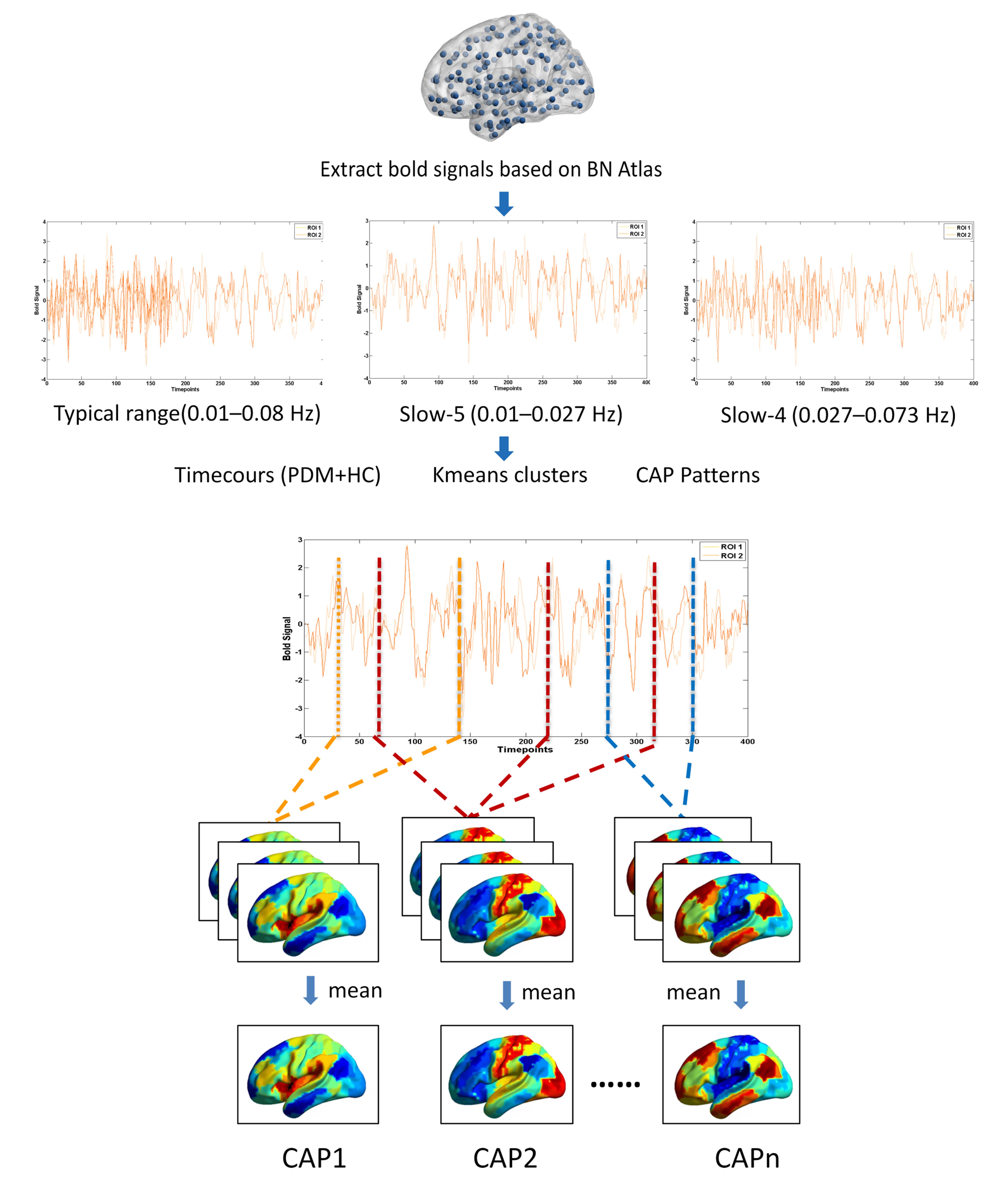
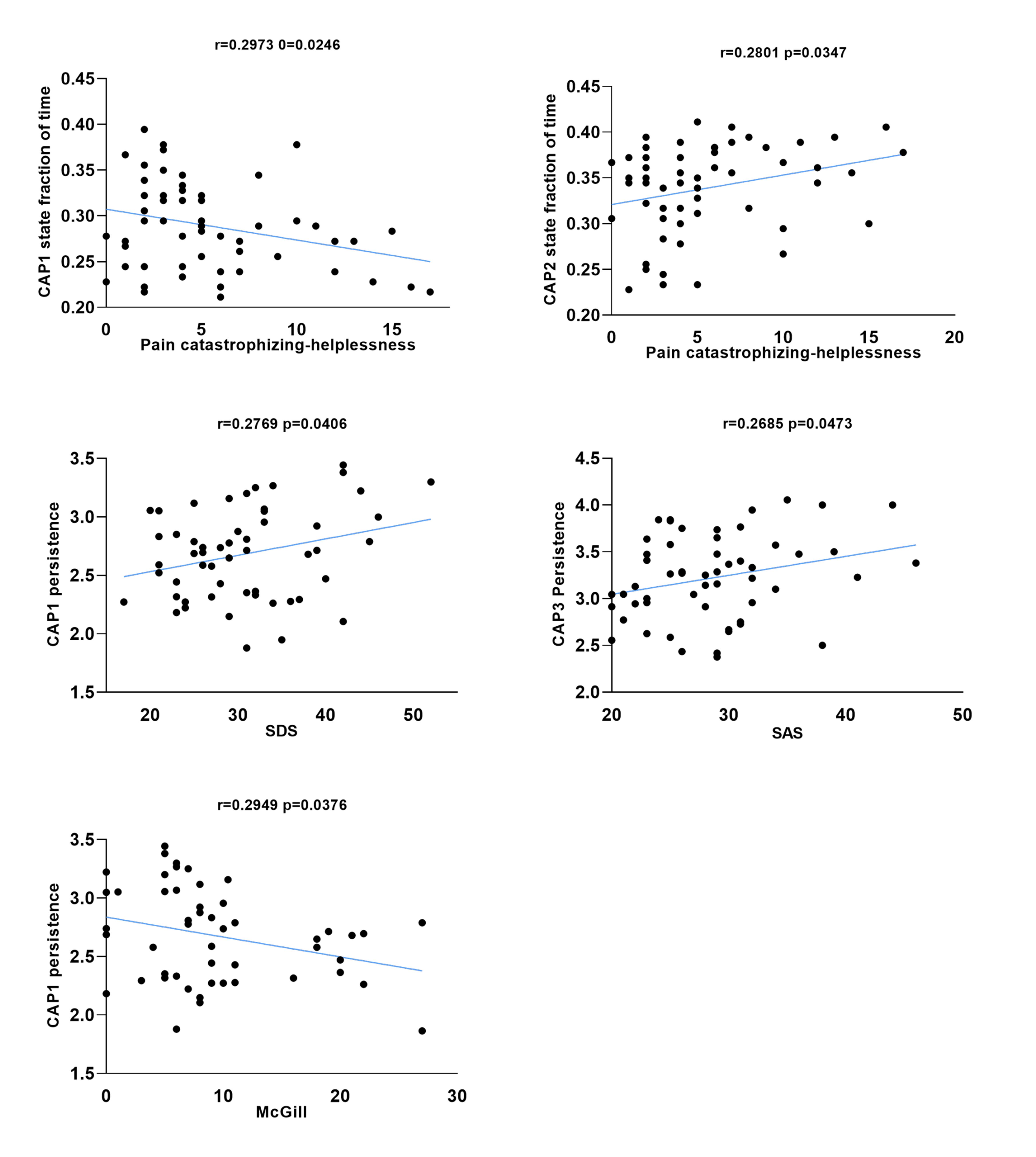
This study was approved by the institutional review board and all participants were provided with informed consent. Rs-fMRI data were acquired using a 3.0-Tesla MRI scanner (GE SIGNA HDxt, Milwaukee, WI, USA) equipped with an 8-channel phase array head coil. Rs-fMRI data was acquired using below parameters: TR/TE: 2,000/30ms, flip angle =90◦, data matrix = 64 × 64, field of view = 240 × 240 mm, and 30 contiguous slices 5 mm thick. MATLAB based SPM12 and Graph Theoretical Network Analysis Toolbox were used for preprocessing rs-fMRI data5. Bandpass filtering was applied to extract fMRI signals in the typical frequency range of 0.01–0.08 Hz, as well as sub-bands including slow-5 (0.01–0.027 Hz) and slow-4 (0.027–0.073 Hz)6. The CAPs analysis pipeline is illustrated in Figure 1. In this study, the CAPs analysis was conducted using custom scripts in MATLAB. In the case of the CAPs metrics, the Wilcoxon rank sum test analysis was conducted. The CAPs metrics were extracted to calculate Pearson’s correlation coefficient using pain-associated factors of PDM patients, the scores of PCS, SDS, and SAS were evaluated at pain-free periovulation, the MPQ were evaluated at painful periovulation.Results
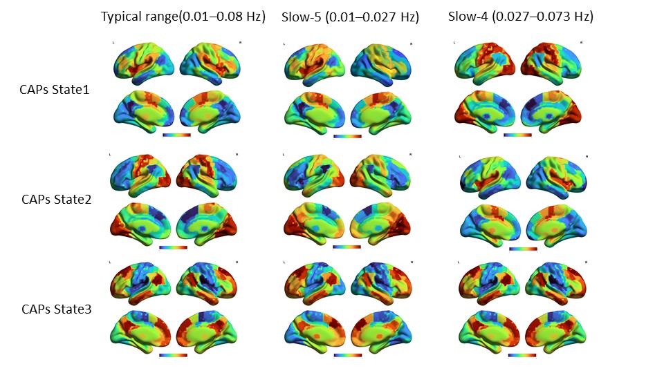

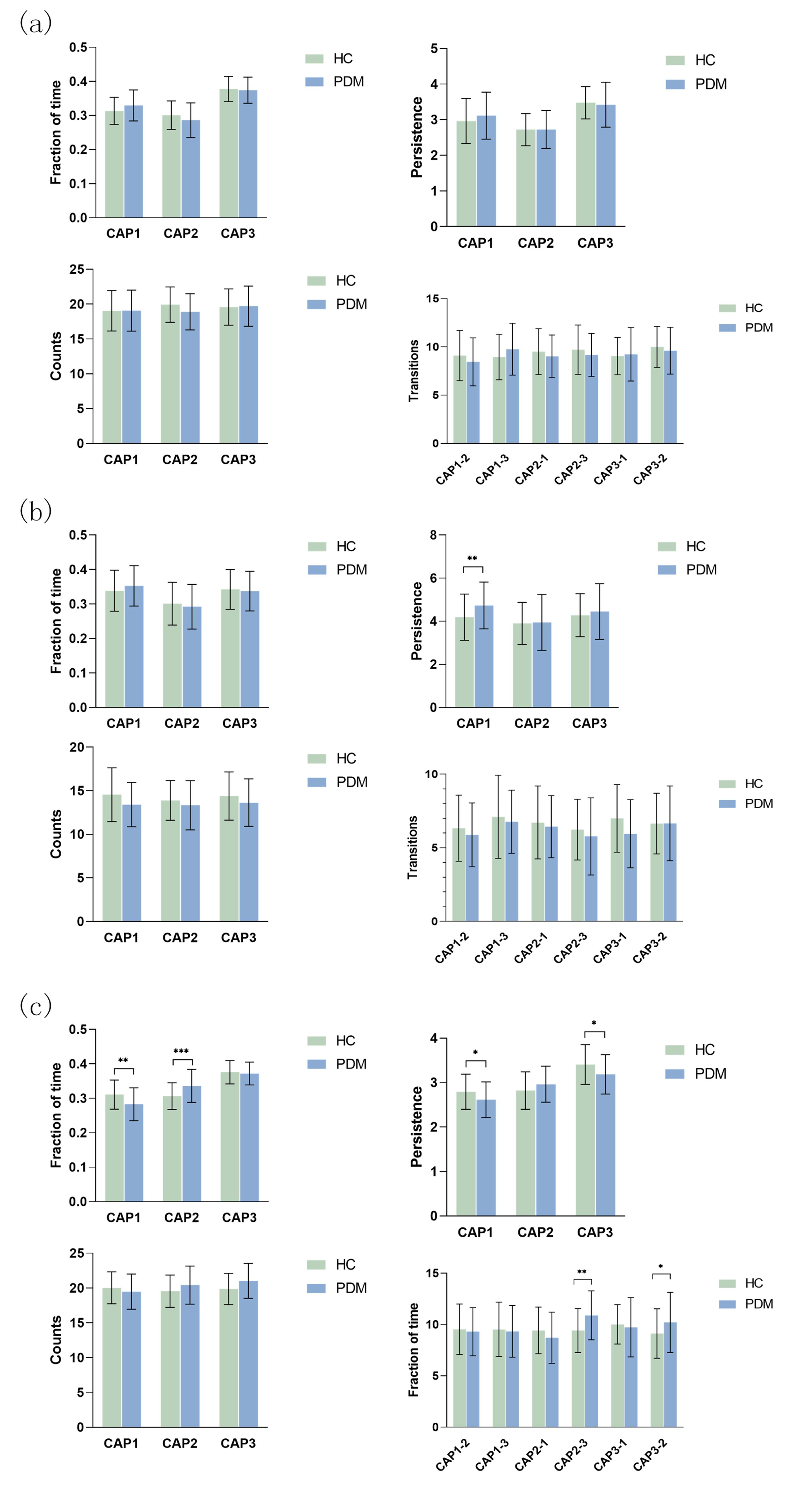
Demographic and Clinical Characteristics of Participants Fifty-seven HC and 59 participants with PDM were included in the final analyses. The groups were similar with respect to sex (see Table 1). Cluster Analysis Yields Three Recurring CAPs States at Different Frequency Bands The CAPs analysis was performed using all subjects with three recurring CAPs states were identified, as shown in Figure 2. The first column showed three CAPs topographies in the typical frequency range. State1 involves the co-activation of the insular gyrus clearly representing a SN. In State 2, co-activation of the Visual Network (VN) and Somatomotor Network (SMN) can be observed. In State 3, DMN co-activation is evident. The spatial similarity of CAPs states between typical frequency range and sub-bands (Slow-5 and Slow-4) is shown in Figure 3. CAPs with high spatial similarity between the typical frequency range and the Slow-5 band maintained the consistent order in terms of the fraction of time. The diagonal correlation coefficient of correlation matrix is the highest as shown in Figure 3(a). The highest correlation coefficients are not distributed on the diagonal of the correlation matrix, as illustrated in Figure 3(b). The results of group comparisons in the typical frequency band and Slow-5 are presented in Figure 4. The results of group comparisons in Slow-4 are shown in Figure 4(c). CAPs Topographies and Clinical Symptoms To investigate the relationship between aberrant brain dynamics and clinical symptoms, significant difference metrics of PDM patients compared to HC were regressed on clinical symptoms of PDM patients. The correlation analysis results in the Slow-4 band are shown in Figure 5.Discussion
This study provides novel insights into the abnormal brain dynamics of individuals with PDM during pain-free periovulation, utilizing the CAP methodology across different frequency bands. Our findings demonstrate the presence of frequency-specific CAPs during the resting-state and highlight disrupted brain dynamic interactions in PDM patients during pain-free periovulation. Additionally, our study identifies a correlation between menstrual pain experienced by individuals with dysmenorrhea and the dynamic functional connectivity (dFC) of their brains during non-pain periods. Notably, women with dysmenorrhea exhibit persistent abnormalities in brain function during non-painful periods, suggesting that prolonged and recurrent menstrual pain has enduring and profound impacts on their brain network connectivity. These findings contribute to a deeper understanding of the neurological mechanisms underlying PDM and its effects on brain function.Conclusion
We found that PDM patients had abnormal dynamic interactions in their brain networks during pain-free ovulation cycles. These results also provide new insights into the neural mechanisms of recurrent chronic pain.Acknowledgements
The authors thank all our study participants for their time, and effort devoted to this study.References
1 Dawood MY. Primary Dysmenorrhea: Advances in Pathogenesis and Management. Obstetrics & Gynecology 2006; 108: 428–441.
2 Treede R-D, Rief W, Barke A, Aziz Q, Bennett MI, Benoliel R et al. Chronic pain as a symptom or a disease: the IASP Classification of Chronic Pain for the International Classification of Diseases (ICD-11). Pain 2019; 160: 19–27.
3 Liu X, Duyn JH. Time-varying functional network information extracted from brief instances of spontaneous brain activity. Proc Natl Acad Sci U S A 2013; 110: 4392–4397.
4 Chen JE, Chang C, Greicius MD, Glover GH. Introducing co-activation pattern metrics to quantify spontaneous brain network dynamics. NeuroImage 2015; 111: 476–488.
5 Wang J, Wang X, Xia M, Liao X, Evans A, He Y. GRETNA: a graph theoretical network analysis toolbox for imaging connectomics. Frontiers in human neuroscience 2015; 9: 386.
6 Zuo XN, Martino A, Kelly C, Shehzad ZE, Gee DG, Klein DF et al. The oscillating brain: Complex and reliable. NeuroImage 2010; 49: 1432–1445.
Figures