4389
Fast SMWI : Optimizing Classifier with FOV reduction simulation1Seoul National University, Seoul, Korea, Republic of, 2Heuron Co.Ltd., Seoul, Korea, Republic of, 3Department of Radiology, Samsung Medical Center, Seoul, Korea, Republic of
Synopsis
Keywords: Parkinson's Disease, Parkinson's Disease
Motivation: SMWI is an advanced SWI method capable of detecting nigral hyperintensity in the substantia nigra. Due to the long scan time, a reduced FOV sequence was proposed to decrease the scan time from 4min 15sec to 2min 45sec, along with the application of denoising techniques. However, when evaluated with the original classifier, the results showed changes, reporting increased FN, which may be a problem in clinic.
Goal(s): Maintaining diagnostic results for the reduced FOV with denoised SMWI images.
Approach: Optimizing classifier to denoised FOV-simulated SMWI images.
Results: The diagnostic result of denoised FOV 64.5% SMWI images is comparable to FOV 100% SMWI images.
Impact: The diagnostic result of denoised FOV 64.5% SMWI images is comparable to FOV 100% SMWI images with the optimizing classifier based on denoised FOV-simulated SMWI images.
Introduction
Susceptibility map-weighted imaging (SMWI)1,3 is an advanced susceptibility weighted imaging (SWI) method for enhancing the visibility of nigral hyperintensity in substantia nigra (SN). Since the conventional SMWI protocol has a long scan time, Fast SMWI method was suggested7 by reducing the field of view (FOV) 100% to 66% and applying a denoising network, cutting scan time from 4 min 15 sec to 2 min 56 sec.However, the diagnostic classifier (mPDia5; Heuron Co.Ltd, Incheon, Korea) showed varied results for the denoised SMWI images with reduced FOV because the classifier was trained with FOV 100% SMWI images. To address this issue, we optimized the classifier for denoised FOV-simulated SMWI images and applied it to the real scan data of denoised FOV 64.5% SMWI images.
Methods
Gradient echo (GRE) images used for SMWI generation were acquired with two different FOV settings: FOV 100% and FOV 64.5%, with scan times of 4 min 15 sec and 2 min 45 sec, respectively1,6,7. SMWI images were generated using the SMWI Toolbox and subsequently denoised using a Coil2Coil2 denoising, which is a pre-trained neural network trained with T1, T2 and Flair images from NYU Fast MRI dataset. These denoised SMWI images were then evaluated using a classifier that was trained on FOV 100% SMWI images. To optimized the classifier for denoised FOV 64.5% SMWI images, we employed a FOV reduction simulation technique, which involved creating simulated datasets from the FOV 100% GRE images using sinc interpolation and noise addition, following the methodology of Fast SMWI7.[Acquisition]
The dataset consisted of scans from 138 subjects (79 HC; 59 PD) acquired using a 3T scanner (Ingenia CS, Philips, Best, Netherlands) at Sinchon Severance Hospital.
[Optimization]
The classifier is equipped with two thresholds: a classification threshold for comprehensive diagnosis based on the evaluation of five slices from the left and right SN region, and a significance threshold for determining the inclusion of each slice in the comprehensive assessment. We optimized the significance threshold for three different datasets: denoised FOV 100% SMWI images, FOV-simulated SMWI images and denoised FOV-simulated SMWI images. Then we optimized the classification threshold to increase the sensitivity while maintaining specificity. The receiver operating characteristic (ROC) curve for optimized classifiers is drawn by controlling the classification threshold.
Results
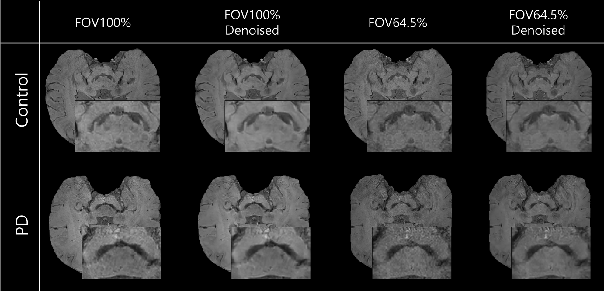
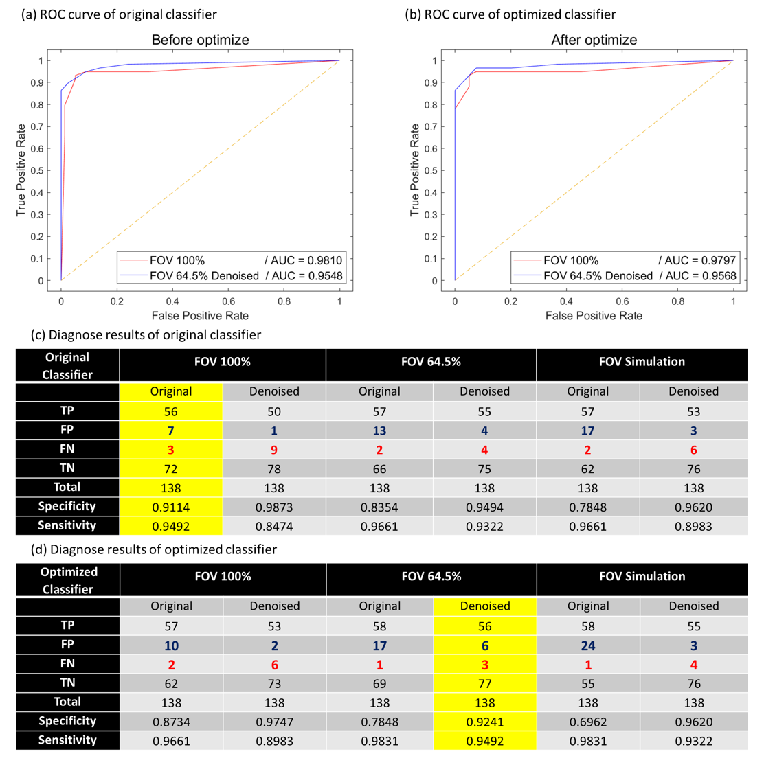
The denoised FOV 65% SMWI images exhibit comparable quality to FOV 100% SMWI images in both control and PD cases (Fig. 1). However, for the reduced FOV images, the classifier’s diagnostic results indicate a slight decrease in specificity from 0.9114 to 0.8354 and an increase in sensitivity from 0.9492 to 0.9661. On the other hand, after denoising, an increase in specificity from 0.8354 to 0.9494 with little change in sensitivity from 0.9661 to 0.9322 are observed (Fig. 2c). After optimizing the classifier for the denoised FOV Simulation SMWI images, the diagnostic results of the denoised FOV 64.5% SMWI images are comparable to those of FOV 100% SMWI images using the original classifier (Fig. 2d). The area under curve (AUC) of the ROC curve was also increased from 0.9548 to 0.9568 after optimizing the classifier. However, when optimized using the FOV-simulated SMWI images, the diagnostic results for the FOV 64.5% SMWI images show reduced sensitivity from 0.9492 to 0.8814 compared to FOV 100% SMWI images. If we optimize for the denoised FOV 100% images, the results are similar to the diagnostic results for FOV 100% using the original classifier where sensitivity is 0.9492 and specificity is 0.9114.Discussion and Conclusion
In this study, we demonstrate comparable diagnostic results between FOV 100% SMWI images and denoised FOV 64.5% SMWI images. Reduced performance in the effectiveness of diagnosis from the original classifier is observed potentially due to SNR change. These findings were previously reported in a study involving the classifier’s performance evaluation in various SNR datasets8. The classifier optimized using the denoised FOV-simulated SMWI images exhibits similar diagnostic results for the denoised FOV 64.5% SMWI images, but the one optimized using the FOV-simulated SMWI images shows a decrease in sensitivity and specificity for FOV 64.5% SMWI images. This highlights the necessity of the denoising protocol to maintain diagnostic results of the reduced FOV SMWI images comparable to the FOV 100% SMWI images.Acknowledgements
This work was supported by the National Research Foundation of Korea(NRF) grant funded by the Korea government(MSIT) (No. 2021M3E5D2A01024795), (No. NRF-2022R1A4A1030579), and Institute of New Media and Communications (INMC), SNU, and Heuron Co., Ltd.
References
[1] Nam, Yoonho, et al. "Imaging of nigrosome 1 in substantia nigra at 3T using multiecho susceptibility map‐weighted imaging (SMWI)." Journal of Magnetic Resonance Imaging 46.2 (2017): 528-536.
[2] Park, Juhyung, et al. "Coil2Coil: Self-supervised MR image denoising using phased-array coil images." arXiv preprint arXiv:2208.07552 (2022).
[3] Sung, Young Hee, et al. "Drug-induced Parkinsonism versus idiopathic Parkinson disease: utility of nigrosome 1 with 3-T imaging." Radiology 279.3 (2016): 849-858.
[4] Noh, Y., et al. "Nigrosome 1 detection at 3T MRI for the diagnosis of early-stage idiopathic Parkinson disease: assessment of diagnostic accuracy and agreement on imaging asymmetry and clinical laterality." American Journal of Neuroradiology 36.11 (2015): 2010-2016.
[5] Shin, Dong Hoon, et al. "Automated assessment of the substantia nigra on susceptibility map-weighted imaging using deep convolutional neural networks for diagnosis of Idiopathic Parkinson's disease." Parkinsonism & Related Disorders 85 (2021): 84-90.
[6] Kim, Eung Yeop, Young Hee Sung, and Jongho Lee. "Nigrosome 1 imaging: technical considerations and clinical applications." The British Journal of Radiology 92.1101 (2019): 20180842.
[7] Youn, Jonghyo, et al. “Fast SMWI via denoising for nigral hyperintensity detection in Parkinson’s disease”. Proceedings of ISMRM&ISMRT ANNUAL MEETING 2023 .
[8] Momeny, Mohammad, et al. "A noise robust convolutional neural network for image classification." Results in Engineering 10 (2021): 100225.
Figures