4387
Association of Asian Parkinson's Disease risk variant rs9638616 with brain structural and functional changes1Research, National Neuroscience Institute; Duke-NUS Medical School, Singapore, Singapore, Singapore, 2Research, National Neuroscience Institute, Singapore, Singapore
Synopsis
Keywords: Parkinson's Disease, Diffusion Tensor Imaging
Motivation: The genetic variant rs9638616, is associated with Parkinson’s Disease (PD) risk in Asian populations.
Goal(s): To provide insight into the neural correlates of rs9638616 in Asian PD, to inform risk models and idiopathic PD aetiology.
Approach: Using imaging and genotyping data from 116 early-PD patients and 57 controls of Chinese ethnicity, we performed voxelwise analyses to assess rs9638616 T-allele association with brain microstructure, morphology and function.
Results: Our results suggest that rs9638616 may confer PD risk in Asian cohorts via lower white matter fractional anisotropy and reduced supplementary motor area functional connectivity.
Impact: In an imaging-genetics analysis, the Asian PD risk variant rs9638616 was associated with altered brain structure/function. This rationalises rs9638616’s role in PD risk, and may be useful in improving PD stratification and risk modeling.
Introduction
The majority of Parkinson’s disease (PD) cases are idiopathic, involving multiple small genetic-risk contributions. An Asian PD GWAS variant, rs9638616, was recently reported [1], and maps to the WBSCR17/GALNT17 gene, which influences synaptic transmission and neurite development. We need to understand the associated brain changes in a race-specific context, to inform PD risk stratification and aetiology [2]. Neuronal changes resulting from rs9638616 may manifest in brain imaging. We assess the relationship between rs9638616 and PD through an imaging-genetics approach. We hypothesised that rs9638616-T would be associated with imaging-derived measures of brain structure and function.Methods
We analysed 3-Tesla MRI and genotyping data from 116 early PD patients (aged 66.8±9.0 years; 39% female; disease duration 1.25±0.71 years) and 57 controls (aged 68.7±7.4 years; 54% female) of Chinese ethnicity. MRI included T1-weighted (TE=0.002s, TR=1.9s, TI=900ms, FA=9°, in-plane voxel size=1x1mm,slice thickness=1mm, matrix=256x256x256), DTI (TE=0.102s,TR=10.118s, FA=90°, in-plane voxel size=1.8x1.8mm, slice thickness=2.5mm,matrix=112x112x55, anterior-posterior phase encoding, 60 diffusion-weighted volumes (30 at b=1000mm/s2 and 30 at 2000mm/s2) with each shell evenly distributed on the unit sphere and 3 volumes with b=0mm/s2) and RS-fMRI (TE=0.030s, TR=3.0s, FA=90°, in-plane voxel size=2.127x2.127mm, slice thickness=3.3mm, matrix=94x94x44, interleaved slice acquisition order, 150 volumes, scan time=7 minutes 30 seconds). We performed voxelwise analyses of rs9638616-T with RS-fMRI (dual regression), morphology (VBM) and fractional-anisotropy (TBSS), with age and gender as covariates, and post-hoc mediation analyses, while controlling forResults
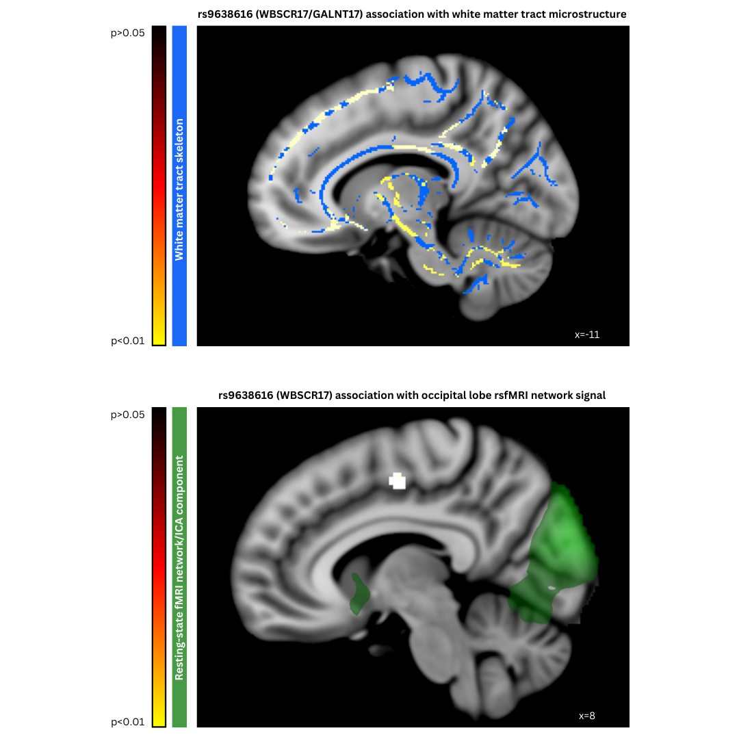
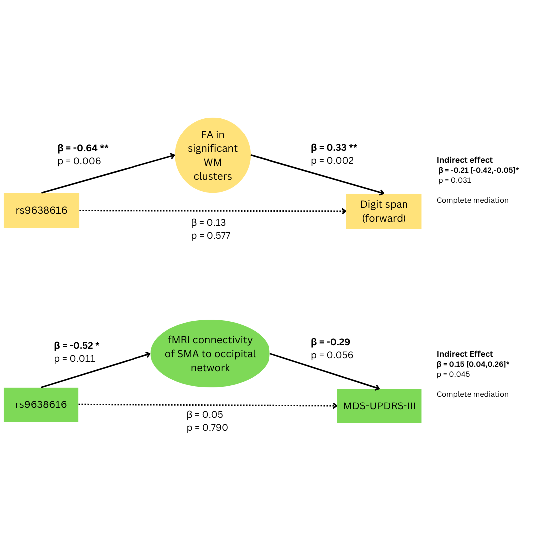
In the absence of significant interaction effects, we used the combined groups’ data. rs9638616-T was negatively associated with lower white matter FA (t=-1.75, p=0.042) distributed broadly across the entire brain, and lower functional connectivity of the occipital lobe network to the right supplementary motor area (SMA) (t=-5.05, p=0.001). We did not observe any significant voxelwise effects for grey matter volumes. These imaging-derived phenotypes significantly mediated the association of rs9638616 to MDS-UPDRS-III reflecting motor severity (indirect effect: β=-0.21 [-0.42,-0.05], p=0.031), and digit span reflecting cognitive performance (indirect effect: β=0.15 [0.04,0.26], p=0.045).Discussion
We identified significant imaging-genetic associations between rs9638616 and two types of voxelwise brain imaging measure, namely reduced widespread white matter FA and reduced functional connectivity of the SMA. The observed imaging-genetic associations are consistent with changes noted in previous PD studies [3, 4]. The associations were not limited to the PD group but extended to the control group, suggesting that the results reflect susceptibility rather than subclinical PD pathological processes. While rs9638616 was not directly associated with working memory or motor function, mediation analyses revealed significant indirect associations through the phenotypes identified, supporting a plausible causal chain where neurologic effects of rs9638616 confer cognitive and/or motor deficits. Thus, our significant mediation findings highlight a potentially important yet unexplored approach for prioritizing future PD GWAS results: through the “intermediate phenotype” concept. It should be noted that rs9638616 confers increased PD risk in Asian [1] but not European [5] populations – as such, our findings may be specific to the Asian PD population, which emphasizes the need to account for ethnic variance in studies of PD genetic risk. Our study was limited in several ways. While genetic factors contribute to PD risk, environmental and epigenetic factors could not be fully accounted for in the analysis. Furthermore, a wider range of disease durations and severities may have provided greater power to detect significant effects.Conclusion
We have shown that a novel GWAS variant which is biologically linked to synaptic transmission is associated with white matter tract and functional connectivity dysfunction in the SMA. These effects are present in both PD and nominally-healthy individuals, and are associated with reduced cognitive and motor functioning, supporting their clinical relevance. This provides pathophysiologic clues linking rs9638616 to PD risk and might be useful for stratification of Asian at-risk individuals.Acknowledgements
No acknowledgement found.References
1. Foo, J.N., et al., Identification of risk loci for Parkinson disease in Asians and comparison of risk between Asians and Europeans: a genome-wide association study. JAMA neurology, 2020. 77(6): p. 746-754.
2. Nalls, M.A., et al., Identification of novel risk loci, causal insights, and heritable risk for Parkinson's disease: a meta-analysis of genome-wide association studies. The Lancet Neurology, 2019. 18(12): p. 1091-1102.
3. Wei, X., et al., White matter abnormalities in patients with parkinson's disease: a meta-analysis of diffusion tensor imaging using tract-based spatial statistics. Frontiers in aging neuroscience, 2021. 12: p. 610962.
4. Tessitore, A., M. Cirillo, and R. De Micco, Functional connectivity signatures of Parkinson’s disease. Journal of Parkinson's disease, 2019. 9(4): p. 637-652.
5. Grover, S., et al., Replication of a novel Parkinson's locus in a European ancestry population. Movement Disorders, 2021. 36(7): p. 1689-1695.
Figures