4383
A preliminary study on the differences in regional changes of neuromelanin and iron in substantia nigra among early parkinsonism1Shandong Provincial Hospital Affiliated to Shandong First Medical University, Jinan, China, 2Shandong University, Jinan, China, 3Department of Radiology, The Affiliated Yantai Yuhuangding Hospital of Qingdao University, Yantai, China, 4Philips Healthcare, Shanghai, China
Synopsis
Keywords: Parkinson's Disease, Parkinson's Disease, neuromelanin
Motivation: Differential diagnosis of parkinsonism is difficult in early stage. Neuromelanin of SN plays an important role in the development of PD, PSP and MSA with iron.
Goal(s): To find more neuroimage biomarkers to differentiate early parkinsonism by altered neuromelanin and iron in the level of SN subregions.
Approach: We applied the 3D-ME-MTC-NM sequence to differentiate subregions based on the distribution of neuromelanin and iron, measured the volume, CR and/or susceptibility of neuromelanin accumulation, iron deposition and overlap regions.
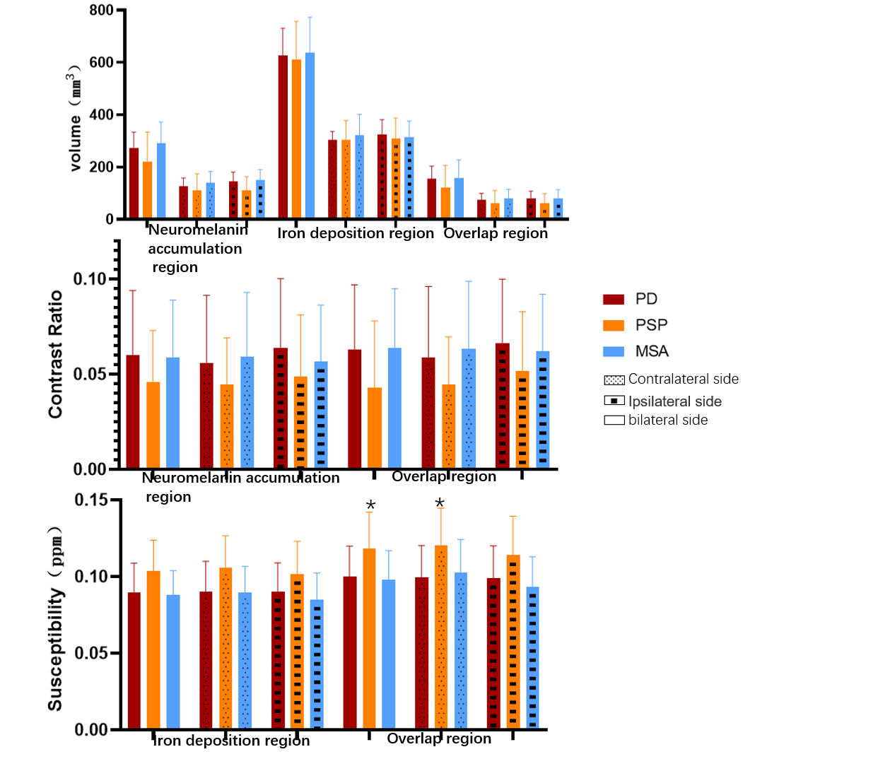
Results: The susceptibility of overlap region increased in early PSP, while no significant difference was seen between PD and MSA.
Impact: The alteration of susceptibility in the overlap region may be helpful to identify characteristic changes in parkinsonism via different pathological proteins.
Introduction
As the manifestation of clinical signs and symptoms among various parkinsonism overlap in high degree 1, differential diagnosis among Parkinson’s Disease (PD), Progressive Supranuclear Palsy (PSP) and Multiple System Atrophy (MSA) is difficult and complex, especially in the early stage2. Neuromelanin plays an important role in the development of parkinsonism with iron. Previous magnetic resonance studies have found that neuromelanin in the substantia nigra is reduced in varying degrees in PD, PSP and MSA, as well as the increased susceptibility of iron deposition3-5. Recently, a few studies have focused on the regional selectivity of neuromelanin and iron in substantia nigra among PD, PSP and MSA 4-6. However, indicators for early diagnosis are still lacking.The 3D multi-echo magnetization-transferred neuromelanin-sensitive sequence (3D ME-MTC-NM) has a superior application prospect as it enables the simultaneous acquisition of neuromelanin images and the generation of quantitative susceptibility mapping (QSM) from multi-echo data7,8.
Therefore, the aim of this study was to apply the 3D ME-MTC-NM sequence to differentiate subregions based on the distribution of neuromelanin accumulation and iron deposition, and measure the volume, contrast ratio and/or susceptibility of neuromelanin accumulation, iron deposition, and overlap regions. We compared the differences of the above measured parameters in the early stage of PD, PSP and MSA to explore the potential role of subregional neuromelanin and iron alterations for differential diagnosis.
Methods
This prospective study included 30 patients with early PD, 18 patients with early PSP, and 17 patients with early MSA who underwent 3D ME-MTC-NM sequence data collection from June 2020 to December 2022. Manual outlining and arithmetic methods were used to obtain neuromelanin accumulation, iron deposition and overlap regions. Covariance analysis was used to compare the differences in volume, contrast ratio, and/or susceptibility among the three regions of interest in early PD, PSP, and MSA. According to the dyskinesia side, the subregions were divided into contralateral side and ipsilateral side. Covariance analysis was used to compare the differences of neuromelanin and iron changes in the three subregions of the contralateral side and ipsilateral side in the three groups of early parkinsonism, and pairwise comparison was performed between the three groups.Results
After multi-comparison correction by false discovery rate (FDR), there was difference in the susceptibility of the overlap regions between the early PD, PSP and MSA groups (P=0.035). In the pairwise comparison between groups, the susceptibility of the overlap regions in the early PSP group was significantly higher than that in the early PD group (0.104±0.0220 ppm, 0.100±0.020 ppm, P=0.016) and the early MSA group (0.104±0.0220ppm, 0.098±0.019ppm, P=0.022). Comparison of the affected contralateral subregion revealed that the susceptibility in the overlap regions differed between the three groups (P=0.038). Comparison of the affected ipsilateral subregion revealed a trend in the difference in volume of the neuromelanin accumulation regions and the susceptibility of the overlap regions between the three groups, while the difference was not statistically significant after FDR correction.Discussion
In this study, we compared the differences in neuromelanin and iron deposition of three substantia nigra subregions in early parkinsonism by a single 3D ME-MTC-NM sequence and found that the susceptibility of overlap region was higher in patients with early PSP, whereas other parameters were not significant among the three groups. This means that altered iron deposition in the overlap region may be valuable in the early identification of PD, PSP and MSA.Although substantia nigra has been regard as a common damaged structure of the three diseases, it degenerates in different way with the specific pathological protein. Since PSP is tightly linked to tau pathology9, MSA and PD are both classified as α-synucleinopathy10,11. Our results confirm that the pattern of iron deposition in early PSP is different from that in PD and MSA, while there is no significant difference between PD and MSA, which is hypothesized to be related to the fact that pathological changes in PSP are distinct from those of α-synuclein diseases.
Conclusion
The susceptibility of the overlap region was elevated in early PSP compared with early PD and MSA, while no significant difference was seen between PD and MSA. The alteration of susceptibility in the overlap region may be helpful to identify characteristic changes in parkinsonism via different pathological proteins and needs to be further validated in future studies with large samples.Acknowledgements
The authors gratefully thank all the patients for their participation and support in the study.References
[1] Erkkinen M G, Kim M-O, Geschwind M D. Clinical neurology and epidemiology of the major neurodegenerative diseases[J]. Cold Spring Harbor Perspectives in Biology, 2018, 10(4): a033118.
[2] Levin J, Kurz A, Arzberger T, et al. The differential diagnosis and treatment of atypical parkinsonism[J/OL]. Deutsches Ärzteblatt International, 2016[2023–03–24]. https://www.aerzteblatt.de/10.3238/arztebl.2016.0061.
[3] Taniguchi D, Hatano T, Kamagata K, et al. Neuromelanin imaging and midbrain volumetry in progressive supranuclear palsy and parkinson’s disease: neuromelanin-mri and midbrain volumetry[J]. Movement Disorders, 2018, 33(9): 1488–1492.
[4] Chougar L, Arsovic E, Gaurav R, et al. Regional selectivity of neuromelanin changes in the substantia nigra in atypical parkinsonism[J]. Movement Disorders, 2022, 37(6): 1245–1255.
[5] Matsuura K, Maeda M, Yata K, et al. Neuromelanin magnetic resonance imaging in parkinson’s disease and multiple system atrophy[J]. European Neurology, 2013, 70(1–2): 70–77.
[6] Mazzucchi S, Frosini D, Costagli M, et al. Quantitative susceptibility mapping in atypical parkinsonisms[J]. NeuroImage: Clinical, 2019, 24: 101999.
[7] Chen Y, Gong T, Sun C, et al. Regional age-related changes of neuromelanin and iron in the substantia nigra based on neuromelanin accumulation and iron deposition[J]. European Radiology, 2023, 33(5): 3704–3714.
[8] He N, Ghassaban K, Huang P, et al. Imaging iron and neuromelanin simultaneously using a single 3d gradient echo magnetization transfer sequence: combining neuromelanin, iron and the nigrosome-1 sign as complementary imaging biomarkers in early stage parkinson’s disease[J]. NeuroImage, 2021, 230: 117810.
[9] Boxer A L, Yu J-T, Golbe L I, et al. Advances in progressive supranuclear palsy: new diagnostic criteria, biomarkers, and therapeutic approaches[J]. The Lancet Neurology, 2017, 16(7): 552–563.
[10] Kalia L V, Lang A E. Parkinson’s disease[J]. The Lancet, 2015, 386(9996): 896–912.
[11] Koga S, Dickson D W. Recent advances in neuropathology, biomarkers and therapeutic approach of multiple system atrophy[J]. Journal of Neurology, Neurosurgery & Psychiatry, 2018, 89(2): 175–184.
Figures