4382
Mapping of iron deposition gradients in the nigrostriatal system in normal aging and Parkinson's disease1Department of Radiology, the Second Affiliated Hospital, Zhejiang University School of Medicine, Hangzhou, China, 2GE Healthcare, Shanghai, China
Synopsis
Keywords: Parkinson's Disease, Parkinson's Disease, Normal aging; Gradient; Quantitative susceptibility mapping; Nigrostriatal
Motivation: The gradient characterization of microenvironment in nigrostriatal system is key to understanding striatal dysfunction in PD.
Goal(s): To investigate the gradients of neurodegeneration in nigrostriatal system in normal aging and PD.
Approach: Quantitative susceptibility mapping (QSM) and spatial method were used to detect the spatial gradient of iron deposition in healthy young people, normal elderly and PD in vivo.
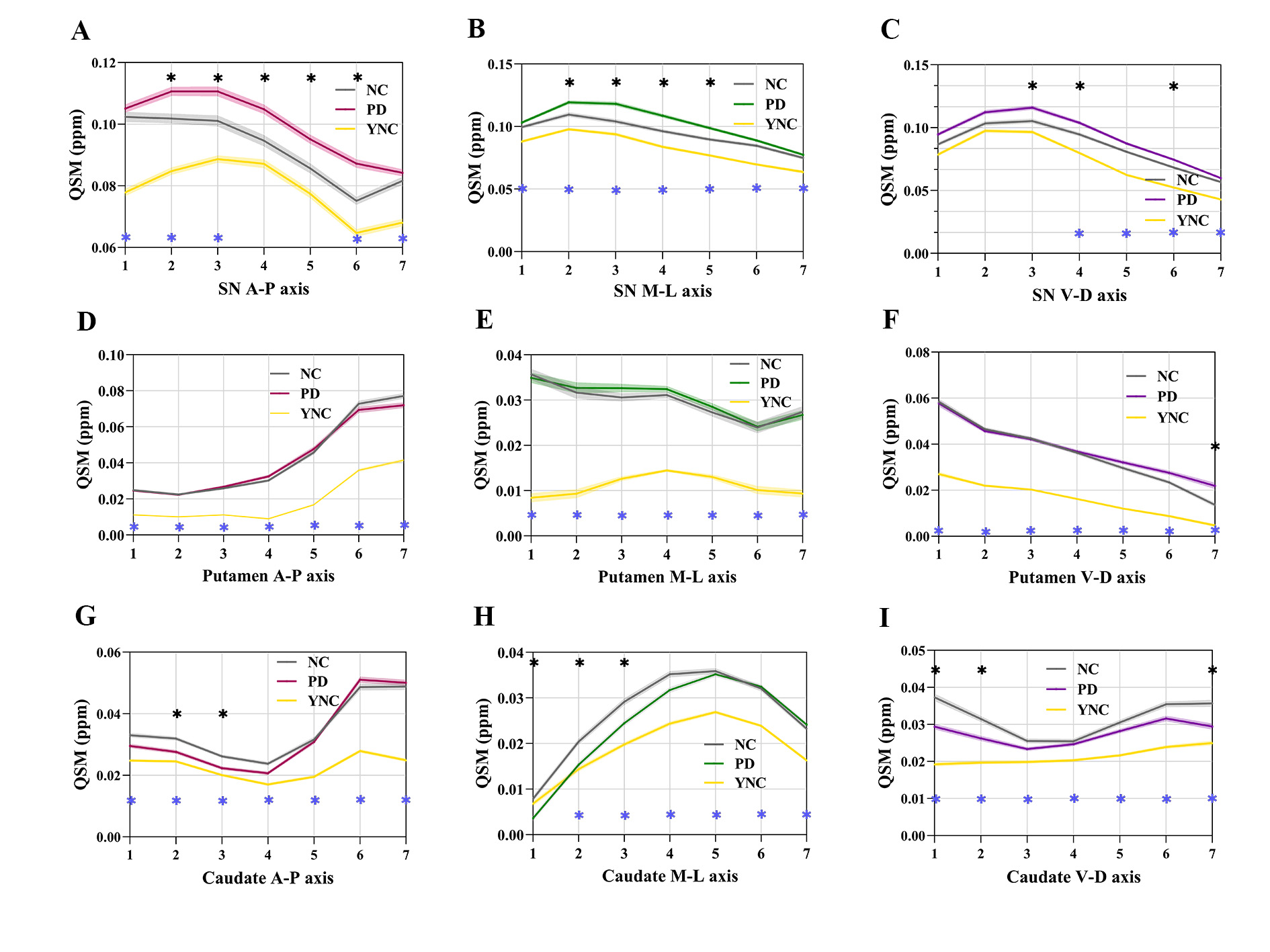
Results: During normal aging, iron deposition was significant in almost all segments of the striatum, and iron content was even higher in some segments of the caudate than in PD. Iron deposition in PD is mainly in the central substantia nigra.
Impact: The present study reveals the spatial gradient of iron deposition in the nigrostriatal system in normal aging and PD, providing more subtle and profound insights into the pathological changes in subcortical nuclei during neurodegeneration.
Background
The neurodegeneration of the nigrostriatal dopaminergic system is the main cause of Parkinson's disease (PD) [1]. Striatal biospatial degeneration in PD is predominantly in the posterior putamen, representing the loss of dopaminergic neurons, leading to a range of dyskinesia [2]. The gradient characteristics of the cellular and neurochemical content levels (i.e., the microenvironment) of the striatum are key to understanding striatal dysfunction in PD [3, 4]. In this study, the spatial changes (i.e., gradients) of iron deposition in substantia nigra (SN) and striatum were mapped to understand the basal ganglia degeneration in normal aging and PD.Materials and Methods
A total of 100 healthy young people, 171 normal elderly and 231 PD patients were enrolled in this study. The brain iron content of all subjects was measured by quantitative susceptibility mapping (QSM). An automatic procedure generate QSM functions along the main axis of a subcortical structure at the individual subject level was used to identify and quantify spatial gradients of iron deposition in individual human brains in vivo [5].Results
During normal aging, iron deposition along the three main axes of the striatum (caudate and putamen) was significant in almost all segments (p<0.05), except for the most medial caudate nucleus (segment 1 of medial-lateral axis); Iron deposition along the three main axes of the SN was more significant in the anterior, posterior, medial, lateral, and dorsal segments (p<0.05). (Figure 1)In PD patients, SN iron deposition was mainly concentrated in the central parts (p<0.05) compared with normal elderly, and putamen iron deposition was found only in the most dorsal part (segment 7 of the ventral-dorsal axis). In addition, the iron content of the anterior, medial, ventral, and dorsal caudate nucleus was higher in normal elderly than in PD patients (p<0.05). (Figure 1)
Conclusion
The preliminary results of our study reveal the spatial gradient of iron deposition in the nigrostriatal system in normal aging and PD in vivo, providing more subtle and profound insights into the pathological changes of subcortical nuclei during neurodegeneration.Acknowledgements
We wish to thank all the participants including patients with Parkinson’s disease and normal volunteers. We also thank the assistance from Department of Neurology in the Second Affiliated Hospital of Zhejiang University School of Medicine.References
1. Redgrave, P., et al., Goal-directed and habitual control in the basal ganglia: implications for Parkinson's disease. Nat Rev Neurosci, 2010. 11(11): p. 760-72.
2. Stormezand, G.N., et al., Intrastriatal gradient analyses of 18F-FDOPA PET scans for differentiation of Parkinsonian disorders. Neuroimage Clin, 2020. 25: p. 102161.
3. Prensa, L., J.M. Giménez-Amaya, and A. Parent, Chemical heterogeneity of the striosomal compartment in the human striatum. J Comp Neurol, 1999. 413(4): p. 603-18.
4. Tian, Y., et al., Topographic organization of the human subcortex unveiled with functional connectivity gradients. Nat Neurosci, 2020. 23(11): p. 1421-1432.
5. Drori, E., S. Berman, and A.A. Mezer, Mapping microstructural gradients of the human striatum in normal aging and Parkinson's disease. Sci Adv, 2022. 8(28): p. eabm1971.
Figures