4379
Association of glymphatic system impairment with aging and cognitive decline in Parkinson's disease1Shandong Provincial Hospital Affiliated to Shandong First Medical University, Jinan, China, 2Department of Radiology, Shandong Provincial Hospital Affiliated to Shandong First Medical University, Jinan, China, 3Department of Radiology, Shandong University, Jinan, China, 4Philips Healthcare, Shanghai, China
Synopsis
Keywords: Parkinson's Disease, Parkinson's Disease, glymphatic system
Motivation: In this study,we aimed to investigate the activity of the glymphatic system in PD using DTI-ALPS method and to further explore the correlation patterns of DTI-ALPS indexes with aging and cognitive impairment.
Goal(s): To explores the correlation between glymphatic system impairment and cognitive impairment.
Approach: Diffusion tensor image analysis along the perivascular space (DTI-ALPS) index offers a noninvasive alternative and the method has been validated well with standard glymphatic MRI.
Results: Our study found that glymphatic system function was impaired in PD as reflected by DTI-ALPS. Glymphatic dysfunction may lead to cognitive impairment,which was affected by aging and disease state.
Impact: Our study explores the correlation between glymphatic system impairment and cognitive impairment.This finding provides new perspectives on cognitive impairment in patients with PD and suggests potential strategies for treating cognitive impairment in such patients.
Introduction
Parkinson’s disease (PD) is a common neurodegenerative disease that manifests with both motor and non-motor symptoms.1The pathological hallmarks of Parkinson's Disease (PD) include dopaminergic neuron degeneration of the substantia nigra pars compacta and the accumulation of cytoplasmic inclusion Lewy bodies.2It is estimated that up to 40% of patients with Parkinson's disease have a mild cognitive impairment( PD-MCI), which is at risk for progressing to PD dementia.3 Post-mortem analysis indicated α-synuclein pathology is the main substrate of dementia in PD.4Recent evidence emphasizes the role of the glymphatic system in eliminating neurotoxic protein aggregations in neurodegenerative diseases, such as α-synuclein in PD.5Several imaging techniques have been proposed for the evaluation of the glymphatic system.6-8 Diffusion tensor image analysis along the perivascular space (DTI-ALPS) index offers a noninvasive alternative and the method has been validated well with standard glymphatic MRI.9In this study,we aimed to investigate the activity of the glymphatic system in PD and to further explore the correlation patterns of DTI-ALPS indexes with aging and cognitive impairment.Methods
Participants and enrollment37 patients with PD and 30 cognitively normal controls(NCs) in this study were recruited between January 2021 and July 2023 from Shandong Provincial Hospital and the surrounding communities. All the patients were diagnosed as idiopathic PD by neurologists. All subjectswere went MRI scans, MMSE(Mini-Mental State Examination) was performed for all patients to evaluate neuropsychology performance .
Image acquisition and processing
Diffusion tensor image (DTI) sequence was acquired using a 32-channel 3T MRI scanner (Ingenia CX, PhilipsHealthcare, Best, Netherlands). Axial images were acquired using the following parameters: TR/TE 2726msec/76msec; 66 axial slices, flip angle 90°, slice thickness 2mm; field of view 224×224 mm2. The total image acquisition time was 6 min.The DTI data using FSL (FMRIB Software Library; http://www.fmrib.ox.ac.uk/fsl) to conduct preprocessing steps,including topup and eddy current correction.Then,We reconstructed images by Q-SpaceDiffeomorphic reconstruction (QSDR) method using DSI STUDIO (DSI Studio; http://dsi-studio.labsolver.org). Four spherical ROIs were manually drawn (diameter, 7 pixels [≈5 mm]) and placed in the areas of bilateral projection and association fibers at the level of lateral ventricle body. Then the diffusivity values of Dxx, Dyy and Dzz of bilateral projection and association fibers were obtained for the ALPS calculation(Fig 1). The DTI-ALPS index was defined as follows: DTI-ALPS=mean (Dxxproj, Dxxassoc)/mean(Dyyproj, Dzzassoc) The DTI-ALPS indexes in both hemispheres were calculated and averaged.
Statistical analysis
Statistical analyses were conducted with GraphPad Prism (version 9, GraphPad Inc., San Diego, CA, USA).Two sample t-test was applied for the comparison of PD with HCs .Pearson correlation analysis were performed to further evaluate the correlation between DTI - ALPS indexes and clinical characteristics,including age,MMSE scores.ROC curve was used for the sensitivity analysis of DTI-ALPS indexes for PD patients.We further explored the exact contribution of glymphatic function to cognition during aging by measuring the mediation effect of DTI-ALPS on age and cognitive function.Results
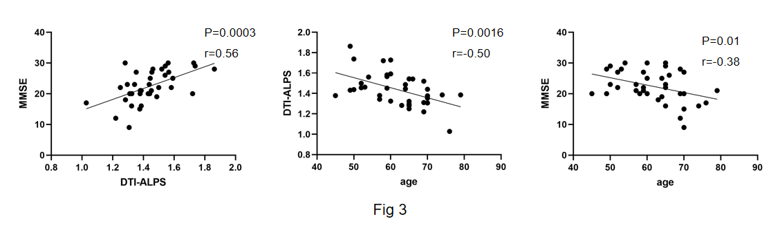
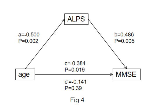
Compared to the NCs group, the PD group exhibited a significantly lower DTI-ALPS index(P<0.0001) (Fig. 2A).ROC curve analysis was further performed to assess the performance of the ALPS index for distinguishing PD group and NCs, and the area under the curve (AUC) for the ALPS index was 0.814 (0.814, 95% confidence interval 0.7100-0.9170) (Fig. 2B). In PD patients, the ALPS index was significantly correlated with the age(r =-0.50; p = 0.0016) and MMSE scores (r =0.56; p = 0.0003).moreover,we found age was significantly correlated with theMMSE scores(r=-0.38 ; p=0.01)(Fig. 3).mediation effect was calculated to further investigate the role of the DTI-ALPS index between age and cognition function in PD groups.In our study,we found in patients with PD, the effects of age on cognition are mediated primarily through DTI-ALPS(Fig. 4).Discussion
In previous study, the glymphatic system was intersection of a broad scope of disorders(such as AD、PD、DLB etc), which share an association with diminished brain fluid clearance.10 Normal aging is also linked to a sharp decline in sleep quality and decreased glymphatic flow. 11In turn, the stagnation of glymphatic flow, and hence that of extracellular proteins, contributes to protein aggregation, with misfolding and seeding, leading to local inflammation, neuronal loss, and ultimately dementia.DTI-ALPS is a recently established non-invasive method to evaluate brain glymphatic function.In this study, we aimed to assess glymphatic system function using DTI-ALPS and to investigate associations of DTI-ALPS with age and cognitive impairment in Parkinson's disease.Our study revealed that glymphatic system was impaired in PD groups.we found that aging and disease state are crucial factor associated with cognitive decline.
Conclusion
Our study found that glymphatic system function was impaired in PD as reflected by DTI-ALPS. Glymphatic dysfunction may lead to cognitive impairment,which was affected by aging and disease state.Acknowledgements
No acknowledgement found.References
1.Postuma RB, Berg D, Stern M, et al. MDS clinical diagnostic criteria for Parkinson's disease. Mov Disord. 2015;30(12):1591-1601.
2.López-Aguirre M, Matarazzo M, Blesa J, et al. Dopaminergic denervation and associated MRI microstructural changes in the nigrostriatal projection in early Parkinson's disease patients. NPJ Parkinsons Dis. 2023;9(1):144. Published 2023 Oct 19.
3.Aarsland D, Brønnick K, Larsen JP, Tysnes OB, Alves G; Norwegian ParkWest Study Group. Cognitive impairment in incident, untreated Parkinson disease: the Norwegian ParkWest study. Neurology. 2009;72(13):1121-1126.
4.Smith C, Malek N, Grosset K, Cullen B, Gentleman S, Grosset DG. Neuropathology of dementia in patients with Parkinson's disease: a systematic review of autopsy studies. J Neurol Neurosurg Psychiatry. 2019;90(11):1234-1243.
5.Iliff JJ, Wang M, Liao Y, et al. A paravascular pathway facilitates CSF flow through the brain parenchyma and the clearance of interstitial solutes, including amyloid β. Sci Transl Med. 2012;4(147):147ra111.
6.Taoka T, Naganawa S. Glymphatic imaging using MRI. J Magn Reson Imaging. 2020;51(1):11-24.
7.Kamagata K, Saito Y, Andica C, et al. Noninvasive Magnetic Resonance Imaging Measures of Glymphatic System Activity [published online ahead of print, 2023 Sep 1]. J Magn Reson Imaging.
8.Eide PK, Ringstad G. MRI with intrathecal MRI gadolinium contrast medium administration: a possible method to assess glymphatic function in human brain. Acta Radiol Open. 2015;4(11):2058460115609635.
9.Zhang W, Zhou Y, Wang J, et al. Glymphatic clearance function in patients with cerebral small vessel disease. Neuroimage. 2021;238:118257.
10.Nedergaard M, Goldman SA. Glymphatic failure as a final common pathway to dementia. Science. 2020;370(6512):50-56.
11.Siow TY, Toh CH, Hsu JL, et al. Association of Sleep, Neuropsychological Performance, and Gray Matter Volume With Glymphatic Function in Community-Dwelling Older Adults. Neurology. 2022;98(8):e829-e838.
Figures
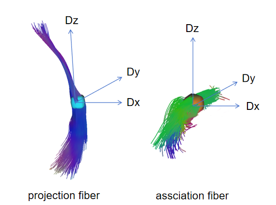
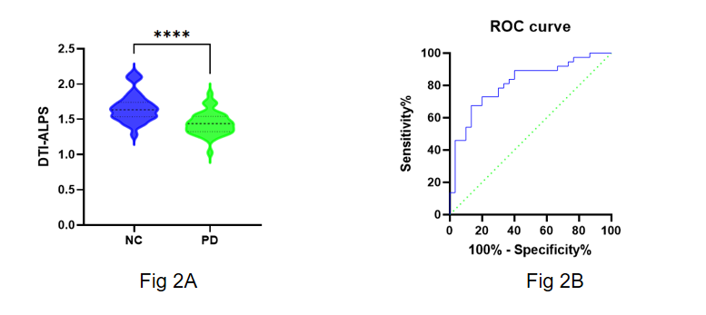
Fig 2A:Comparison of the DTI-ALPS between the PD and NC groups.
Fig 2B:Receiver operating characteristic (ROC) curves for diagnosing PD using DTI-ALPS index.