4376
Gender-Specific Brain Morphological and Network Differences in Parkinson's Disease Patients with Rapid Eye Movement Sleep Behavior Disorder1Second Clinical School, Lanzhou University, lanzhou, China, 2Department of Magnetic Resonance, Lanzhou University Second Hospital, lanzhou, China, 3Gansu Province Clinical Research Center for Functional and Molecular Imaging, lanzhou, China, 4Philips Healthcare, Xi’an, China
Synopsis
Keywords: Parkinson's Disease, Parkinson's Disease
Motivation: The influence of gender on the brain morphology of PD-RBD patients remains unclear.
Goal(s): We aimed to investigate gender differences in PD-RBD patients in terms of cortical morphology and individual structural covariance network.
Approach: We firstly conducted volume- and surface-based morphometry analyses, followed by further exploration of the topological characteristics of individual level morphological similarity networks based on Kullback-Leibler Divergence.
Results: Male patients presented decreased cortical indicators in salience, along with increased volume, cortical complexity and sulcus depth increase. Furthermore, in the individual morphological networks, we found significant differences between male and female patients in terms of both global and nodal properties.
Impact: PD-RBD patients exhibit significantly gender-specific differences in brain morphology and covariant patterns, which may reflect distinct clinical treatment needs and disease progression patterns. Further exploration is needed to enhance clinical management efficiency.
Introduction
Parkinson's disease (PD) is highly heterogeneous, especially with significant clinical and brain differences between different genders. Rapid eye movement sleep behavior disorder (RBD) is associated with a more severe presentation of PD1,2. However, the influence of gender on the brain morphology of PD-RBD patients remains unclear. This study aims to conduct an integrated analysis of Voxel- and Surface-Based Morphometry (VBM and SBM), subsequently construct individual-level morphological similarity networks.Methods
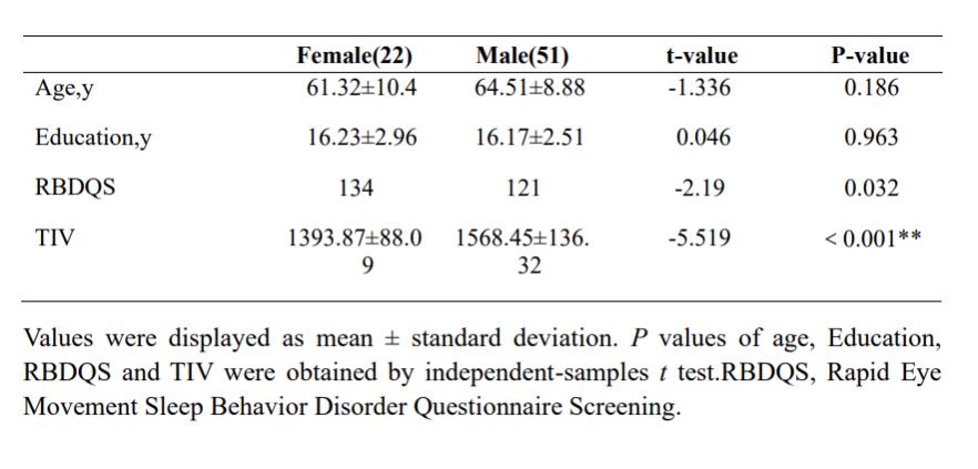
Baseline structural MRI data and RBD Questionnaire Screening (RBDQS) scores were obtained from 73 PD patients with comorbid RBD (51 males and 22 females), sourced from the Parkinson's Progression Markers Initiative dataset3(Table1).3D-T1WI data were acquired using a 3.0 Tesla MRI scanner. VBM and SBM analyses were performed using CAT12 through standard procedures. In VBM analysis, GM maps were smoothed using an 8mm Gaussian kernel. In SBM analysis, fractal dimension (FD), gyrification index (GI), sulcal depth (SD), and cortical thickness (CT) and Torsion GI (toroGI) were obtained4, with a 12mm smoothing kernel applied to CT images and a 20mm smoothing kernel applied to the remaining images. In the morphological network,68brain regions were defined based on the DK40 atlas5.Symmetric similarity measures based on KL divergence were computed as edge weights6. The topological properties of each cortical network were calculated using GRETNA (sparsity threshold range:0.063–0.4, step size0.01)7. Both global properties (Small-worldness and Efficiency) and nodal properties (Efficiency (Ne), Degree Centrality (Dc), and Betweenness Centrality (Bc)) were computed. Inter-group comparisons of VBM and SBM maps were conducted using two-sample t-tests (FWE, voxel/vertex level P<0.001, cluster level P<0.05), and group differences in graph theory metrics were assessed using non-parametric permutation tests (10,000 permutations) with FDR correction for nodal metrics. Age and education were considered as covariates, and total intracranial volume was included as a covariate in VBM analysis.Results
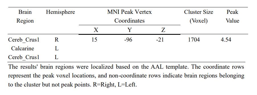
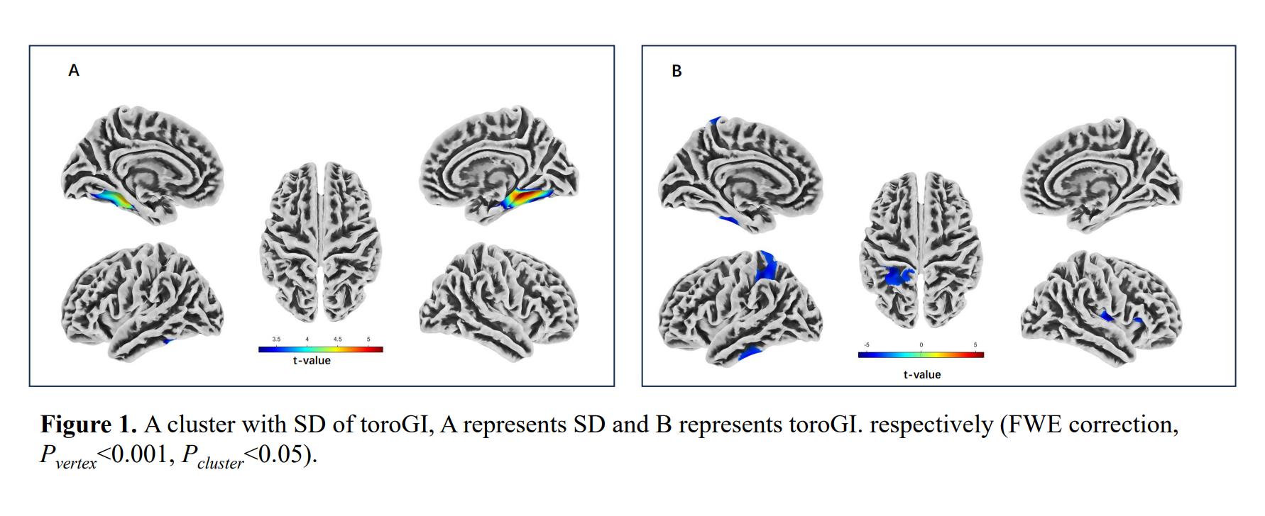
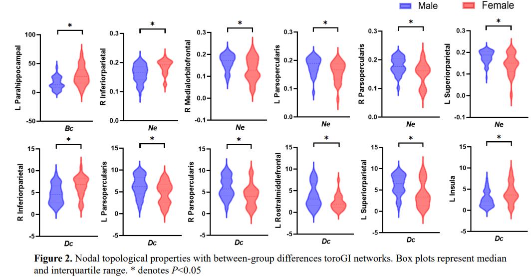
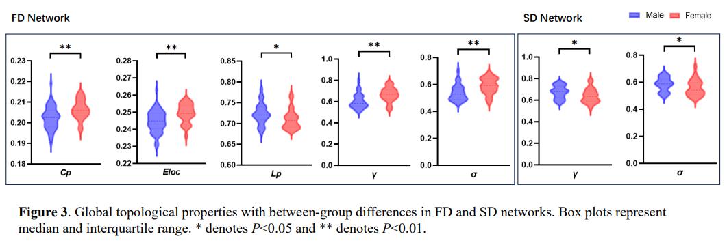
In PD-RBD patients, VBM analysis revealed that males exhibited increased grey matter volume (GMV) in the bilateral Cerebellum_Crus1and the left Calcarine regions compared to females (Table2). SBM analysis showed that males had larger SD in the bilateral fusiform, inferior temporal, parahippocampal, lingual, and the right entorhinal gyrus than females. Higher toroGI in the bilateral postcentral, right insula, precentral, pars opercularis, and pars triangularis, and the left superior parietal, paracentral, precuneus, and inferior temporal gyrus were found in male patients (Figure1). Global property analysis indicated that in the FD network, males exhibited reduced local efficiency (Eg), σ, γ, and clustering coefficient (Cp) with increased path length (Lp). In the SD network, males had higher σ and γ. Differences in nodal properties were observed in the toroGI network. Specifically, males had higher Dc and Ne in the bilateral pars opercularis, the left superior parietal, rostral middle frontal, and the right medial orbitofrontal regions compared to females. Conversely, males exhibited larger Dc and Ne in the right inferior parietal, and the left insula, and parahippocampal regions compared to females (Figure2-3).Discussion
This study primarily focused on the influence of gender on the brain morphology and structural covariance network topology in PD-RBD patients. In PD-RBD male patients, higher GMV in bilateral Cerebellum Crus1 and the left Calcarine region may reflect adaptations related to motor dysfunction8. Compared to female patients, male patients exhibited greater SD and toroGI in SBM, involving regions such as parahippocampal, lingual, insula, superior parietal, and entorhinal gyrus, which play crucial roles in emotion and cognition. While bilateral postcentral, precentral, paracentral, and precuneus regions are mainly involved in patients' motor function. Cortical changes revealed by SBM are associated with both motor and memory functions, also consistent with previous researches9,10. Male and female patients exhibited differences in global properties in the FD and SD networks. Wherein the FD is related to cortical complexity patterns and the SD is associated with neural fiber bundles. Changes in the integration of morphological similarity networks may further reflect deep white matter impairments. Male patients showed stronger integration in the FD network, while in female patients, it was found in the SD network. These results suggest that FD and SD are sensitive to network changes in male and female patients, respectively. Significant differences also existed in nodal properties of toroGI network. ToroGI, as an improved parameter of GI, may help identify local disruptions in patients of different genders, warranting further exploration of its potential value in disease differentiation and predication.Conclusion
There are significant gender-specific morphological differences in the motor and cognition-related regions of PD-RBD patients, which may further reflect the neuropathologic basis of clinical symptoms and be related to disease progression and treatment needs. FD, SD, and toroGI may be sensitive in revealing gender differences in PD-RBD patients.Acknowledgements
This work was supported by the Natural Science Foundation of China (No. 81960309), Gansu Province Clinical Research Center for Functional and Molecular Imaging (No. 21JR7RA438) and Gansu Provincial Science and Technology Program Projects (Key Research and Development Program No. 23YFFA0041).References
1. Chahine LM, Xie SX, Simuni T et al (2016) Longitudinal changes in cognition in early Parkinson’s disease patients with REM sleep behavior disorder. Parkinsonism Relat Disord.;27:102–106. doi:10.1016/j.parkreldis.2016.03.006
2. Trout J, Christiansen T, Bulkley MB et al (2020) Cognitive impairments and self-reported sleep in early-stage Parkinson’s disease with versus without probable REM sleep behavior disorder. Brain Sci. doi:10.3390/brainsci10010009
3. Marek K, Jennings D, Lasch S, et al (2011) The Parkinson progression marker initiative (PPMI) Prog Neurobiol. 95:629–635. doi: 10.1016/j.pneurobio.2011.09.005.
4. Lai K L, Niddam D M, Fuh J L et al (2020) Cortical morphological changes in chronic migraine in a Taiwanese cohort: Surface- and voxel-based analyses. Cephalalgia 40:575-585. doi:10.1177/0333102420920005
5. Desikan R S, Ségonne F, Fischl B et al (2006) An automated labeling system for subdividing the human cerebral cortex on MRI scans into gyral based regions of interest. Neuroimage 31:968-980.
6. Lai H, Kong X, Zhao Y et al (2022) Patterns of a structural covariance network associated with dispositional optimism during late adolescence. Neuroimage 251:119009. doi:10.1016/j.neuroimage.2022.119009
7. Boucetta S, Saliimi A A, Dadar M et al (2016) Structural Brain Alterations Associated with Rapid Eye Movement Sleep Behavior Disorder in Parkinson's Disease Scientific reports,6:26782.
8. Huang W, Hu W, Zhang P et al (2022) Early Changes in the White Matter Microstructure and Connectome Underlie Cognitive Deficit and Depression Symptoms After Mild Traumatic Brain Injury[J]. Front Neurol. 30;13:880902. DOI:10.3389/fneur.2022.880902.
9. Liu R, Umbach D M, Peddada S D et al (2015) Potential sex differences in nonmotor symptoms in early drug-naive Parkinson disease. Neurology.84:2107–2115. doi:10.1212/WNL.0000000000001609
10. Bakeberg M C, Gorecki A M, Kenna J E et al (2021) Differential effects of sex on longitudinal patterns of cognitive decline in Parkinson’s disease. Neurol. doi:10.1007/s00415-020-10367-8.
Figures