4375
Putaminal spontaneous activity characterizes and predicts wearing-off in Parkinson’s disease1The Second Affiliated Hospital, Zhejiang University School of Medicine, Hangzhou, Zhejiang, China
Synopsis
Keywords: Parkinson's Disease, Parkinson's Disease
Motivation: Delayed detection of wearing off (WO) in Parkinson’s disease (PD) has a negative impact on quality of life. However, there is currently no method for WO prediction prior to treatment.
Goal(s): To investigate whether resting-state functional MRI (rs-fMRI) could aid in WO prediction.
Approach: Fractional amplitude of low-frequency fluctuation (fALFF) was quantified from rs-fMRI to determine whether spontaneous activity dysfunction could be a predisposing vulnerability related to WO.
Results: Putaminal fALFF reduction was the independent predictor of WO development.
Impact: Detection of putaminal fALFF reduction prior to treatment provides a quantitative metric to facilitate WO prediction and improve prognosis.
Background
The wearing off (WO) develops as the benefit of dopaminergic treatment in Parkinson’s disease (PD) wanes. WO is a predictable re-emergence of symptom deterioration. Delayed detection of WO results in worse quality of life and limited treatment options. Neuronal activity dysfunction could be a predisposing vulnerability related to WO and provide valuable information for early prediction.Material and methods
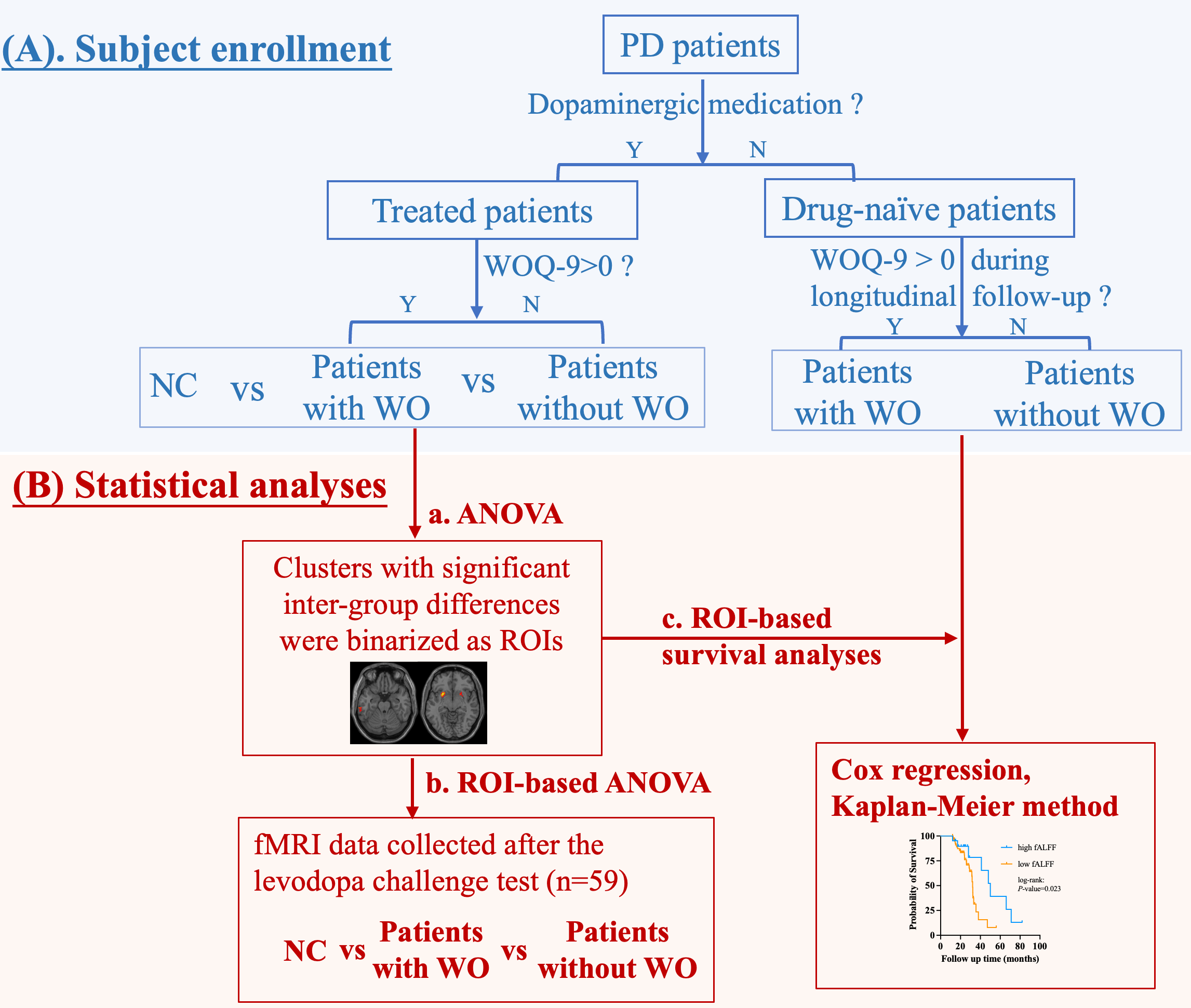
A total of 67 normal controls (NCs), 104 drug-managed patients under stable dopamine replacement therapy, and 81 drug-naïve patients were enrolled. Clinical assessments and MRI scans were performed before (OFF-state) and after (ON-state) levodopa challenge test. Drug-managed patients were stratified into patients with and without WO according to the wearing-off questionnaire. Drug-naïve patients have been longitudinally followed up since they initiated dopamine replacement therapy, and the time point when they developed WO was recorded. The fractional amplitude of low-frequency fluctuations (fALFF) was quantified using the resting-state functional MRI to reflect spontaneous brain activities. Voxel-based analysis was carried out to detect fALFF differences among NCs, patients with WO, and patients without WO in OFF state. The significant clusters were binarized as regions of interest (ROIs). Averaged fALFF values within the ROIs were extracted and compared before and after the levodopa challenge test to explore how levodopa modulated spontaneous activity in patients with WO. Additionally, fALFF values within the ROIs were extracted in drug-naïve patients at baseline as candidate factors for predicting WO development.Results
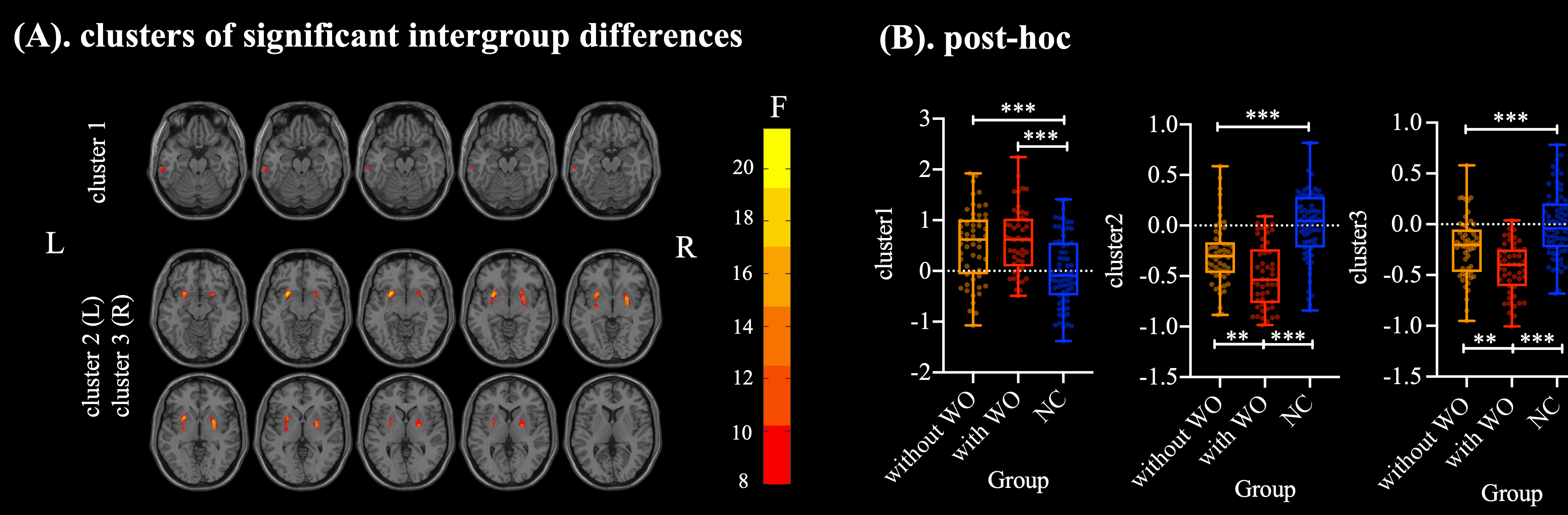
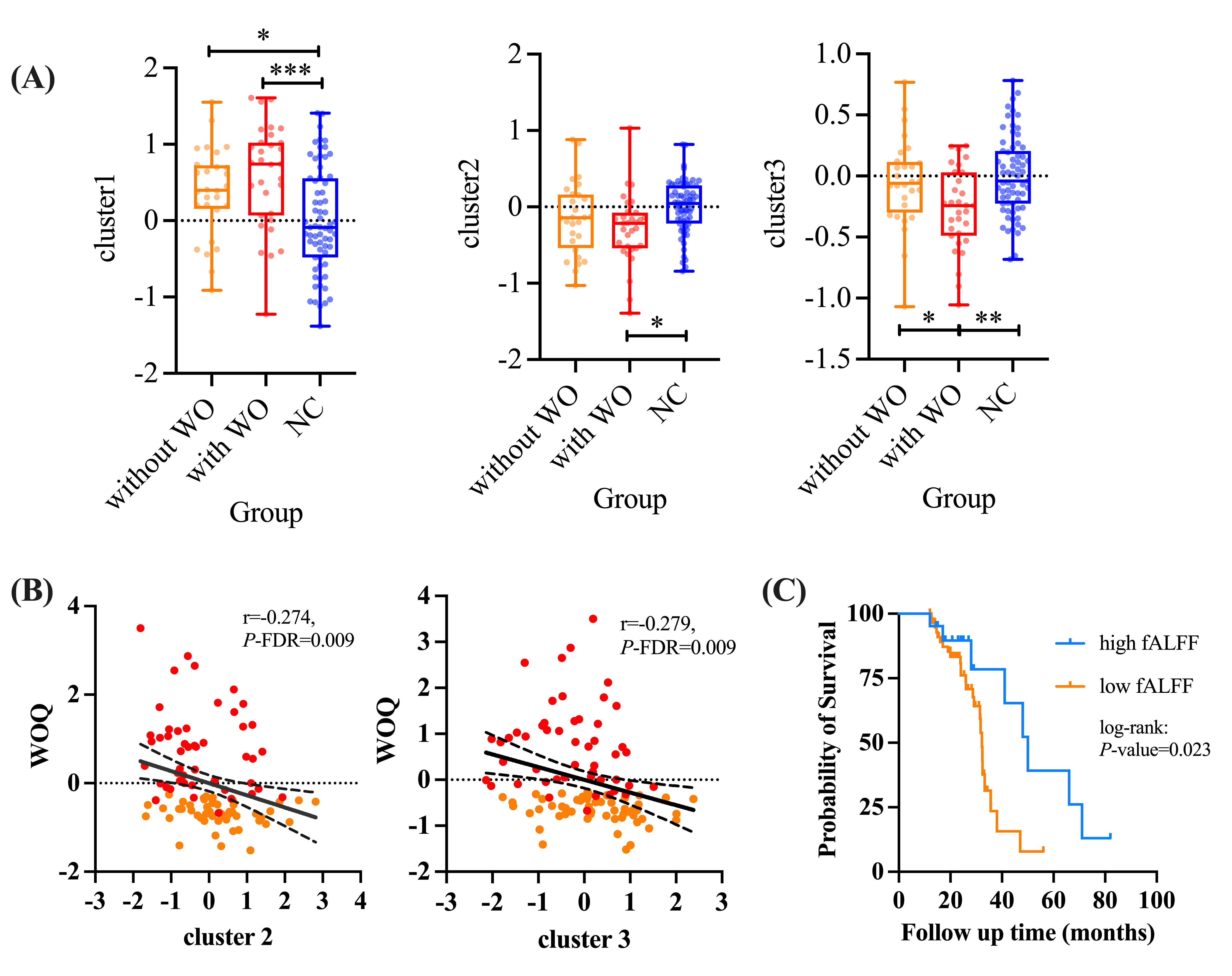
There were three clusters that demonstrated significant fALFF differences among NCs, patients with WO, and patients without WO. These clusters primarily comprised voxels located in the left inferior temporal gyrus (ITG) and bilateral putamen. In posthoc analyses, patients with and without WO both exhibited significantly increased fALFF in the left ITG and declined fALFF in bilateral putamen (P-value < 0.001) than NC. Additionally, patients with WO demonstrated significant fALFF reduction in bilateral putamen (P-value < 0.050) when compared to those without WO. WO severity was negatively associated with fALFF in bilateral putamen (R-value left=0.274, R-value right=0.279, P-FDR=0.009) in all patients. After the levodopa challenge test, patients with WO still exhibited a significant fALFF reduction in the bilateral putamen (P-value < 0.050) when compared to NC. However, there were no measurable fALFF differences in bilateral putamen between patients without WO and NC (P-value > 0.050). Multivariate Cox analyses confirmed that right putaminal fALFF was the independent predictor of WO development (HR=0.133, P-value=0.003).Conclusion
Putaminal fALFF reduction was a predisposing vulnerability related to WO and provided valuable information for early prediction.Acknowledgements
No acknowledgement found.References
1. Lewitt PA. Levodopa for the treatment of Parkinson's disease. N Engl J Med. 2008;359(23):2468-76.
2. Zappia M, Oliveri RL, Montesanti R, Rizzo M, Bosco D, Plastino M, et al. Loss of long-duration response to levodopa over time in PD: implications for wearing-off. Neurology. 1999;52(4):763-7.
3. Bhidayasiri R, Hattori N, Jeon B, Chen R-S, Lee MK, Bajwa JA, et al. Asian perspectives on the recognition and management of levodopa ‘wearing-off’ in Parkinson’s disease. Expert Review of Neurotherapeutics. 2015;15(11):1285-97.
4. Farzanehfar P, Woodrow H, Horne M. Assessment of Wearing Off in Parkinson's disease using objective measurement. J Neurol. 2021;268(3):914-22.
5. Reichmann H, Emre M. Optimizing levodopa therapy to treat wearing-off symptoms in Parkinson's disease: focus on levodopa/carbidopa/entacapone. Expert Rev Neurother. 2012;12(2):119-31.
6. Warren Olanow C, Kieburtz K, Rascol O, Poewe W, Schapira AH, Emre M, et al. Factors predictive of the development of Levodopa-induced dyskinesia and wearing-off in Parkinson's disease. Mov Disord. 2013;28(8):1064-71.
7. Lee J, Zhu WM, Stanic D, Finkelstein DI, Horne MH, Henderson J, et al. Sprouting of dopamine terminals and altered dopamine release and uptake in Parkinsonian dyskinaesia. Brain. 2008;131(Pt 6):1574-87.
8. Rajput AH, Fenton ME, Di Paolo T, Sitte H, Pifl C, Hornykiewicz O. Human brain dopamine metabolism in levodopa-induced dyskinesia and wearing-off. Parkinsonism Relat Disord. 2004;10(4):221-6.
Figures