4373
Effect of Nicotinamide riboside supplementation on cerebral NAD+ levels in vivo1CAMIPM, Radiology, Perelman School of Medicine at The University of Pennsylvania, Philadelphia, PA, United States, 2Psychiatry, Perelman School of Medicine at The University of Pennsylvania, Philadelphia, PA, United States, 3Biostatistics, Epidemiology, and Informatics, Perelman School of Medicine at The University of Pennsylvania, Philadelphia, PA, United States, 4Bioengineering, School of Engineering and Applied Sciences, University of Pennsylvania, Philadelphia, PA, United States, 5Neurology, Perelman School of Medicine at The University of Pennsylvania, Philadelphia, PA, United States, 6Physiology, Institute for Diabetes, Obesity and Metabolism, Perelman School of Medicine at The University of Pennsylvania, Philadelphia, PA, United States
Synopsis
Keywords: Aging, Translational Studies, Brain, Nicotinamide adenine dinucleotide, NAD+, Nicotinamide ribosome, NR, 1H MRS, Aging
Motivation: To determine if acute nicotinamide riboside (NR) supplementation increases cerebral nicotinamide adenine dinucleotide (NAD+) levels in the human brain.
Goal(s): To measure cerebral NAD+ levels before and after nicotinamide riboside supplementation using downfield 1HMRS at 7T MRI in ten healthy volunteers.
Approach: First MR scan was performed in each healthy volunteer after overnight fasting to obtain baseline NAD+ levels. In the second scan on the following day, the same protocol was repeated, but with NR supplements administered orally 4 hours before the scan.
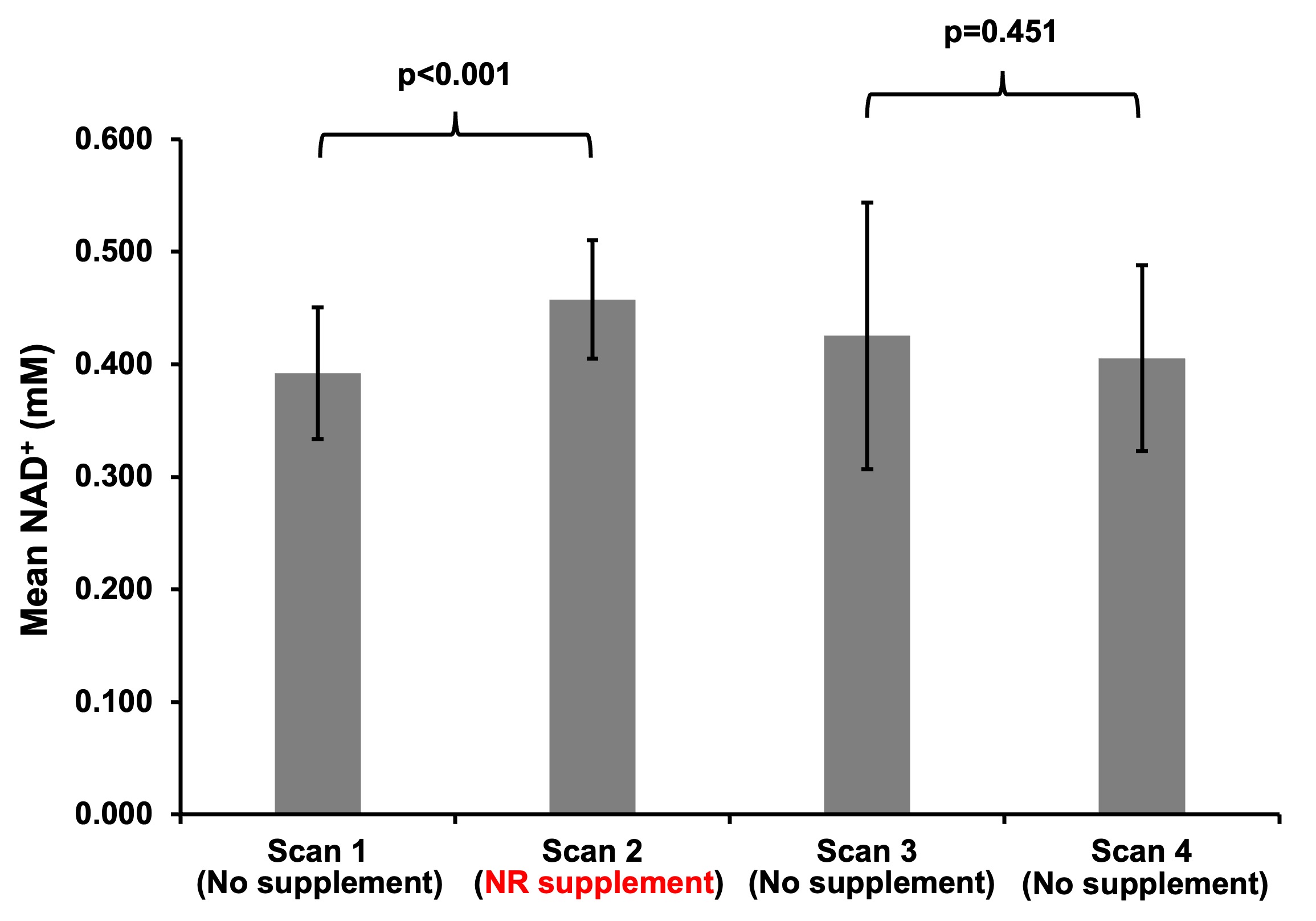
Results: An increase in mean NAD+ concentration was observed with NR supplementation, compared to the baseline (0.458±0.053 vs 0.392±0.058mM; p<0.001).
Impact: The preliminary results from this study show that oral NR supplementation increases NAD+ levels in brain and demonstrates the potential of downfield 1HMRS for noninvasive quantification of cerebral NAD+ and monitoring the effects of NR supplementation on cerebral NAD+ levels.
Introduction
Nicotinamide adenine dinucleotide (NAD+) in the oxidized form is an important coenzyme involved in many vital processes such as DNA repair, aging, apoptosis, mitochondrial function, etc.1-6 A decline in cerebral NAD+ concentrations with aging has been implicated in various aging-associated neurodegenerative and neurological disorders.7,8 NAD+ precursors such nicotinamide riboside (NR) have been steadily gaining popularity as a nutritional supplement for neurogenerative and neurological disorders. Several preclinical studies in different disease models with NR supplementation have shown improved cerebral NAD+ levels,9-11 but a few studies have demonstrated this in humans. Clinical studies in human subjects showing NAD+ levels were limited to measuring changes in blood plasma. One study also measured NAD+ levels invasively from cerebrospinal fluid.12 Thus, there remains a need for methods to non-invasively detect and quantify cerebral NAD+ levels in vivo. In the current study, we utilized downfield (DF) 1HMRS13,14 along with a large voxel with a total scan time of ~7min to detect and quantify the changes in cerebral NAD+ levels in healthy human subjects with pre- and post-NR supplementation.Methods
Ten healthy volunteers (n=10; 5males; 5females; 31.44±10.32Y) participated in the study after reviewing the study protocol and obtaining the informed consent. The volunteers were asked to fast overnight after dinner and refrain from exercise as well as consuming any breakfast or drinks (except water) before the scan day (Scan1). For the second scan (Scan2) on the following day, the same protocol was followed except that 900mg (three 300mg capsules) of nicotinamide riboside (NR) supplements (TRUNIAGEN 300mg capsules; purchased from https://www.truniagen.com) were consumed 4 hours before the scan. After few weeks, two more scans were performed on the same volunteers on back-to-back days (Scan3 and Scan4) following the same paradigm both without NR supplementation to assess for retest effects. All the MRI acquisitions were performed using a single-channel volume transmit, and 32-channel receive proton phased array head radiofrequency coil (Nova Medical, Wilmington, MA, USA) on a 7T scanner (MAGNETOM Terra, Siemens Healthcare, Erlangen, Germany). The study protocol consisted of a localizer, anatomical image acquisition with MPRAGE, and single-voxel STEAM MRS without water suppression for reference voltage calibration followed by DF 1HMRS acquisition as described recently.13,14 Total acquisition time for DF 1HMRS alone including a water reference was ~7min (Voxel-size=204-to-300mm3, TR=1000ms, TE=18ms, Averages=256). For scans 2, 3, 4, ImScribe15 was utilized to reproduce the spectroscopy voxel placement. The water reference and NAD+ H2 resonance at 9.33 ppm were quantified using the custom-written software based on the HSVD method.13,16,17 Paired-sample t-tests and one-way ANOVA were used to assess cerebral NAD+ levels across different scans. All statistical analyses were conducted in R (version 4.3.1) with two-sided tests and a significance level of 0.05.Results
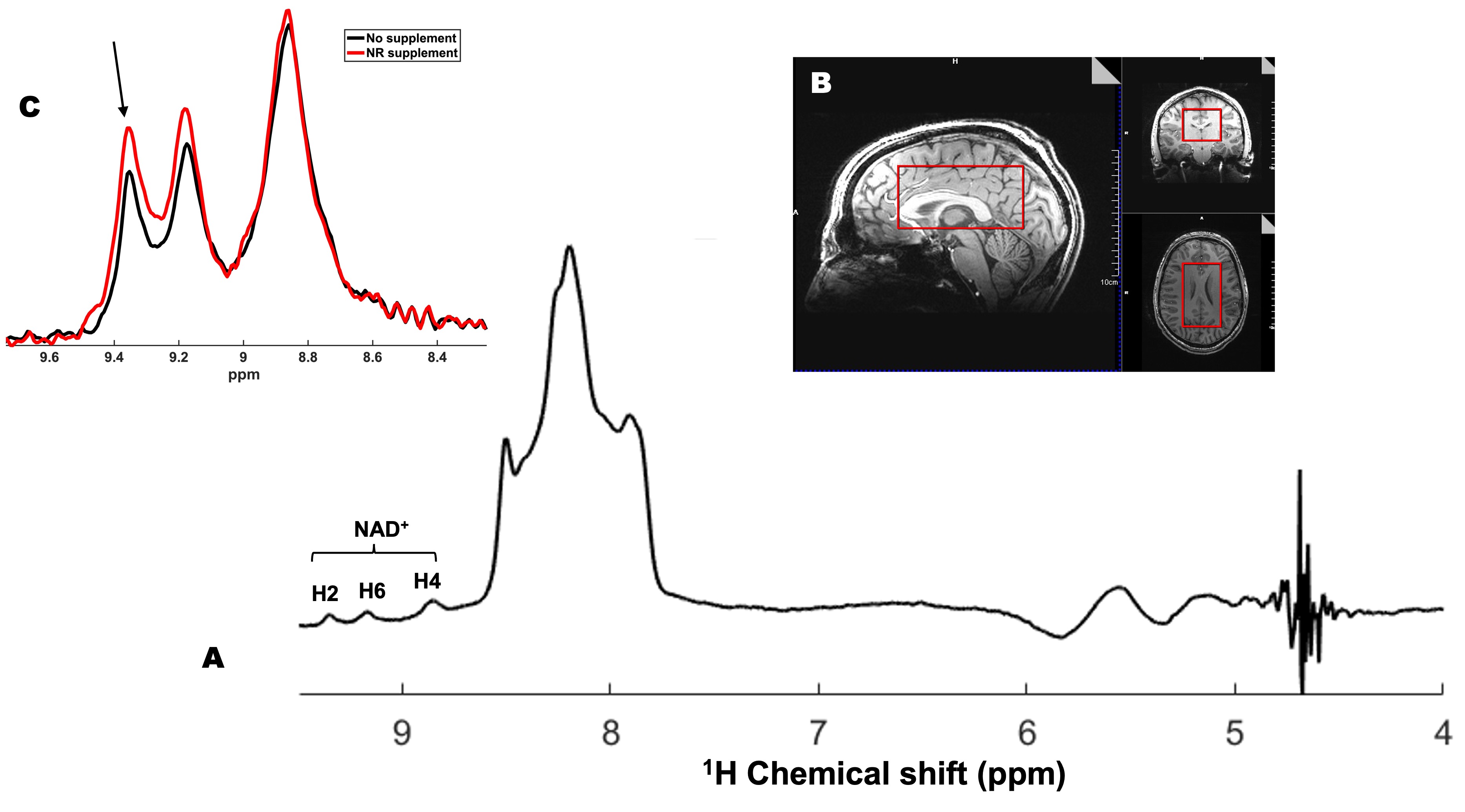
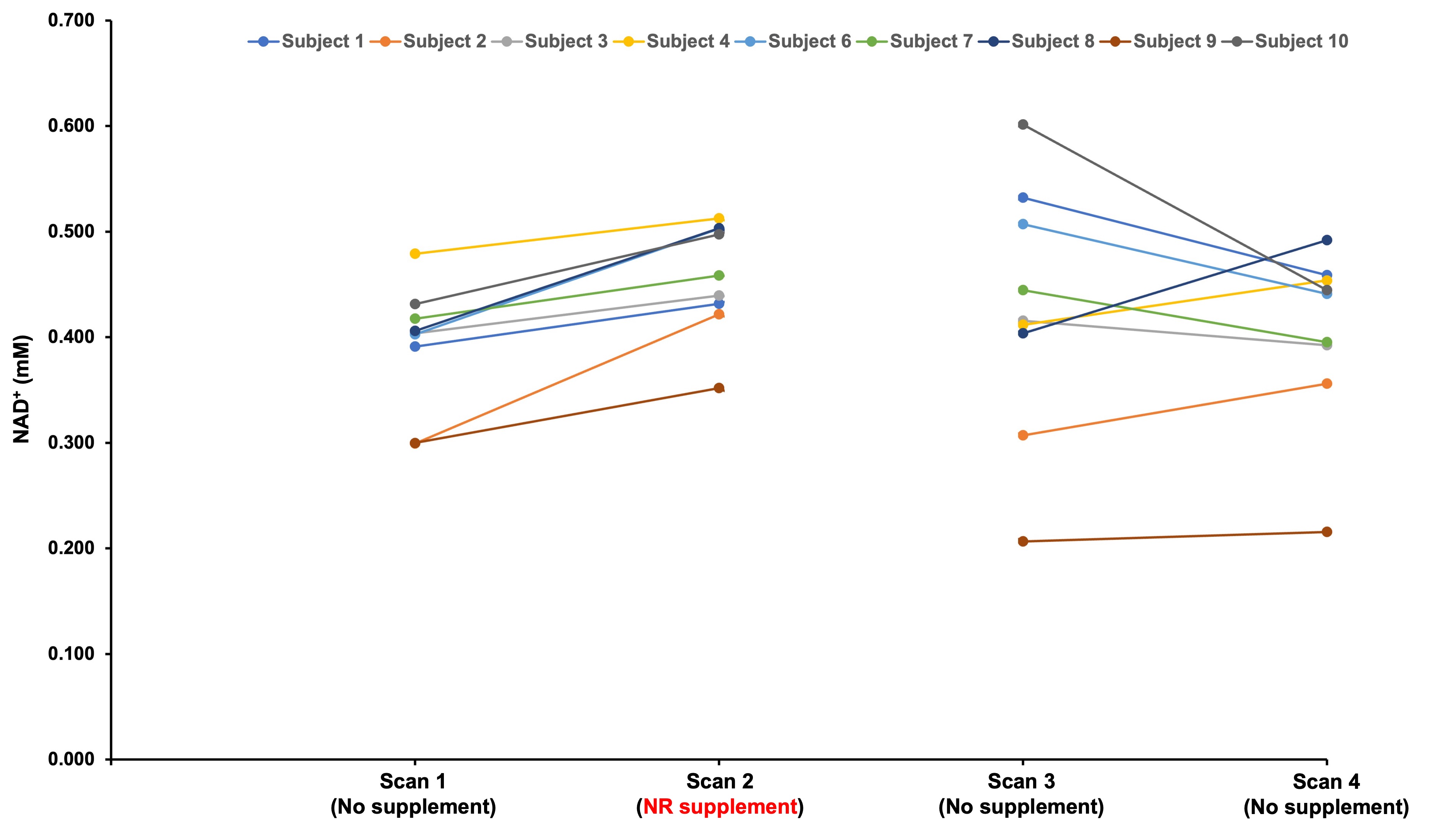
Data from one volunteer was omitted due to excessive motion artifacts. A representative NAD+ spectrum (1A) along with the voxel location superimposed on the anatomical image (1B) is shown in Figure 1. Also, shown in Figure 1C are the NAD+ spectra from Scan 1 (No supplementation) and Scan 2 (with NR supplementation) showing an increase in NAD+ levels with NR supplementation. Cerebral NAD+ levels from all the subjects are shown in Figure 2. There was a significant difference in cerebral NAD+ levels before (Mean±SD=0.392±0.058mM) and after taking NR supplements (Mean±SD=0.458±0.053mM); p<0.001 as shown in Figure 3. On average, cerebral NAD+ levels were 0.065 mM (or 16.6%) higher after taking NR supplements. There was no significant difference in cerebral NAD+ levels between the third scan (Mean±SD=0.425±0.118mM) and the fourth scan (Mean±SD=0.405±0.082mM); p=0.451. Additionally, the cerebral NAD+ levels across the three scans without NR supplementation were also not statistically different (p=0.443).Discussion
Our results demonstrate that oral NR supplementation increases NAD+ levels in the brain and demonstrates the potential of downfield 1HMRS for noninvasive quantification of cerebral NAD+ and for monitoring the effects of NR supplementation on cerebral NAD+ levels. Recently, phosphorous (31P) MRS was utilized to measure the cerebral NAD+ levels in vivo.18 But this study relies heavily on the one half of the overlapping quartet of NAD+ resonance with NADH and suffers from poor sensitivity and requires longer acquisition time (~15min).18 In contrast, downfield 1HMRS has higher sensitivity and spectral resolution as well as shorter acquisition time (~7min) compared to 31P MRS.Conclusion
The preliminary results from this study in a small sample show an increase in cerebral NAD+ levels in healthy volunteers following acute supplementation of NR and demonstrates the utility of downfield 1HMRS for robust detection of cerebral NAD+ levels in vivo.Acknowledgements
This work was supported by the National Institute of Biomedical Imaging and Bioengineering of the National Institutes of Health under award Number P41EB029460 and by the National Institute of Aging of the National Institutes of Health under award numbers R56AG062665, R01AG071725 and R01AG063869. This work is also supported by the National Heart Lung and Blood Institute under award number NIH HL137984.References
1. Stein LR, Imai S. The dynamic regulation of NAD metabolism in mitochondria. Trends Endocrinol Metab. 2012;23(9):420-8. Epub 20120721. doi: 10.1016/j.tem.2012.06.005. PubMed PMID: 22819213; PubMed Central PMCID: PMC3683958.
2. Bai P, Cantó C. The role of PARP-1 and PARP-2 enzymes in metabolic regulation and disease. Cell Metab. 2012;16(3):290-5. Epub 20120823. doi: 10.1016/j.cmet.2012.06.016. PubMed PMID: 22921416.
3. Gupte R, Liu Z, Kraus WL. PARPs and ADP-ribosylation: recent advances linking molecular functions to biological outcomes. Genes Dev. 2017;31(2):101-26. doi: 10.1101/gad.291518.116. PubMed PMID: 28202539; PubMed Central PMCID: PMC5322727.
4. Cohen MS, Chang P. Insights into the biogenesis, function, and regulation of ADP-ribosylation. Nat Chem Biol. 2018;14(3):236-43. doi: 10.1038/nchembio.2568. PubMed PMID: 29443986; PubMed Central PMCID: PMC5922452.
5. Houtkooper RH, Pirinen E, Auwerx J. Sirtuins as regulators of metabolism and healthspan. Nat Rev Mol Cell Biol. 2012;13(4):225-38. Epub 20120307. doi: 10.1038/nrm3293. PubMed PMID: 22395773; PubMed Central PMCID: PMC4872805.
6. Nikiforov A, Kulikova V, Ziegler M. The human NAD metabolome: Functions, metabolism and compartmentalization. Crit Rev Biochem Mol Biol. 2015;50(4):284-97. Epub 20150402. doi: 10.3109/10409238.2015.1028612. PubMed PMID: 25837229; PubMed Central PMCID: PMC4673589.
7. Katsyuba E, Romani M, Hofer D, Auwerx J. NAD. Nat Metab. 2020;2(1):9-31. Epub 20200120. doi: 10.1038/s42255-019-0161-5. PubMed PMID: 32694684.
8. McReynolds MR, Chellappa K, Baur JA. Age-related NAD. Exp Gerontol. 2020;134:110888. Epub 20200222. doi: 10.1016/j.exger.2020.110888. PubMed PMID: 32097708; PubMed Central PMCID: PMC7442590.
9. Xie X, Gao Y, Zeng M, Wang Y, Wei TF, Lu YB, et al. Nicotinamide ribose ameliorates cognitive impairment of aged and Alzheimer's disease model mice. Metab Brain Dis. 2019;34(1):353-66. Epub 20181206. doi: 10.1007/s11011-018-0346-8. PubMed PMID: 30523581.
10. Hou Y, Wei Y, Lautrup S, Yang B, Wang Y, Cordonnier S, et al. NAD + supplementation reduces neuroinflammation and cell senescence in a transgenic mouse model of Alzheimer’s disease via cGAS–STING. Proc Natl Acad Sci U S A. 2021;118(37):1-12. doi: 10.1073/pnas.2011226118. PubMed PMID: 34497121; PubMed Central PMCID: PMC8449423.
11. Hou Y, Lautrup S, Cordonnier S, Wang Y, Croteau DL, Zavala E, et al. NAD + supplementation normalizes key Alzheimer’s features and DNA damage responses in a new AD mouse model with introduced DNA repair deficiency. Proc Natl Acad Sci U S A. 2018;115(8):E1876-E85. Epub 20180205. doi: 10.1073/pnas.1718819115. PubMed PMID: 29432159; PubMed Central PMCID: PMC5828618.
12. Damgaard MV, Treebak JT. What is really known about the effects of nicotinamide riboside supplementation in humans. Sci Adv. 2023;9(29):eadi4862. doi: 10.1126/sciadv.adi4862. Epub 2023 Jul 21. PubMed PMID: 37478182; PubMed Central PMCID: PMC10361580.
13. Bagga P, Hariharan H, Wilson NE, Beer JC, Shinohara RT, Elliott MA, Baur JA, Marincola FM, Witschey WR, Haris M, Detre JA, Reddy R. Single-Voxel 1 H MR spectroscopy of cerebral nicotinamide adenine dinucleotide (NAD+) in humans at 7T using a 32-channel volume coil. Magn Reson Med. 2020 Mar;83(3):806-814. doi: 10.1002/mrm.27971. Epub 2019 Sep 10. PMID: 31502710; PMCID: PMC6879788.
14. Nanga RPR, Elliott MA, Swain A, Wilson N, Swago S, Soni ND, Witschey WR, Reddy R. Identification of l-Tryptophan by down-field 1 H MRS: A precursor for brain NAD+ and serotonin syntheses. Magn Reson Med. 2022 Dec;88(6):2371-2377. doi: 10.1002/mrm.29414. Epub 2022 Aug 25. PMID: 36005819; PMCID: PMC10165892.
15. Wolf DH, Satterthwaite TD, Loughead J, Pinkham A, Overton E, Elliott MA, et al. Amygdala abnormalities in first-degree relatives of individuals with schizophrenia unmasked by benzodiazepine challenge. Psychopharmacology (Berl). 2011;218(3):503-12. Epub 20110521. doi: 10.1007/s00213-011-2348-7. PubMed PMID: 21603892; PubMed Central PMCID: PMC3962022.
16. Barkhuijsen H, De Beer R, Van Ormondt D. Improved algorithm for non iterative time-domain model fitting to exponentially damped magnetic resonance signals. Journal of Magnetic Resonance. 1987;73:553-7.
17. Pijnappel WWF, van den Boogaart A, de Bee R, van Ormondt D. SVD-based quantification of magnetic resonance signals. Journal of Magnetic Resonance (1969). 1992;97(1):122-34.
18. Brakedal B, Dölle C, Riemer F, Ma Y, Nido GS, Skeie GO, Craven AR, Schwarzlmüller T, Brekke N, Diab J, Sverkeli L, Skjeie V, Varhaug K, Tysnes OB, Peng S, Haugarvoll K, Ziegler M, Grüner R, Eidelberg D, Tzoulis C. The NADPARK study: A randomized phase I trial of nicotinamide riboside supplementation in Parkinson's disease. Cell Metab. 2022 Mar 1;34(3):396-407.e6. doi: 10.1016/j.cmet.2022.02.001. PMID: 35235774.
Figures