4369
Correlation analysis between flortaucipir tau PET and quantitative susceptibility mapping in progressive supranuclear palsy1Department of Neurology, Mayo Clinic, Rochester, MN, United States, 2Department of Radiology, Mayo Clinic, Rochester, MN, United States
Synopsis
Keywords: Other Neurodegeneration, Neurodegeneration, tau
Motivation: To improve understanding of underlying mechanisms of flortaucipir tau PET uptake.
Goal(s): To clarify the relationship between flortaucipir uptake and iron deposition in progressive supranuclear palsy (PSP).
Approach: We performed a correlation analysis between flortaucipir PET and quantitative susceptibility mapping across subcortical regions in PSP.
Results: Positive correlations between flortaucipir uptake and susceptibility were found in most subcortical regions in PSP, suggesting the possibility that some flortaucipir uptake is associated with iron in these regions.
Impact: The positive correlation between flortaucipir PET uptake and magnetic susceptibility suggests that some flortaucipir uptake in subcortical structures may be associated with iron deposition in PSP. This finding improves our understanding of the underlying mechanisms of flortaucipir PET.
Introduction
Quantitative susceptibility mapping (QSM) has been widely used to study abnormal iron deposition in neurodegenerative diseases1. Several studies have suggested that iron is involved in tau aggregation in tauopathies such as Alzheimer’s disease (AD) and progressive supranuclear palsy (PSP)2, 3. Previous studies have investigated the relationship between QSM and tau PET in AD and found positive correlations in cortical and subcortical regions4. However, it remains unknown whether the relationship between these two modalities exists in other tauopathies such as PSP. Since the patterns of QSM and tau PET uptake are topographically similar in PSP; both are increased in the subcortical regions such as the basal ganglia and midbrain1, 5, we hypothesized that they would be strongly associated in these subcortical regions. The aim of this study is to investigate the relationships between these two modalities in PSP.Methods
SubjectsFifty PSP patients (26 females) were prospectively recruited and diagnosed according to the Movement Disorder Society (MDS)-PSP diagnostic criteria6. Of the 50 patients, 19 were diagnosed with PSP Richardson syndrome (PSP-RS), 22 with subcortical clinical variants of PSP (including parkinsonism, gait freezing and postural instability variants) and nine with cortical variants of PSP (including frontal, corticobasal and speech-language variants).
MRI and PET processing
All patients underwent 3 Tesla MRI and tau PET scans using standardized protocols. The Siemens MRI scan included a five-echo gradient echo (GrE) and magnetization-prepared rapid acquisition with gradient echo (MPRAGE) sequences7. QSM images were reconstructed from the GrE images by using MATLAB and STI suites8. Brain atlases for regional analysis were nonlinearly registered from the template space to each subject space by using Advanced Normalization Tools. A 20-minute PET acquisition was performed 80 minutes after the injection of approximately 370 MBq flortaucipir. Gray-matter cerebellar crus was used as the reference region to calculate standardized uptake value ratio (SUVR) images. Both QSM and SUVR images were spatially normalized and smoothed after rigid registration to MPRAGE images for the voxel-based analysis.
Regional and voxel-based correlation analysis
For regional analysis, mean susceptibility and SUVR were calculated in seven subcortical regions, including caudate, putamen, pallidum, subthalamic nucleus, substantia nigra, red nucleus, and cerebellar dentate, using brain atlases as previously reported9. Spearman's partial correlation coefficients between susceptibility and SUVR were calculated with age and sex as covariates in these seven regions. Voxel-based correlation analysis was performed by using VoxelStats software10. This software fits a linear regression in each voxel to assess the association between two modalities. Age and sex were again included as covariates in the linear model (SUVR ~ QSM + age + sex). Surviving regions were presented using the p<0.001 threshold and random field theory based multiple comparison correction10.
Results
In the regional analysis, magnetic susceptibility and flortaucipir SUVR were positively correlated in most subcortical regions, including putamen (r=0.37), pallidum (r=0.36), subthalamic nucleus (r=0.43), red nucleus (r=0.59), and cerebellar dentate (r=0.42) (FDR corrected p<0.05) (Figure 1), while no significant correlation was observed in caudate (r=0.23) and substantia nigra (r=0.19). The results of the voxel-based analysis were consistent with those of the regional analysis (Figure 2). A positive correlation between two signals was observed in the caudate, putamen, pallidum, thalamus, subthalamic nucleus, and red nucleus. In addition, a significant positive correlation was observed in the bilateral frontal white matter and the right fusiform gyrus.Discussion
We found a positive correlation between magnetic susceptibility and flortaucipir SUVR in most subcortical regions. These results suggest that some flortaucipir PET uptake is related to iron, given that estimated susceptibility is strongly correlated with iron concentration in the subcortical regions11. The positive correlations may be due to several reasons, including off-target binding of iron or iron-related proteins12 or the coexistence of iron and tau2, although this study cannot address the cause of the correlation. Voxel-based correlation analysis supported the regional analysis and additionally found a positive correlation in the bilateral frontal white matter. These frontal regions are consistent with the regions where a previous diffusion tensor study found white matter degeneration in PSP13. This positive correlation in the frontal white matter may be indicative of the relationship between tau uptake and white matter degeneration, given that myelin loss is likely to be associated with increased susceptibility. No correlation was found in the substantia nigra in either analysis, which may be confounded by other off-target substances such as neuromelanin14.Conclusion
Flortaucipir tau PET and QSM signals were positively correlated in subcortical regions in PSP, suggesting the possibility that some flortaucipir uptake is associated with iron.Acknowledgements
We would like to acknowledge AVID Radiopharmaceuticals for provision of AV-1451 precursor, chemistry production advice and oversight, and FDA regulatory cross-filing permission and documentation needed for this work. This study is supported by NIH grants R01-NS89757, R01-DC12519, R01-DC14942 and RF1-NS112153.References
1. Ravanfar P, Loi SM, Syeda WT, et al. Systematic review: Quantitative susceptibility mapping (QSM) of brain iron profile in neurodegenerative diseases. Front Neurosci. 2021;15:618435.2. Pérez M, Valpuesta JM, De Garcini EM, et al. Ferritin is associated with the aberrant tau filaments present in progressive supranuclear palsy. Am J Pathol. 1998;152(6):1531.
3. Yamamoto A, Shin RW, Hasegawa K, et al. Iron (III) induces aggregation of hyperphosphorylated tau and its reduction to iron (II) reverses the aggregation: implications in the formation of neurofibrillary tangles of Alzheimer's disease. J Neurochem. 2002;82(5):1137-1147.
4. Cogswell PM, Fan AP. Multimodal comparisons of QSM and PET in neurodegeneration and aging. Neuroimage. 2023;273:120068.
5. Whitwell JL, Lowe VJ, Tosakulwong N, et al. [(18) F]AV-1451 tau positron emission tomography in progressive supranuclear palsy. Mov Disord. 2017;32(1):124-133.
6. Hoglinger GU, Respondek G, Stamelou M, et al. Clinical diagnosis of progressive supranuclear palsy: The movement disorder society criteria. Mov Disord. 2017;32(6):853-864.
7. Satoh R, Arani A, Senjem ML, et al. Spatial patterns of elevated magnetic susceptibility in progressive apraxia of speech. Neuroimage Clin. 2023;38:103394.
8. Li W, Wang N, Yu F, et al. A method for estimating and removing streaking artifacts in quantitative susceptibility mapping. Neuroimage. 2015;108:111-122.
9. Satoh R, Weigand SD, Pham NTT, et al. Magnetic Susceptibility in Progressive Supranuclear Palsy Variants, Parkinson's Disease, and Corticobasal Syndrome. Mov Disord. 2023; in press.
10. Mathotaarachchi S, Wang S, Shin M, et al. VoxelStats: A MATLAB package for multi-modal voxel-wise brain image analysis. Front Neuroinform. 2016;10:20.
11. Langkammer C, Schweser F, Krebs N, et al. Quantitative susceptibility mapping (QSM) as a means to measure brain iron? A post mortem validation study. Neuroimage. 2012;62(3):1593-1599.
12. Choi JY, Cho H, Ahn SJ, et al. Off-target (18)F-AV-1451 binding in the basal ganglia correlates with age-related iron accumulation. J Nucl Med. 2018;59(1):117-120.
13. Whitwell JL, Schwarz CG, Reid RI, Kantarci K, Jack CR, Jr., Josephs KA. Diffusion tensor imaging comparison of progressive supranuclear palsy and corticobasal syndromes. Parkinsonism Relat Disord. 2014;20(5):493-498.
14. Lowe VJ, Curran G, Fang P, et al. An autoradiographic evaluation of AV-1451 Tau PET in dementia. Acta Neuropathol Commun. 2016;4(1):58.
Figures
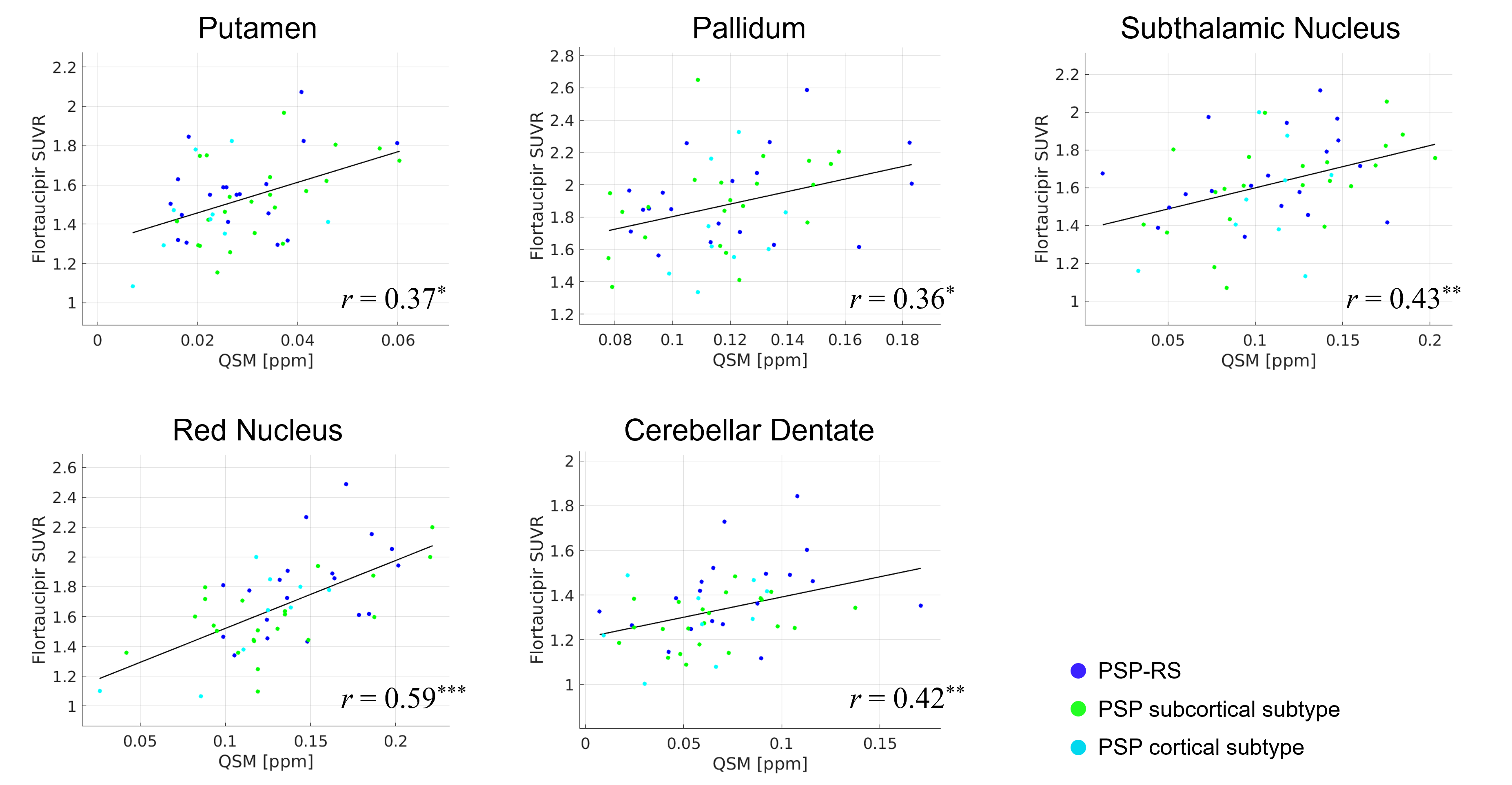
Figure 1. Positive correlations between magnetic susceptibility and flortaucipir SUVR in the subcortical regions. Asterisks indicate the level of statistical significance where p-values were adjusted for multiple comparisons of seven regions: ***p<0.001; **p<0.01; *p<0.05.
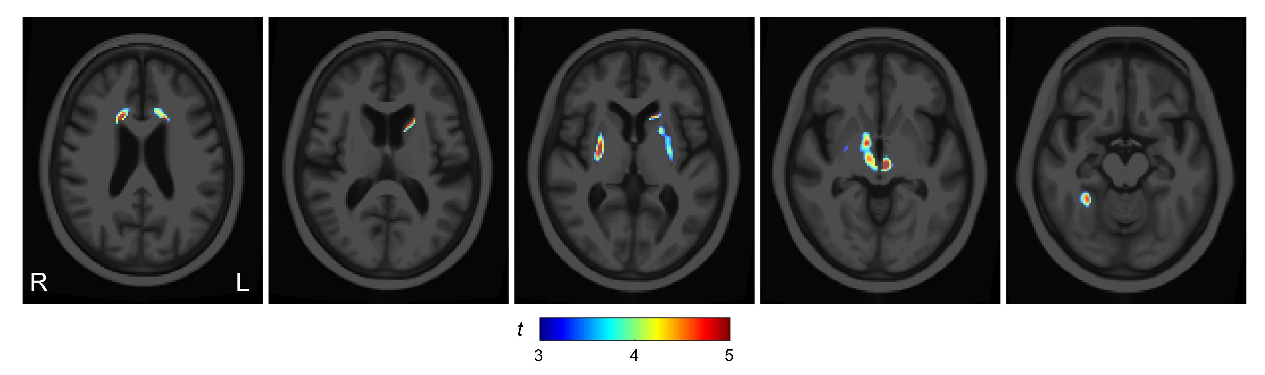
Figure 2. T-value map showing positive correlation (p<0.001) between magnetic susceptibility and flortaucipir SUVR in the voxel-based analysis. Surviving regions were shown after correction for multiple comparisons based on random field theory.