4364
Asymmetric impairment of hippocampus in patients with type 2 diabetes mellitus related with cognitive function1Shaanxi Provincial People's Hospital, Xi'an, China, 2Shaanxi University of Chinese Medicine, Xianyang, China, 3Xi'an Medical University, Xi'an, China, 4Philips Healthcare, Xi'an, China, 5Philips Healthcare, Shanghai, China
Synopsis
Keywords: Other Neurodegeneration, Arterial spin labelling
Motivation: To explore the relationship between hippocampal perfusion asymmetry and hippocampal enlarged perivascular spaces (H-EPVS) and cognitive function in patients with T2DM.
Goal(s): To explore the relationship between hippocampal perfusion asymmetry and H-EPVS and cognitive function in patients with T2DM.
Approach: CBF in hippocampus,H-EPVS and cognitive function were compared between HCs and patients with T2DM .The relationship between CBF in hippocampus, H-EPVS counts and cognitive measurement was analyzed.
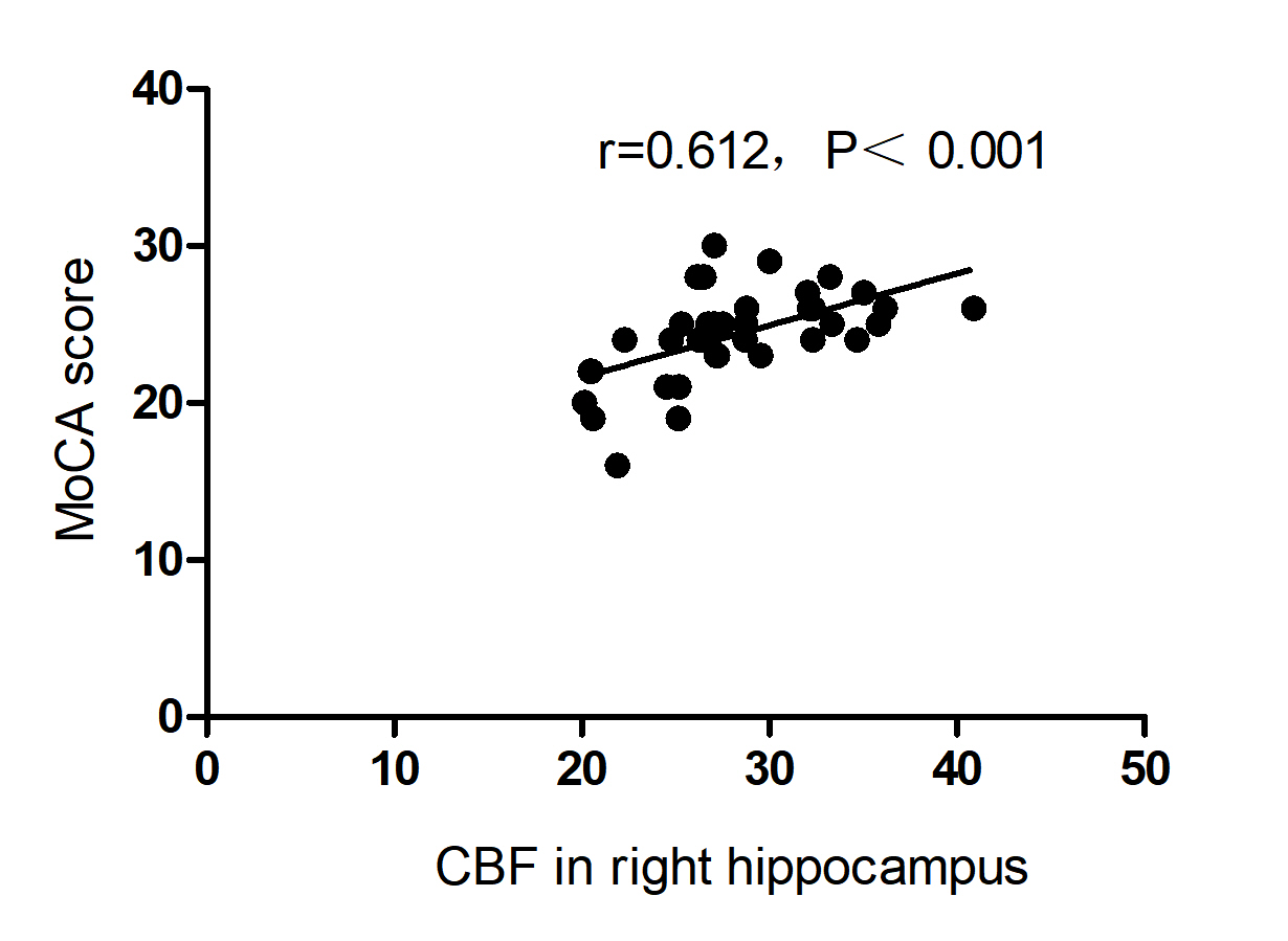
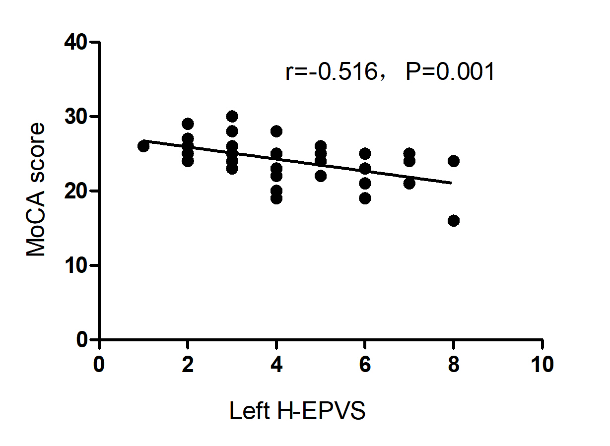
Results: In patients with T2DM, hippocampal perfusion was reduced and H-EPVS counts were higher on the left side. Decreased right hippocampal perfusion and increased left H-EPVS count were associated with cognitive impairment in patients with T2DM .
Impact: Asymmetric impairment of hippocampal in patients with T2DM , and the asymmetry may be caused by a variety of mechanisms and may contribute to cognitive impairment.
Introduction
Type 2 diabetes mellitus (T2DM) is an independent risk factor for cognitive impairment1. The underlying neuropathological mechanism of cognitive impairment in patients with T2DM remains unclear, and the decoupling of neurovascular units caused by abnormal cerebral blood perfusion is considered to be one of the mechanisms of cognitive impairment in patients with T2DM2. Enlarged perivascular spaces (EPVS) is considered to be a key anatomic marker of impaired neurovascular function3. Hippocampus is an important target of early cognitive impairment in patients with T2DM4. The aim of the study was to investigate the relationship between cerebral blood flow (CBF) in hippocampus, enlarged perivascular space in hippocampus (H-EPVS), and cognitive function in patients with T2DM using pseudo-continuous arterial spin labeling (pCASL).Methods
The study was approved by the local institutional Review Board. The study included 8 patients with T2DM and 31 age - and sex-matched healthy controls (HCs). All participants accept MRI and Montreal Cognitive Assessment (MoCA) . Routine MRI and pCASL are performed on a 3.0T scanner (Ingenia, Philips Healthcare, Netherlands) with a 32-channel head coil. The pCASL protocol is: TR/TE=250/16ms, turnover Angle =40◦, FOV=240mm×240mm, matrix size =80×80, slice thickness =6mm, total scanning time =248s. The CBF in left and right hippocampus was calculated using in-house software developed by Philips. For H-EPVS, the numbers of H-EPVS on the left and right sides were calculated in the sections with largest number of EPVS in the T2WI image.The CBF in hippocampus, H-EPVS count, clinical and cognitive function were compared between the two groups. With age and sex as covariables, multiple linear regression was used to analyze the correlation betweenl the CBF in hippocampus and H-EPVS and cognitive function. Multiple comparisons were corrected using Bonferroni correction. All tests were significant with P<0.05.Results
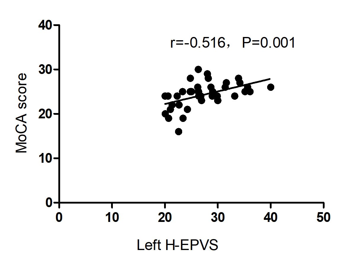
Compared with the HCs, the CBF in left hippocampus (P=0.037) and CBF in right hippocampus (P=0.037) were lower than those in patients with T2DM, and the H-EPVS count on the left side of patients with T2DM (P=0.009) was higher than that in the HCs. No significant difference was found in MoCA scores and other clinical data between the two groups (P > 0.05). Correlation analysis showed that left hippocampal CBF was negatively correlated with left H-EPVS (r=-0.567, P < 0.001), and CBF in right hippocampus was negatively correlated with right H-EPVS (r=-0.434, P=0.006). MoCA score was positively correlated with CBF in left hippocampus (r=0.529, P=0.001) and CBF in right hippocampus (r=0.612, P < 0.001), and negatively correlated with H-EPVS of the left hippocampus (r=-0.516, P=0.001). Using age and sex as covariables, multiple linear regression analysis showed that MoCA score was still correlated with CBF in right hippocampus (β=0.698, P=0.037) and H-EPVS of the left side (β=-0.329, P=0.033), but was no longer correlated with CBF of the left side.Discussion
The study showed that the patients with T2DM had lower CBF in the left and right hippocampus than HCs, and had higher left H-EPVS count.There was a correlation between them.CBF and H-EPVS in the left and right hippocampus were correlated with global cognitive function. Multiple linear regression showed that CBF in right hippocampus and left H-EPVS were still correlated with global cognitive function, while CBF in left hippocampus was no longer correlated. This may represent an asymmetry in hippocampal perfusion associated with cognitive dysfunction in patients with T2DM , and abnormal right hippocampal vascular perfusion are more closely related to cognitive function. This difference may be related to anatomical asymmetry, with perfusion in the right hemisphere of the brain higher than the left hemisphere in healthy adults 5. The difference between the left and right H-EPVS count and cognitive function in patients with T2DM may be related to hippocampal atrophy. Previous studies have found that patients with cognitive dysfunction show more significant brain atrophy in the left hemisphere 6, and the left hippocampus atrophy in patients with AD is also more significant than the contralateral one7,8. Brain atrophy itself is an independent risk factor for EPVS 9. Therefore, the left hippocampal atrophy may be more obvious in patients with T2DM with cognitive function impairment, and the H-EPVS count on the left side is higher than that on the contralateral side, which is worthy of further exploration and verification in future studies.Conclusion
The asymmetric impairment of hippocampus appeared in patients with T2DM.The CBF in right hippocampus and left H-EPVS were independently associated with cognitive dysfunction in T2DM patients. In T2DM patients, cognitive function is more sensitive to reduced right hippocampal perfusion. Left H-EPVS may be used as an imaging marker for early cognitive dysfunction in patients with T2DM .Acknowledgements
The authors thank all participants who volunteered for this study as well as the researchers who participated in this project.References
1. Shang Y, Fratiglioni L, Vetrano DL, et al. Not Only Diabetes but Also Prediabetes Leads to Functional Decline and Disability in Older Adults[J]. Diabetes Care, 2021, 44 (3): 690-698.
2.Jansen JF, van Bussel FC, van de Haar HJ, et al. Cerebral blood flow, blood supply, and cognition in Type 2 Diabetes Mellitus[J]. Sci Rep, 2016, 6 (1): 10.
3.Troili F, Cipollini V, Moci M, et al. Perivascular Unit: This Must Be the Place. The Anatomical Crossroad Between the Immune, Vascular and Nervous System[J]. Front Neuroanat, 2020, 14: 17.
4.Gupta M, Pandey S, Rumman M, et al. Molecular mechanisms underlying hyperglycemia associated cognitive decline[J]. IBRO Neurosci Rep, 2023, 14: 57-63.
5.Li QQ, Chen F, Zhong JG, et al. [Application of multiple post labeling delay time arterial spin labeling imaging in the quantitative blood flow analysis of brain subregions in healthy adults][J]. Zhonghua Nei Ke Za Zhi, 2022, 61 (8): 908-915.
6.Ezzati A, Katz MJ, Zammit AR, et al. Differential association of left and right hippocampal volumes with verbal episodic and spatial memory in older adults[J]. Neuropsychologia, 2016, 93 (Pt B): 380-385.
7.Li X, Li D, Li Q, et al. Hippocampal subfield volumetry in patients with subcortical vascular mild cognitive impairment[J]. Sci Rep, 2016, 6: 20873.
8.Dhikav V, Duraisamy S, Anand KS, et al. Hippocampal volumes among older Indian adults: Comparison with Alzheimer's disease and mild cognitive impairment[J]. Ann Indian Acad Neurol, 2016, 19 (2): 195-200.
9. Zhang X, Ding L, Yang L, et al. Brain Atrophy Correlates with Severe Enlarged Perivascular Spaces in Basal Ganglia among Lacunar Stroke Patients[J]. PLoS One, 2016, 11 (2): e0149593.
Figures