4361
Optimization of Deep Learning-Accelerated 3D-T1-MPRAGE and Quantitative Assessment of Regional Brain Volumes Compared to Wave-CAIPI MPRAGE1Siemens Medical Solutions, Boston, MA, United States, 2Department of Radiology, Massachusetts General Hospital, Boston, MA, United States, 3Siemens Healthcare GmbH, Erlangen, Germany
Synopsis
Keywords: Other Neurodegeneration, MR Value, AI/ML Image Reconstruction, Brain, Translational studies, Neurodegeneration
Motivation: Deep-learning-accelerated MPRAGE holds potential to enhance image resolution, reduce acquisition times, and improve diagnostic precision. However, there is currently a lack of clinical validation regarding its performance in neuroimaging.
Goal(s): To optimize and assess the performance of deep-learning-accelerated MPRAGE in comparison to Wave-CAIPI MPRAGE for non-contrast T1-weighted volumetric brain imaging.
Approach: In this prospective clinical study, we systematically optimized and implemented a novel deep-learning-accelerated MPRAGE sequence and compared against Wave-CAIPI MPRAGE, a state-of-the-art acceleration method.
Results: Deep-learning-accelerated MPRAGE enhances resolution and grey-white matter differentiation compared to Wave-CAIPI MPRAGE, with equivalent volumetric estimation in most brain regions.
Impact: Deep-learning-accelerated MPRAGE yields sharper, higher-resolution images while preserving equivalent volumetric estimations. This technique holds significant potential for deploying deep learning across various medical imaging disciplines, potentially enabling faster and more precise disease characterization.
Introduction
The 3D T1-weighted MPRAGE sequence has been widely used for assessing regional brain volume in patients with neurodegenerative diseases. Recent advances in acquisition techniques such as Wave-CAIPI have provided 2-3 fold acceleration for non-contrast1,2 while preserving diagnostic quality.Deep learning reconstruction has emerged as a powerful approach for accelerating image acquisition and improving image quality in MRI3,4. The application of these techniques in clinical settings especially for 3D sequences like MPRAGE remains underexplored.
The goal of this study was to optimize and evaluate a novel deep-learning-accelerated MPRAGE (DL-MPRAGE) sequence for volumetric brain MRI relative to state-of-the-art Wave-CAIPI MPRAGE.
Methods
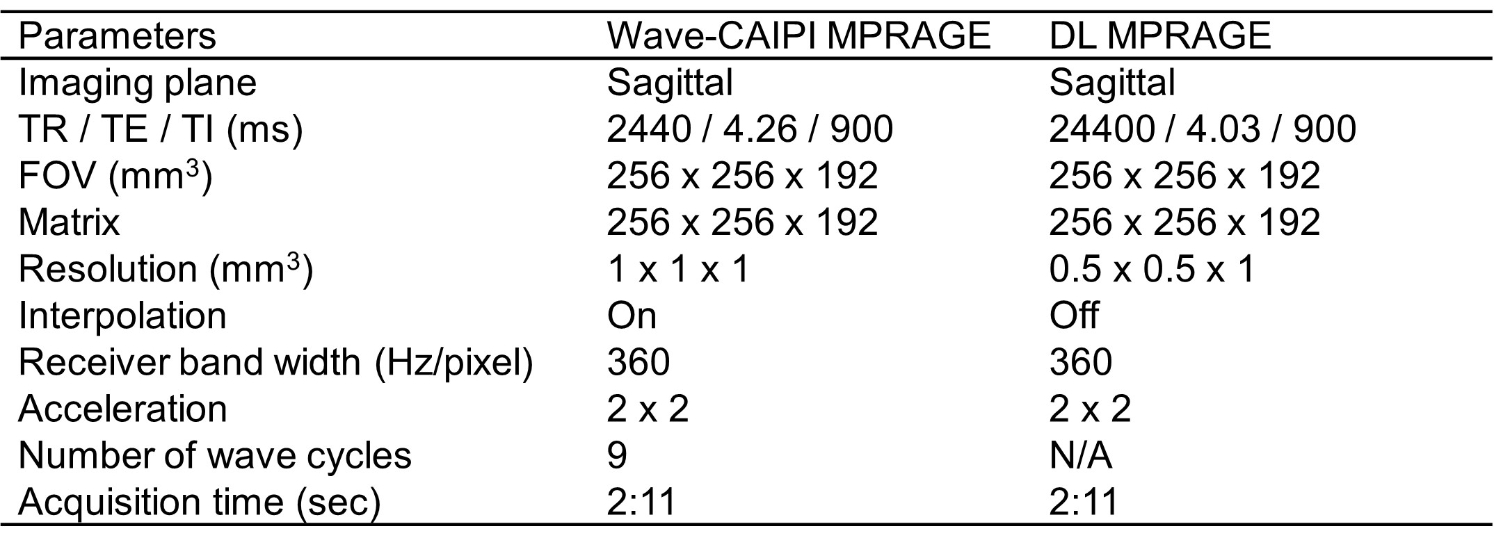
Data acquisitionThis prospective study was approved by the Mass General Brigham Institutional Review Board. A healthy adult volunteer was scanned with non-contrast 3D-T1-MPRAGE for optimization of DL settings. The optimized DL protocol was then implemented on three clinical 3T MRI scanners (MAGNETOM Vida, Siemens Healthineers, Erlangen, Germany) and run alongside Wave-CAIPI MPRAGE in an outpatient imaging facility. Adult patients undergoing brain MRI exams for evaluation of memory loss were recruited during October 2023. Acquisition parameters for Wave-CAIPI and DL-MPRAGE are provided in Table 1.
Image reconstruction
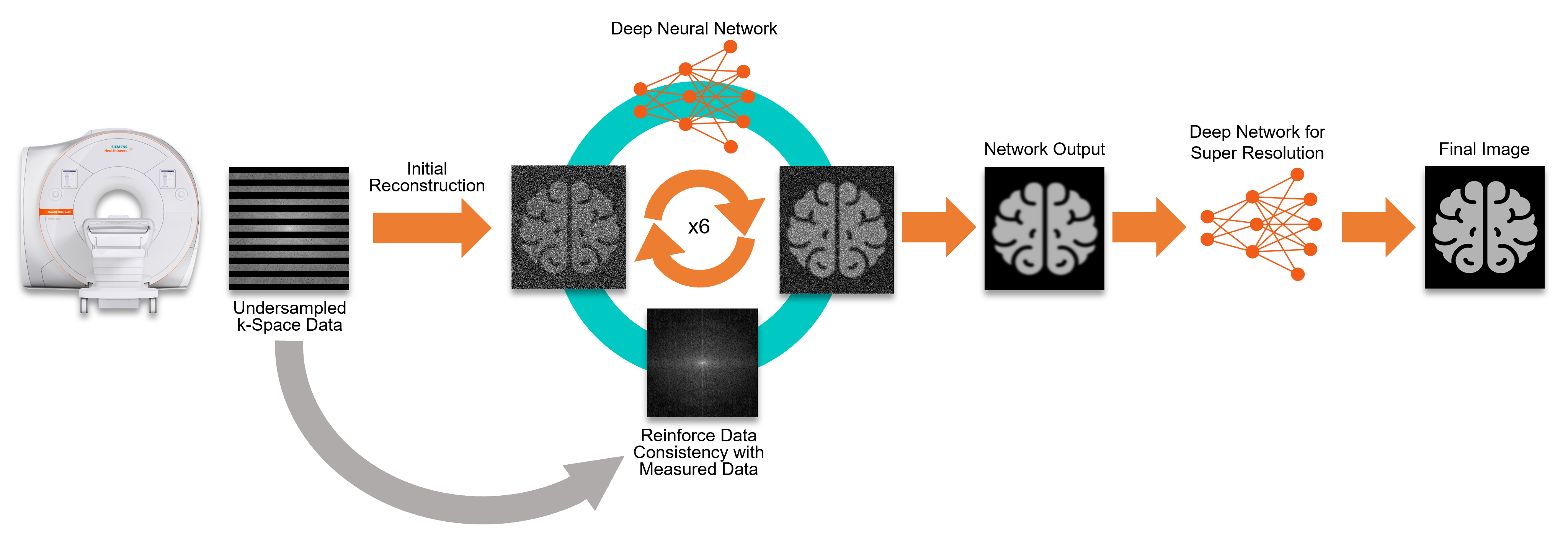
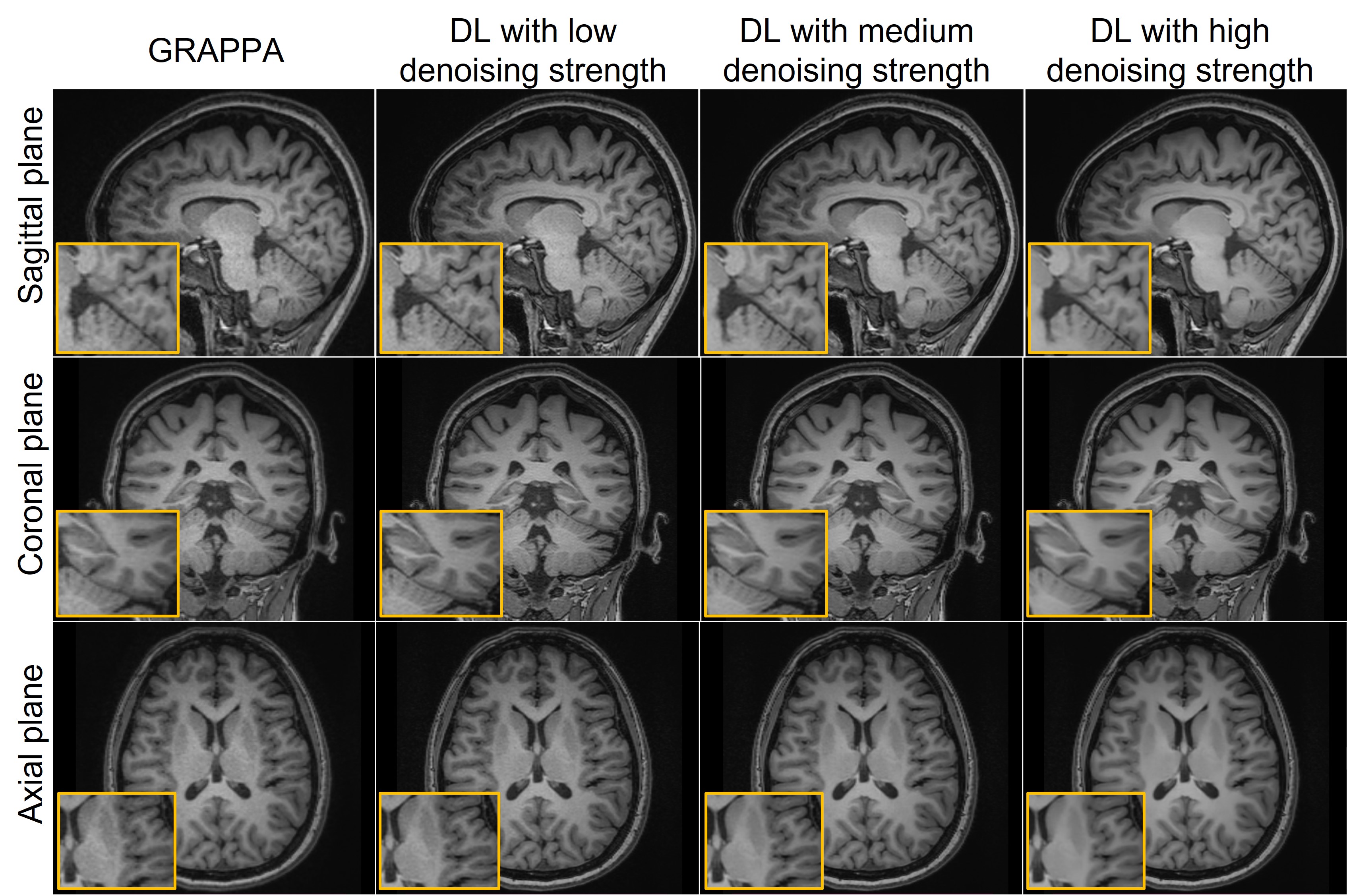
The research application DL-MPRAGE uses a two-step deep-learning-based image reconstruction (Figure 1). The first step, inspired by variational networks5, reconstructed images from undersampled k-space data and coil sensitivity maps. This process involved six iterations alternating between data consistency updates and neural network evaluations. Implementation in PyTorch and training with 5000 pairs of 500 fully sampled 3D datasets from healthy volunteers was carried out on 1.5 and 3T scanners (MAGNETOM scanners, Siemens Healthineers, Erlangen, Germany). The resulting network was integrated into the scanner's reconstruction pipeline using ONNX Runtime, processing a single volume in approximately 15 seconds. The second step involved a super-resolution algorithm for further enhancement6,7. Both steps were integrated into a research application for prospective use in the scanner's reconstruction pipeline. The research application was tested in healthy adult volunteer data and retrospectively reconstructed with conventional GRAPPA and DL-MPRAGE with mild, moderate, and strong denoising strength levels (Figure 2).
Quantitative evaluation – Non-contrast MPRAGE
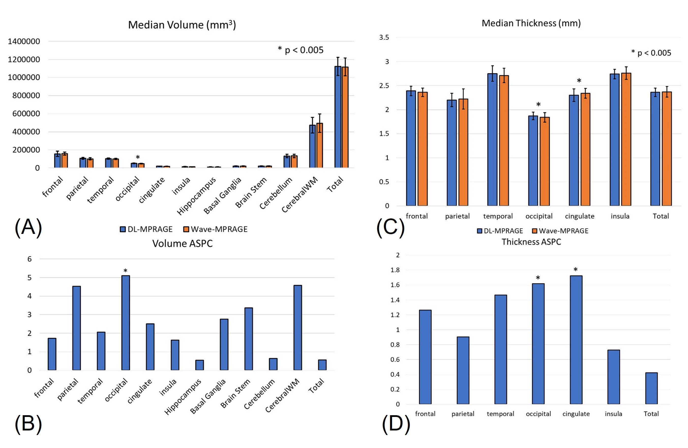
Cortical volume and thickness measurements were performed on non-contrast DL and Wave-CAIPI MPRAGE images using the longitudinal FreeSurfer pipeline8,9. Differences in cortical volume between the two sequences were quantified across patients using the absolute symmetrized percent change (ASPC)8. Significance for all comparisons was assessed using paired Wilcoxon rank sum tests with Bonferroni correction, with a significance threshold set at p < 0.005.
Results and Discussion
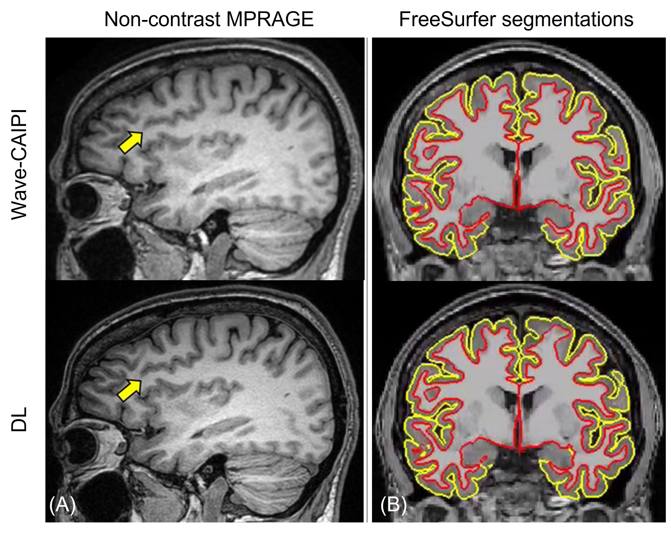
In the volunteer study, DL-MPRAGE at different denoising levels showed better overall image quality and clearer gray-white interface delineation as compared to conventional GRAPPA (Figure 2). The optimized protocol with low denoising strength and super-resolution was subsequently evaluated in a clinical setting.Nine patients (4F/5M, 60±21 years old) undergoing evaluation for memory loss were scanned with non-contrast DL and Wave-CAIPI MPRAGE. Automated FreeSurfer segmentations were qualitatively similar for both sequences across all subjects (Figure 3). Cortical volumes from DL and Wave-CAIPI MPRAGE showed no significant differences (p > 0.005), except in the occipital lobe where DL-MPRAGE demonstrated higher volumes (Figure 4). ASPC values averaged around 2.5%, peaking at 5.1% in the occipital lobe. Similarly, cortical thicknesses exhibited no significant differences (p > 0.005), except in the occipital lobe and cingulate gyrus. Here, DL-MPRAGE showed higher thicknesses in the occipital lobe and lower in the cingulate gyrus. ASPC values for thicknesses were lower than for volumes, with peak values at 1.6% and 1.7% in the occipital lobe and cingulate gyrus, respectively. The discrepancies observed in the occipital lobe and cingulate gyrus may be due to sharper delineation of the gray-white interface on the DL-MPRAGE images, which has been observed in other super-resolution reconstruction methods10.
Conclusion
The newly developed and optimized DL-MPRAGE acquisition demonstrates superior resolution and enhanced gray-white matter differentiation compared to state-of-the-art Wave-CAIPI MPRAGE. This technique also maintains equivalent volumetric measurements, indicating its potential to enable precise characterization of regional brain volumes, ultimately benefiting a broad spectrum of patients undergoing evaluation for neurodegenerative disorders. Separate studies are underway evaluating the diagnostic quality of DL-MPRAGE for assessment of intracranial enhancing lesions11.Acknowledgements
No acknowledgement found.References
1. Polak D, Setsompop K, Cauley SF, Gagoski BA, Bhat H, Maier F, et al. Wave-CAIPI for highly accelerated MP-RAGE imaging. Magn Reson Med. 2018;79:401-6.
2. Longo MGF, Conklin J, Cauley SF, Setsompop K, Tian Q, Polak D, et al. Evaluation of Ultrafast Wave-CAIPI MPRAGE for Visual Grading and Automated Measurement of Brain Tissue Volume. AJNR Am J Neuroradiol. 2020;41:1388-96.
3. Herrmann J, Koerzdoerfer G, Nickel D, Mostapha M, Nadar M, Gassenmaier S, Kuestner T, Othman AE. Feasibility and Implementation of a Deep Learning MR Reconstruction for TSE Sequences in Musculoskeletal Imaging. Diagnostics. 2021; 11(8):1484.
4. Herrmann, J., Wessling, D., Nickel, D., Arberet, S., Almansour, H., Afat, C, et al. Comprehensive clinical evaluation of a deep learning-accelerated, single-breath-hold abdominal HASTE at 1.5 T and 3 T. Academic Radiology, 2021, 30(1), 93-102.
5. Hammernik K, Klatzer T, Kobler E, et al. Learning a variational network for reconstruction of accelerated MRI data. Magn Reson Med 2018; 79: 3055–3071.
6. Afat S, Wessling D, Afat C et al (2022) Analysis of a Deep Learning-Based Superresolution Algorithm Tailored to Partial Fourier Gradient Echo Sequences of the Abdomen at 1.5 T: Reduction of Breath-Hold Time and Improvement of Image Quality. Invest Radiol 57:157-162
7. Wessling D, Herrmann J, Afat S, Nickel D, Almansour H, Keller G, Othman AE, Brendlin AS, Gassenmaier S. Application of a Deep Learning Algorithm for Combined Super-Resolution and Partial Fourier Reconstruction Including Time Reduction in T1-Weighted Precontrast and Postcontrast Gradient Echo Imaging of Abdominopelvic MR Imaging. Diagnostics (Basel). 2022 Sep 29;12(10):2370.
8. Reuter M, Schmansky NJ, Rosas HD, Fischl B. Within-subject template estimation for unbiased longitudinal image analysis. Neuroimage. 2012;61:1402-18.
9. Fischl B. FreeSurfer. Neuroimage. 2012;62:774-81.
10. Tian Q, Bilgic B, Fan Q, Ngamsombat C, Zaretskaya N, Fultz NE, et al. Improving in vivo human cerebral cortical surface reconstruction using data-driven super-resolution. Cereb Cortex. 2021;31:463-82.
11. Tabari A et al. Clinical Validation of Deep Learning-Accelerated vs. Wave-CAIPI Post-Contrast 3D-T1 MPRAGE for Evaluation of Intracranial Enhancing Lesions. Proc ISMRM. 2024 (submitted).
Figures