4360
Year-by-year White Matter Hyperintensity Probabilistic Atlases based on China Aging Cohort and its Application to Cognitive Decline Evaluation1School of Biomedical Engineering, ShanghaiTech University, Shanghai, China, 2The Second Affiliated Hospital, Zhejiang University School of Medicine, Hangzhou, China, 3Shanghai Clinical Research and Trial Center, Shanghai, China
Synopsis
Keywords: Aging, Aging, White matter hyperintensity; Cerebral small-vessel disease
Motivation: As frequently observed in older populations, white matter hyperintensity (WMH) appears a risk factor for cognitive decline.
Goal(s): To construct normative WMH probabilistic atlases for quantifying its spatiotemporal progression and for disease detection.
Approach: We released the first set of such atlases and related trajectories, based on a large cohort of the Chinese typical aging population, with AI techniques.
Results: We validated its clinical usefulness by comparing cognitive impairment groups with the atlases, as a new tool for early Alzheimer’s disease evaluation. We also found that the WMH in deep, rather than periventricular, white matter moderated aging-induced general cognitive ability reduction.
Impact: Given normative curves summarizing WHM progression in Chinese typical aging population, clinicians can better assess abnormalities related to cerebral small-vessel diseases, detect high-risk individuals who will develop cognitive decline in the future, and conduct large-scale community screening for older population.
Introduction
White matter hyperintensity (WMH) is a brain imaging sign of cerebral small-vessel diseases (CSVD). Presence of mild WMH can be found in most older adults, while moderate-to-severe WMH has been suggested an indication of cognitive impairments1. WMH is also independent prediction factor of stroke, and presents in populations with Alzheimer’s disease (AD) and its continuum spectrum2. Determining the threshold of pathological WMH is crucial for separating patients from normal older individuals, but unfortunately, lacking both normative references (e.g., atlases, trajectories) and clinical effectiveness evaluations. Building WMH probabilistic atlases will enable clinicians to quantify WMH load and spatial distribution by comparing the WMH lesions with normal ranges at each age3, thus facilitating risk assessment4. Existing WMH probabilistic atlases were mostly based on western populations5, and the first Asian WMH probabilistic atlases were established based on the South Korean population6, no such atlas was built based on the Chinese population. Due to differences in genetic and environmental factors, characterizing Chinese WMH progression and its effect on cognitive decline is in pressing need. To fill in this blank and explore the effect of WMH on cognition, we tried to construct the WMH probabilistic atlases based on the Chinese typical aging population, and to use the moderation effect to explain the effect of WMH.Method
To fill in the aforementioned research gap, we utilized the data of typical Chinese aging population from a large multicenter multimodal MRI database namely Chinese Brain Molecular and Functional Mapping (CBMFM). We used 735 MRI data from healthy aging older subjects (M/F, 341/394, age 20-79, no neurological diseases or psychological disorders) with paired T1w, T2-FLAIR images (acquired with uMR790, United Imaging) and intelligent quotient (IQ, Wechsler Intelligence Scale7, for measuring general cognitive function) to construct WMH probabilistic atlases.We trained a deep learning-based WMH segmentation model8 according to expert delineation. After validating of our model on 32 additional subjects, including 20 health and 12 AD patients, we applied it to all subjects. The atlases were built by averaging lesion maps within each age group, after aligning to the standard space using ANTs. We further divided total WMH into periventricular hyperintensity (PVH) and deep WMH (DWMH). The PVH was defined as those occurred within 10mm of the ventricular area9.
To quantify WMH progression during aging, exponential curves were adopted. The moderation effect of total WMH, PVH, and DWMH measurement on age-dependent IQ reduction was examined with PROCESS in SPSS. Of note, all WMH measures were adjusted considering individual variability in total intracranial volume (TIV, calculated with CAT12 based on T1w)9.
We also used a patient cohort, including 68 mild cognitive impairment (MCI) and 54 AD patients (age 40-79), to test the clinical effectiveness of our atlases and normative curves. A z-test was conducted between each patient and age-corresponding typical aging subjects, followed by a Chi-square test comparing the z-scores (i.e., abnormality level) of MCI group and typical aging group, and the AD group and typical aging group.
Results
Our WMH segmentation model reached 0.791 in testing dice accuracy. Fig. 1 shows the constructed WMH atlases that grow in both probability and spatial extent with aging.Fig. 2 delineates age-dependent total WMH, PVH, and DWMH curves in Chinese population, all grow exponentially with aging.
Fig. 3 depicts the moderation analysis result, confirming that aging-induced general cognitive function reduction is moderated by the severity of DWMH. No such moderation effect was observed for total WMH and PVH.
As shown in Table 1, 70-90% AD patients, and 50-70% MCI patients had more severe WMH compared with typical aging subjects at the same ages. Only 26-32% typical aging subjects had more severe WMH compared with other typical aging subjects. All patient groups had significantly higher probabilities of having more severe WMH for all WMH types, compared to the typical aging group.
Discussion
The WMH probabilistic atlases of the Chinese population and its progression trajectory were similar to the findings of other ethnic populations3,5,6. Our result indicates that DWMH might accelerate aging-related general cognitive ability decline, suggesting that more attention should be paid to the abnormal progression of DWMH to prevent cognitive impairment. Our WMH atlases and normative curves are crucial to clinical evaluation of early AD and large-scale early AD screening for community older populations. The entire procedure is automatic, quantitative, effective, warranting its promising future in intelligent medicine.Conclusions
We presented the first WMH spatiotemporal progression patterns for Chinese typical aging population. This normative model is informative for early detection of abnormal aging such as cognitive impairment and AD. The entire procedure is automatic, quantitative, and effective.Acknowledgements
This work is partially supported by the STI 2030—Major Projects (2022ZD0209000), Shanghai Zhangjiang National Innovation Demonstration Zone Special Funds for Major Projects “Human Brain Research Imaging Equipment Development and Demonstration Application Platform” (ZJ2018-ZD-012), Shanghai Pilot Program for Basic Research—Chinese Academy of Science, Shanghai Branch (JCYJ-SHFY-2022-014), Open Research Fund Program of National Innovation Center for Advanced Medical Devices (NMED2021ZD-01-001), Shenzhen Science and Technology Program (KCXFZ20211020163408012), and Shanghai Pujiang Program (21PJ1421400).References
1. Prins, N. D. & Scheltens, P. White matter hyperintensities, cognitive impairment and dementia: an update. Nature Reviews Neurology 11, 157-165 (2015).
2. Debette, S., Schilling, S., Duperron, M.-G., Larsson, S. C. & Markus, H. S. Clinical significance of magnetic resonance imaging markers of vascular brain injury: a systematic review and meta-analysis. JAMA neurology 76, 81-94 (2019).
3. Ryu, W.-S. et al. Grading and interpretation of white matter hyperintensities using statistical maps. Stroke 45, 3567-3575 (2014).
4. Qi, X. et al. White matter hyperintensities predict cognitive decline: a community-based study. Canadian Journal of Neurological Sciences 46, 383-388 (2019).
5. Enzinger, C. et al. Lesion probability maps of white matter hyperintensities in elderly individuals: results of the Austrian stroke prevention study. Journal of neurology 253, 1064-1070 (2006).
6. Kim, J. S. et al. Construction and validation of a cerebral white matter hyperintensity probability map of older Koreans. NeuroImage: Clinical 30, 102607 (2021).
7. Gong, Y.-x. Revision of Wechsler's Adult Intelligence Scale in China. Acta psychologica sinica (1983).
8. Isensee, F., Jaeger, P. F., Kohl, S. A. A., Petersen, J. & Maier-Hein, K. H. nnU-Net: a self-configuring method for deep learning-based biomedical image segmentation. Nat Methods 18, 203-211 (2021).
9. Griffanti, L. et al. Classification and characterization of periventricular and deep white matter hyperintensities on MRI: a study in older adults. Neuroimage 170, 174-181 (2018).
Figures
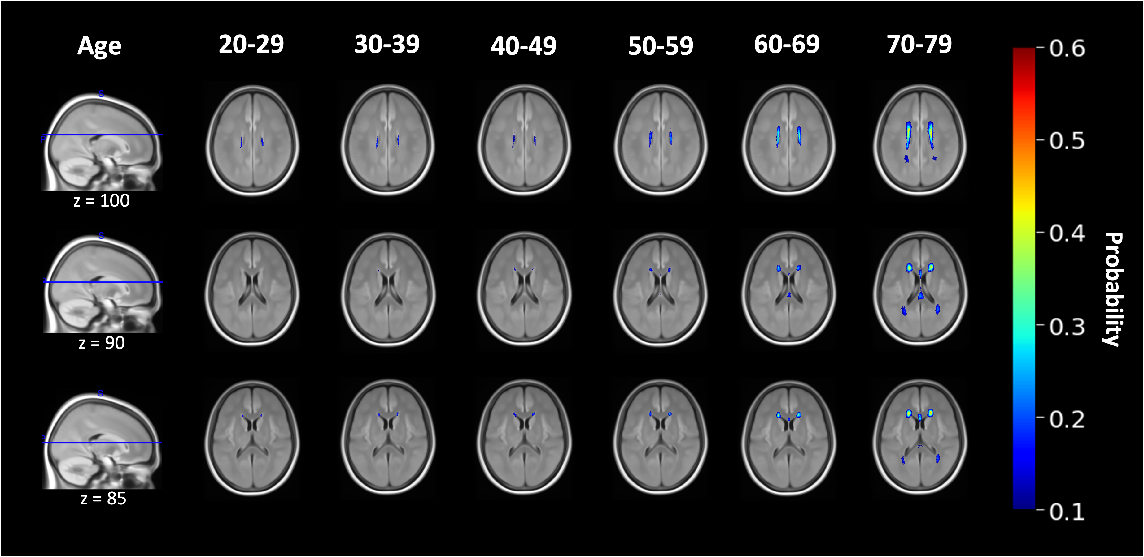
Fig 1. The constructed WMH atlases. WMH of high probability is mainly located near the ventricles.
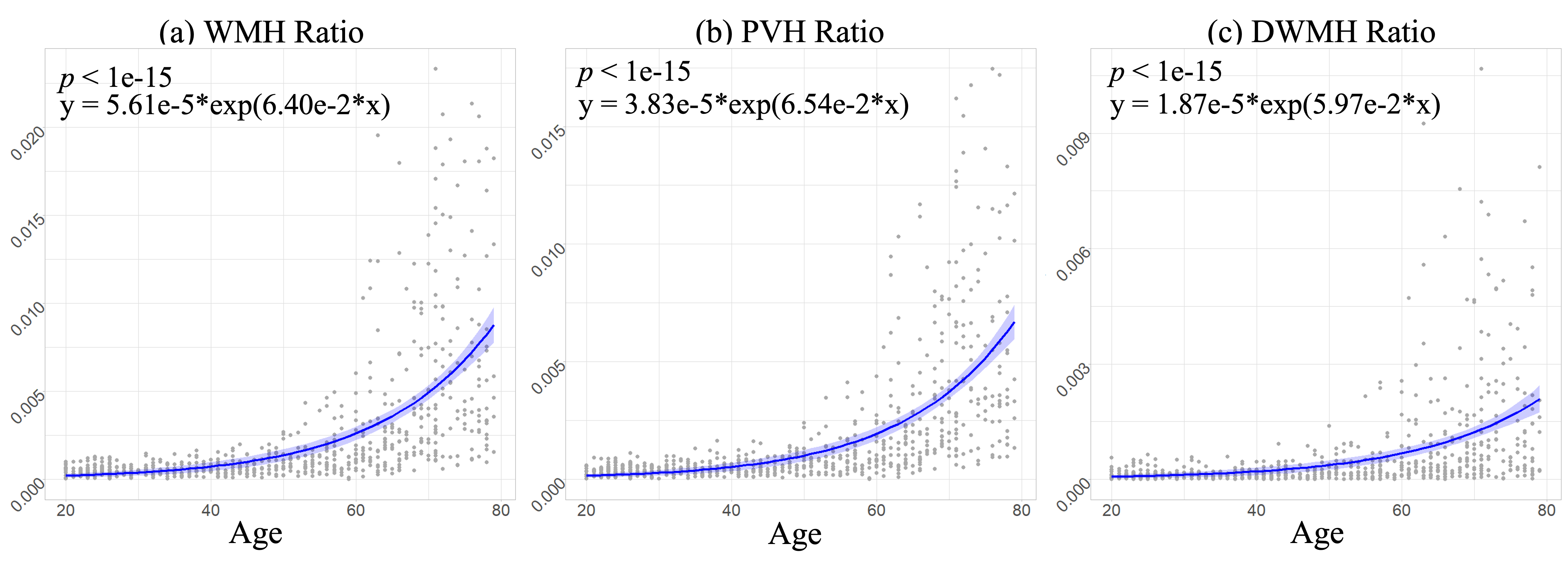
Fig 2. The WMH developmental curve with aging (shade indicates 95% C.I.).
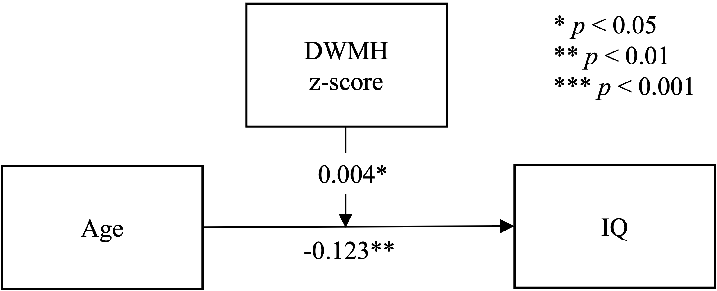
Fig 3. The result of moderation analysis. Sex, educational experience, and scanning sites are considered as covariates. Significant moderation effect is observed, where the z-score of the DWMH positively moderate the relationship between age and IQ.
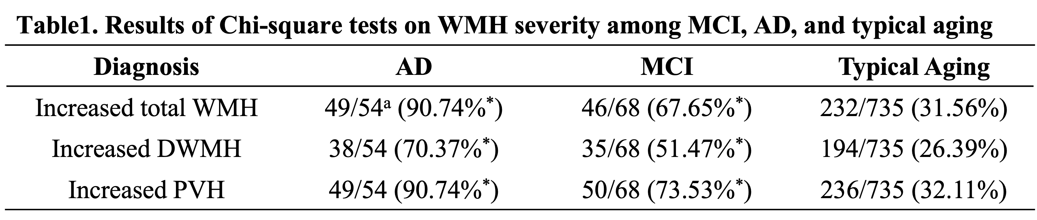
a49/54 (90.74%) means 49 out of 54 (90.74%) AD patients had significantly abnormal WMH as evaluated by our WMH normative reference (atlases and curves). * The WMH severity is significantly higher than that of the typical aging group (p <0.001).