4359
Wide-spectrum framework in Disorders of Arousal: combined resting state connectivity and metabolic study of cingulate cortex1IRCCS Istituto delle Scienze Neurologiche di Bologna, Bologna, Italy, 2Department of Medicine and Surgery, University of Parma, IT, Parma, Italy, 3Department of Biomedical and NeuroMotor Sciences, University of Bologna, IT, Bologna, Italy, 4Department of Medical and Surgical Sciences, University of Bologna, IT, Bologna, Italy, 5Department for Life Quality Studies, University of Bologna, IT, Bologna, Italy
Synopsis
Keywords: Other Neurodegeneration, Brain, Disorder of Arousal, Biomarkers, Brain Connectivity, fMRI (resting state), Functional connectivity, Metabolism, Neuroscience, Spectroscopy
Motivation: A comprehensive framework for the characterization of Disorders of Arousal (DoA) in adulthood is lacking, and the brain metabolic and functional mechanisms underlying this disorder are still unknown.
Goal(s): To fill this gap, we aimed to quantitatively investigate the cingulate cortex in DoA through advanced brain MRI, combined with thorough neuropsychological evaluations.
Approach: We collected and compared resting-state, 1H-MRS and clinical data from adult patients diagnosed with DoA and a matched group of healthy controls.
Results: We identified metabolic and functional alterations in regions involving the limbic system and changes in the connectivity of the sensorimotor network crucial for understanding the pathology.
Impact: Through a combined MR imaging investigation and psychological assessments, we extracted results in the form of brain metabolic and functional quantitative markers that advance our knowledge on the brain alterations underlying Disorders of Arousal.
Introduction
Disorders of Arousal (DoA) are parasomnias characterized by involuntary motor and emotional behaviors arising out of non-REM sleep. Psychological distress could be a precipitating factor although the relationship with DoA episodes is controversial1. In this study of adult DoA patients, we investigated both brain functional networks using resting state fMRI techniques, and metabolism in the cingulate cortex (CC), a key region in emotion and behavior regulation2, by means of 1H-MRS.Methods
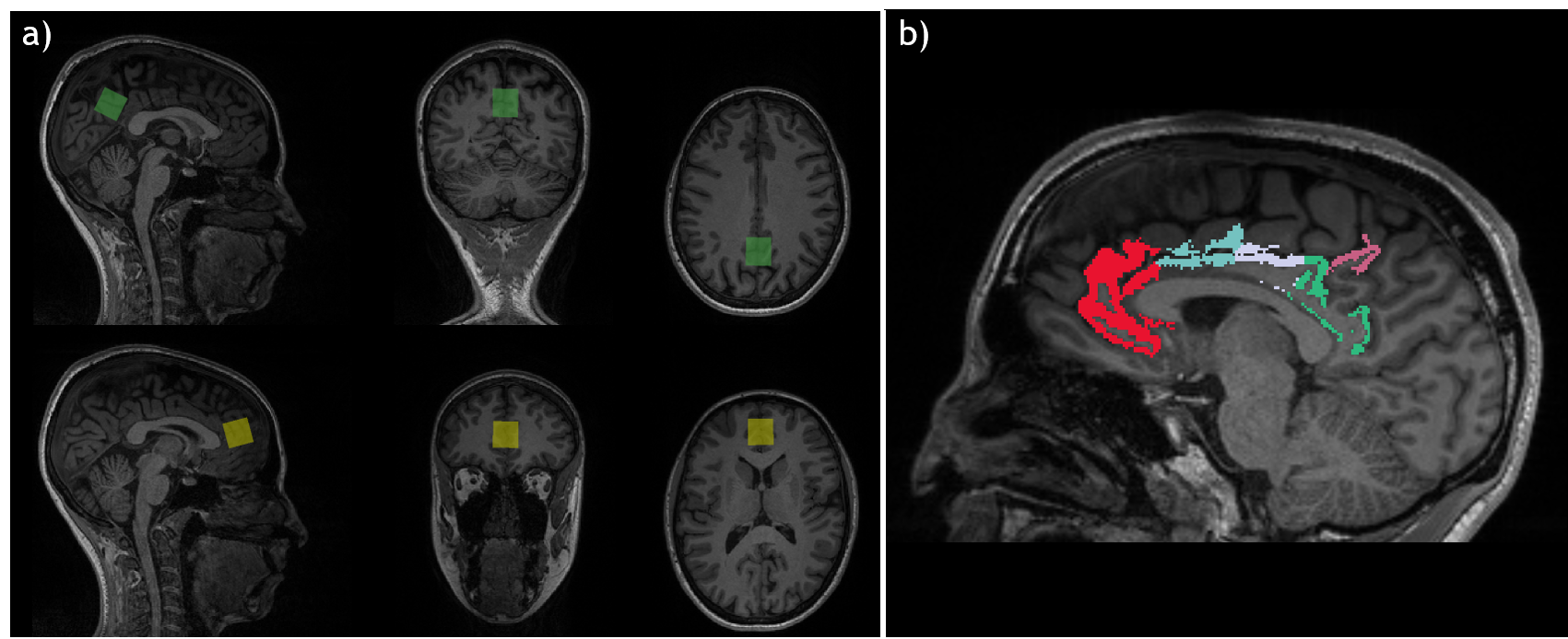
Fifteen adult patients with DoA (8F; 26.9±7.8yo, range 19-47yo) and 15 healthy controls (HC, 7F; 25.6±2.7yo, 21-30yo) were recruited. The same MRI and neuropsychological assessment protocol was administered to each participant. The MRI protocol (Siemens MAGNETOM Skyra 3T scanner with a head–neck high-density 64 channels array coil) included a 1-mm isotropic 3D MPRAGE-T1-weighted sequence, a resting-state fMRI (rs-fMRI) sequence and two single-voxel 1H-MRS PRESS sequences (TR/TE=2000/30,128 averages, 8 ml volume) within the anterior (ACC) and posterior cingulate cortices (PCC) (Figure 1a).FreeSurfer 5.3 was used to extract volumetric data in the sub-parcellations of the CC defined by the Destrieux atlas3(Figure 1b). Metabolite content was quantified with LCModel 6.3: N-acetyl-aspartate (NAA), choline (Cho), myo-Inositol (mI), glutamate (Glu), glutamine (Gln), and glutamate complex (Glx) related to creatine (Cr) or mI as internal reference were measured. The rs-fMR images were analysed via FSL4 tools using Seed-Based Analysis (SBA) of the FreeSurfer sub-parcellation of the CC and Independent Component Analysis (ICA) to study resting-state networks involving this region.
The normality of sample distributions was tested with Shapiro-Wilk test. Student’s t-test or Mann-Whitney tests were executed for neuropsychological assessments, ANCOVA was performed for morphometric data (covariates: total intracranial volume), and to compare metabolite ratios (covariates: sex, age). Dual regression coupled with general linear model was used to compare rs-fMRI maps (covariates: sex, age). Pearson/Spearman’s partial correlations, regressing out age, sex and education, were calculated between clinical scales and functional connectivity and spectroscopic results. Statistical significance was set at p<0.05 after false discovery rate or family-wise error (for rs-fMRI) correction for multiple comparisons.
Results
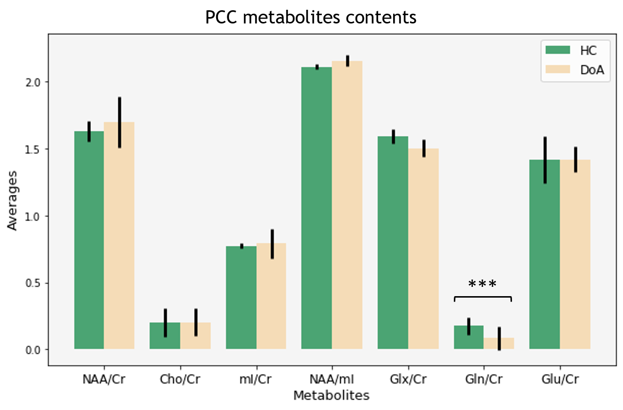
On neuropsychological testing, DoA patients displayed increased stress levels (P=0.03), a higher overall score of emotion dysregulation (P=0.04) and a borderline significance on some subitems of childhood trauma questionnaire (P=0.05).The CC sub-parcellation was not significantly different between DoA and HC. Patient metabolic profiles in the PCC (Figure 2) were characterized by a significant decrease of the Gln (P-adjusted<0.001), but not Glx. In the ACC no metabolic differences emerged. In the patient group, spectroscopic data did not correlate with clinical scales after correction for multiple comparisons. Considering the entire sample (DoA+HC), Glx significantly correlated with the DERS Impulse Control Difficulties scale (P-adjusted=0.008).
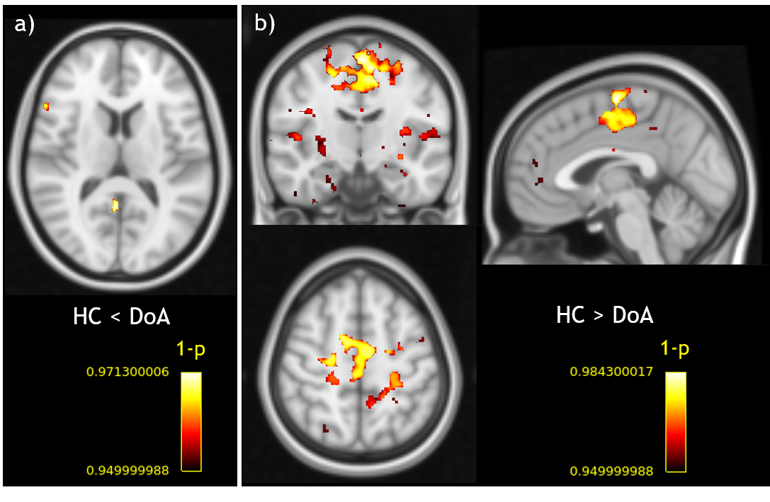
Using ICA, significantly increased connectivity between the sensory-motor network (SMN) with opercular cortex, praecuneus, occipital pole and lingual gyrus was found in patients (P-adjusted<0.05, Figure 3a). Moreover, lower PCC connectivity was found in extensive regions (Figure 3b), including motor areas, ACC, putamen, hippocampus and thalamus (P-adjusted<0.05). Among patients, no correlation with clinical data survived multiple comparison. In the full sample, SMN connectivity correlated with emotion regulation, sleep and depression scales (P-adjusted<0.04); posterior cingulate connectivity correlated with depression scores (P-adjusted<0.05).
Discussion
Cingulate cortex represents a key structure in the deep complexity of limbic system5, PCC appears as one of the most important multi-sensorial integration areas, involved in arousal and awareness processes. Our study showed metabolic alterations of PCC through 1H-MRS and disrupted functional connectivity of PCC and SMN. As glutamine is involved in excitatory and inhibitory activity, Gln deficiency in patients might account for functional alterations of stress control and, overall, an unbalanced emotion regulation. The metabolic alterations in PCC, not previously investigated by means of spectroscopy techniques, are consistent with the connectivity changes of this central hub, as SBA showed decreased connectivity of left dorsal PCC with several cortical and subcortical areas. The increased connectivity of SMN with frontal operculum and other regions of interest in resting-state condition may be related to a cortical “facilitation” of motor circuits, especially under unaware conditions. No volumetric alterations previously found in PCC6, and ACC metabolic variations have been confirmed in this study, possibly due to the small study sample and CC parcellation.Conclusions
New findings in DoA mechanisms, obtained combining MRI data and specific clinical assessments, revealed complex processes underlying arousal and awareness. The ACC normal metabolic profile might be useful to discriminate between subjects with parasomnias and those with similar motor signs such as frontal nocturnal epilepsy7. The increased connectivity of the SMN and the crucial role of the PCC in emotional dysregulation8 contribute to characterize DoA patients’ pathophysiology.Acknowledgements
No acknowledgement found.References
- Castelnovo, Anna, et al. "Behavioural and emotional profiles of children and adolescents with disorders of arousal." Journal of sleep research 30.1 (2021): e13188.
- Ramm, Markus, et al. "Increased behavioral inhibition trait and negative stress coping in non–rapid eye movement parasomnias." Journal of Clinical Sleep Medicine 16.10 (2020): 1737-1744.
- Destrieux, Christophe, et al. "Automatic parcellation of human cortical gyri and sulci using standard anatomical nomenclature." Neuroimage 53.1 (2010): 1-15.
- Jenkinson, Mark, et al. "Fsl." Neuroimage 62.2 (2012): 782-790.
- Vogt, Brent A. "The cingulate cortex in neurologic diseases: history, structure, overview." Handbook of Clinical Neurology 166 (2019): 3-21.
- Heidbreder, Anna, et al. "Gray matter abnormalities of the dorsal posterior cingulate in sleep walking." Sleep Medicine 36 (2017): 152-155.
- Naldi, Ilaria, et al. "Proton MR spectroscopy in patients with sleep-related hypermotor epilepsy (SHE): evidence of altered cingulate cortex metabolism." Sleep 40.9 (2017): zsx115.
- Gibbs, Steve A., et al. "Sleep-related epileptic behaviors and non-REM-related parasomnias: insights from stereo-EEG." Sleep Medicine Reviews 25 (2016): 4-20.
Figures