4358
Association of carotid stiffness and pulsatility using single-slice oblique-sagittal PC-MRI with cognitive impairment in elderly adults1Department of Radiology, Northwestern University, Chicago, IL, United States, 2Department of Biomedical Engineering, Northwestern University, Chicago, IL, United States, 3Department of Neurology, University of Southern California, Los Angeles, CA, United States, 4USC Mark and Mary Stevens Neuroimaging and Informatics Institut, University of Southern California, Los Angeles, CA, United States
Synopsis
Keywords: Dementia, Dementia
Motivation: Arterial stiffening and pulsatility serve as important markers of vascular dysfunction. Oblique-sagittal PC-MRI (OS PC-MRI) is a technique that provides a one-stop-shop approach for multiple vascular metrics.
Goal(s): This study aims to investigate the association of carotid vascular metrics measured by OS PC-MRI with cognitive impairment and cerebral perfusion in an elderly cohort.
Approach: OS PC-MRI data were collected on 40 elderly participants, who also underwent cognitive tests. Cerebral perfusion was measured using 3D pCASL. Pulse wave velocity(cPWV), pulsatility index(PI), and damping factor(DF) were calculated.
Results: Our results showed increased cPWV and reduced cDF were associated with cognitive impairment and reduced cerebral perfusion.
Impact: OS PC-MRI measures multiple vascular metrics including cPWV, PI, and cDF within two minutes, which shows strong associations with cognitive impairment and cerebral perfusion, consistent with previous findings. This study suggests OS PC-MRI could be promising to study vascular dysfunction.
Introduction
Arterial stiffening and pulsatility serve as important markers of vascular dysfunction1. Elevated carotid stiffness is associated with cognitive impairment and dementia. Increased carotid pulsatility quantified by pulsatility index (PI) is closely linked to aging, small vessel disease, and white matter hyperintensities2,3,4. Separate MRI scans at different carotid segments are typically performed to measure pulse wave velocity (PWV), which is prone to cardiac variations. Recent work has introduced a fast and robust PC-MRI technique that simultaneously imagesnarterial velocity waveforms along the common carotid artery (CCA) and the internal carotid artery (ICA) using a single-slice oblique-sagittal phase-contrast MRI (OS PC-MRI)5. OS PC-MRI provides multiple vascular metric measures, including carotid PWV (cPWV), PI of both ICA and CCA, and CCA-ICA damping factor (cDF). This study aims to investigate the association of these carotid vascular metrics measured by OS PC-MRI with cognitive impairment and cerebral perfusion in aged subjects.Methods
Participants and clinical assessmentsForty elderly participants (22 female,73.3±7.7 years) were enrolled in the study after providing written informed consent. Among them, 29 participants had Clinical Dementia Rating(CDR), 29 received Mini-Mental State Exam(MMSE), and 40 received Montreal Cognitive Assessment(MoCA).
MRI experiments
The MRI experiments were conducted on a Siemens Prisma 3T MRI scanner using a 20-channel head/neck coil. A 2min TOF MRA was performed to localize carotid arteries including CCA and ICA. The 3D MR angiogram was reformatted to determine an oblique slice to maximally cover both CCA and ICA segments as shown in Figures 1a and b. A single-slice retrospectively gated 2D OS PC-MRI with a single in-plane velocity encoding (CCA to ICA) was performed on each participant to acquire blood velocity waveforms along the CCA-ICA segment. Imaging parameters include spatial resolution=1x1x1mm3, VENC=80cm/s, TE/TR=4.32/14.22ms, flip angle=10°, real temporal resolution=14.22ms, 70-90 phases across a cardiac cycle, scan time was 1 to 2min depending on the heart rate. pCASL with background suppressed 3D GRASE was performed on 25 participants.
Image processing and statistical analysis
A reference waveform was calculated as the average of velocity waveforms obtained from all axial locations along ICA-CCA. The transit time between each waveform and reference waveform was calculated using time-to-foot (TTF) method5. cPWV was calculated as inverse slope of the line fitted to the transit time versus distance along the vessel (Figure 1c). PI was calculated from the average velocity waveforms along each segment i.g., CCA, ICA (Figure 1e). cDF was calculated as ratio of the average PI values between CCA and ICA (Figure 1f). According to the results of the normality test, the correlations of carotid vascular metrics with cognitive measures and CBF were calculated using Pearson or Spearman's correlation. Age, gender, and education were considered as covariances using partial correlation.
Results and Discussion
Table 1 lists the demographic information and clinical assessments of participants. cPWV showed significant negative correlations with MoCA and MMSE (cPWV vs. MoCA: r = -0.4, p = 0.01; cPWV vs. MMSE: r = -0.57, p = 0.008), and the correlations remained significant after controlling for age, gender, years of education (cPWV vs. MoCA: r = -0.36, p = 0.03; cPWV vs. MMSE: r = -0.53, p = 0.005). Furthermore, the participants with CDR>0 (n=19) showed higher cPWV values compared to those with CDR=0 (n=21) (p=0.0045). These results provide convergent evidence that elevated cPWV is strongly associated with cognitive decline.No significant correlations were found between the average PI values at both ICA and CCA with cognitive measures, which may be caused by the variations along vessel segments during the PI measurements. However, a significant negative correlation between cDF and MoCA was observed (p = 0.036) (Figure 3). This finding indicates cDF could be a sensitive vascular marker for cognitive impairment.
Elevated cPWV was strongly associated with CBF in gray matter (p = 0.02) and multiple regions, such as hippocampus (p = 0.007) (Figure 4), suggesting carotid arterial stiffening and increased pulsation may cause cerebral microvascular and neurological dysfunctions. A similar trend was observed on cDF, although there was no significance (p=0.5, 0.097).
Conclusion
OS PC-MRI provides multiple vascular metrics including cPWV, PI, and cDF. Our results showed that both elevated carotid PWV and DF were strongly associated with cognitive impairment and downstream cerebral perfusion in elderly adults using the OS PC-MRI, further supporting that arterial stiffening with reduced damping leads to excessive transmission of pulse energy to downstream vasculature resulting in microcirculation dysfunction and thus cognitive impairment. This study also indicates that OS PC-MRI could be a promising imaging tool to assess intracranial arterial stiffness and pulsatility, which can serve as sensitive imaging markers for cognitive impairment and dementia.Acknowledgements
This work was partly supported by National Institute of Health (NIH) grants R01NS118019, RF1AG072490, and BrightFocus Foundation A20201411S.References
1. Van Sloten, Thomas T., et al. "Carotid stiffness is associated with incident stroke: a systematic review and individual participant data meta-analysis." Journal of the American College of Cardiology 66.19 (2015): 2116-2125.
2. Lau, Kui Kai, et al. "Age and sex-specific associations of carotid pulsatility with small vessel disease burden in transient ischemic attack and ischemic stroke." International Journal of Stroke 13.8 (2018): 832-839.
3. Aribisala, Benjamin S., et al. "Blood pressure, internal carotid artery flow parameters, and age-related white matter hyperintensities." Hypertension 63.5 (2014): 1011-1018.
4. Shi, Yulu, et al. "Small vessel disease is associated with altered cerebrovascular pulsatility but not resting cerebral blood flow." Journal of Cerebral Blood Flow & Metabolism 40.1 (2020): 85-99.
5. Heidari Pahlavian S, Cen SY, Bi X, Wang DJJ, Chui HC, Yan L. Assessment of carotid stiffness by measuring carotid pulse wave velocity using a single-slice oblique-sagittal phase-contrast MRI. Magn Reson Med. 2021;86(1):442-455. doi:10.1002/mrm.28677
Figures
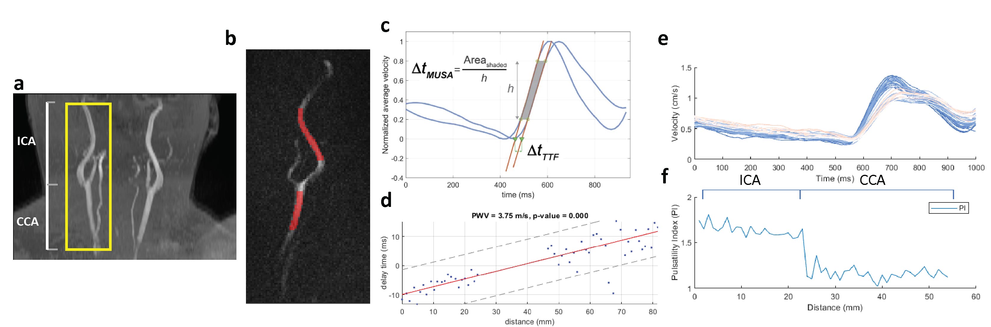
Figure 1. a. 3D time-of-flight angiogram. A yellow box indicates the image plane of OS PC-MRI covering the ICA and CCA segments; b. The OS PC-MRI image with the CCA-ICA ROI marked in red; c. Transit time calculation by the TTF method; d. The scatter plots of transit time (or delay time) versus vessel distance which generates the cPWV value; e. Averaged velocity curves along CCA-ICA f. Caluclated PI along CCA-ICA. DF is the ratio of the average PI in CCA and ICA segment.
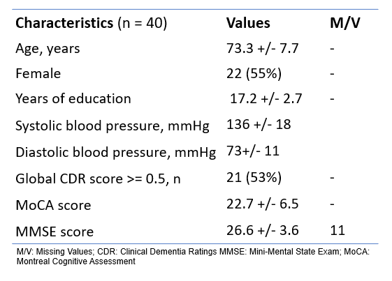
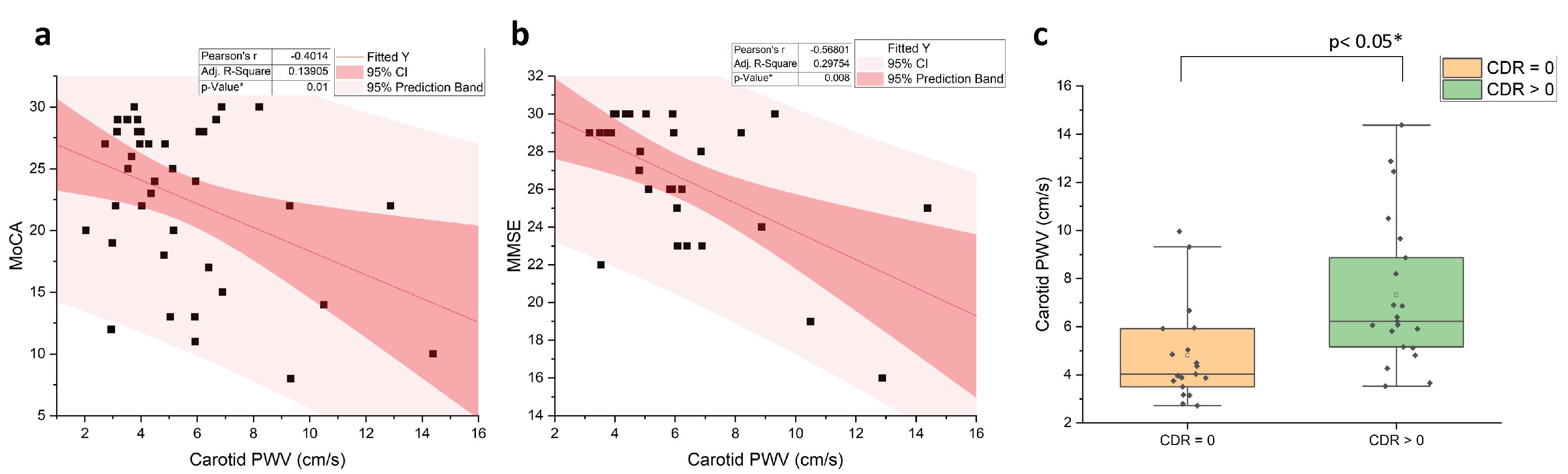
Figure 2. Scatter plots of PWV values with MoCA scores (a) and MMSE scores (b) across subjects; c. Box plot shows group comparison between CDR = 0 and CDR > 0.
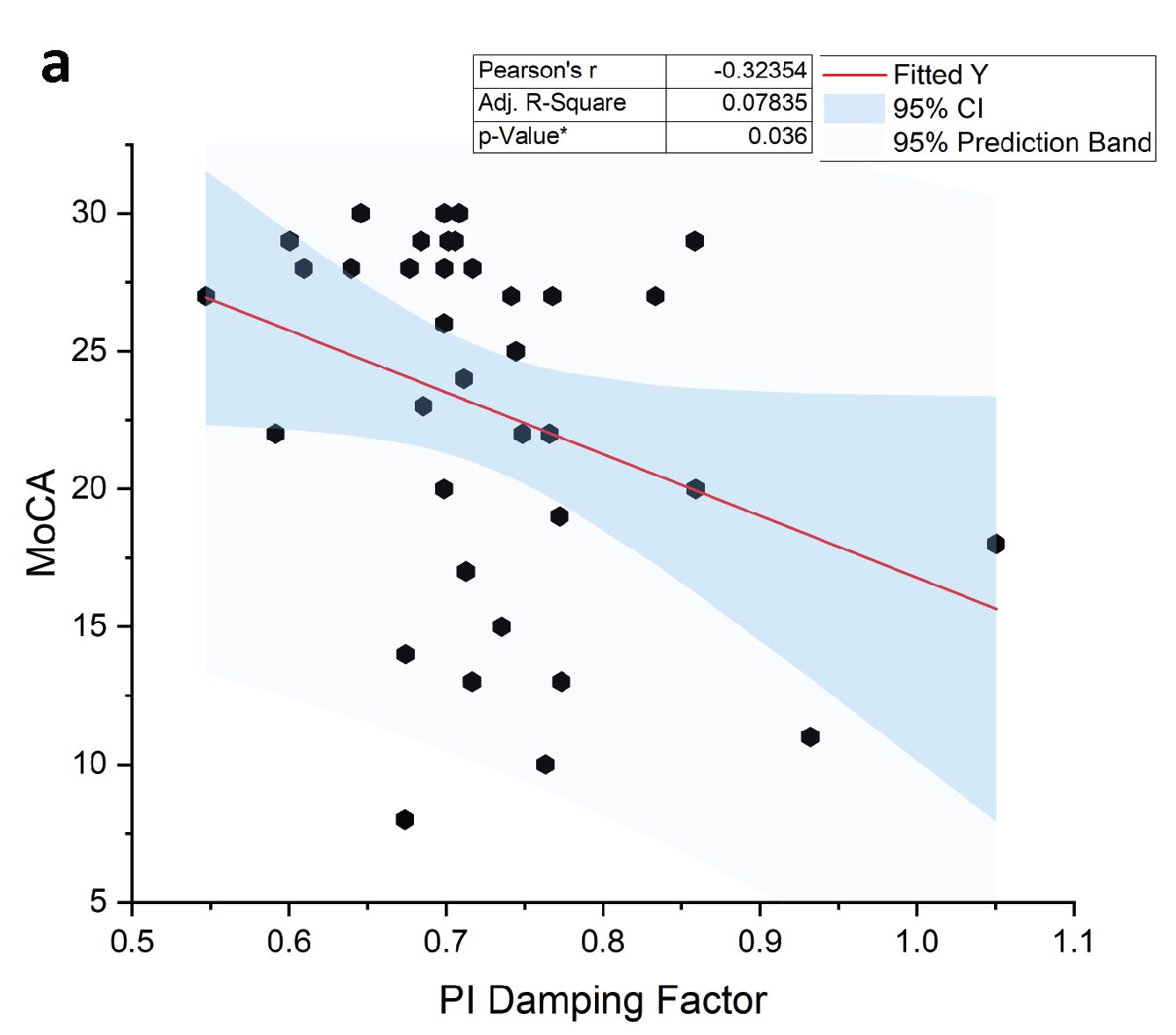
Figure 3. Scatter plot of cDF with MoCA scores.
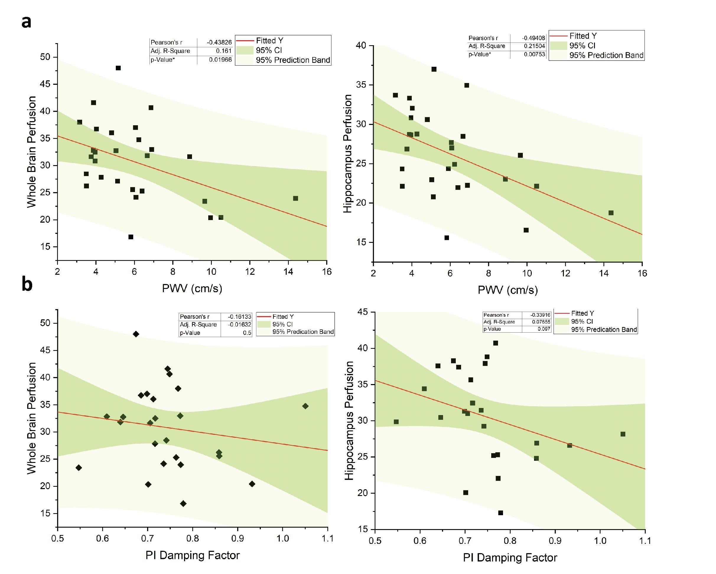
Figure 4. Scatter plots of PWV (a) and cDF (b) with global perfusion (left) and hippocampus perfusion (right), respectively.