4353
Reduced FOV Neuromelanin Imaging at 7T using Saturation Bands1Mayo Clinic Florida, Jacksonville, FL, United States, 2Siemens Medical Solutions USA, Jacksonville, FL, United States
Synopsis
Keywords: Parkinson's Disease, Parkinson's Disease, 7T; Neuromelanin imaging; reduced FOV; 3D GRE; SNc; LC; MT; Saturation band
Motivation: GRE based neuromelanin (NM) imaging at 7T is sensitive to B0 field disturbance by patient breathing or even mild head motion.
Goal(s): A reduced FOV method is proposed for 7T NM imaging using 3D GRE with three saturation bands for creating MT contrast and reducing ghosting artifacts.
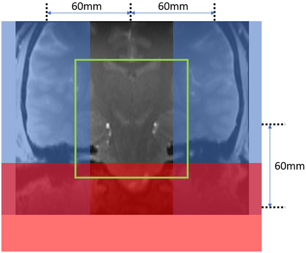
Approach: The sequence has bilateral sagittal saturation bands placed in close proximity to the SNc for artifacts and FOV reduction, and the 3rd axial saturation band placed below the SNc for arterial blood suppression.
Results: The method yields higher resolution, enhanced NM contrast, and a reduction in artifacts.
Impact: The reduced FOV neuromelanin (NM) imaging method using three saturation bands offers higher resolution NM imaging at 7T with improved NM contrast and image quality.
Introduction
GRE based Neuromelanin (NM) imaging at 7T is sensitive to variation of B0 field disturbed by patient breathing or even mild head motion. The ghosting artifacts arising from field variation and blood flow pulsation can significantly impact the accuracy of quantitative analysis of NM images. In our 7T Parkinson disease (PD) protocol development, we noticed that placing saturation bands near the midbrain can effectively mitigate the aforementioned artifacts. Furthermore, these saturation bands offer better magnetization transfer (MT) effects than the default MT pulse. Previous study at 3T also suggested positioning superior and inferior saturation bands near the NM imaging slab can improve the contrast between NM-containing tissue and its surrounding structures compared to the default MT pulse[1]. In this work, in addition to assess the MT effects by the saturation bands, we propose a reduced FOV method with three saturation bands for NM imaging: bilateral sagittal saturation bands placed in close proximity to the midbrain to reduce image FOV and ghost artifacts in the phase encoding direction, and a third inferior band to suppress the incoming arterial blood signal.Purpose
To evaluate the reduced FOV method for 7T NM imaging using saturation bands for creating MT contrast and reducing ghosting artifacts.Method
In compliance with local institutional regulations, volunteer scans were performed on a Siemens 7T MAGNETOM Terra (Siemens Healthcare, Erlangen, Germany) equipped with Nova 8Tx/32Rx head coil (Nova Medical Inc, Wilmington MA, USA) under TrueForm B1 shimming mode. The proposed reduced FOV 3D GRE sequence has: FOV=80×80mm, slice thickness=1.5mm, TR/TE=81/2.28ms, FA=15°, interpolated resolution= 0.2×0.2mm2, iPAT factor=3(PE), 56 slices with slab selective excitation. Imaging FOV center was placed at substantia nigra pars compacta (SNc). Bilateral sagittal saturation bands were placed with 60mm distance to the SNc (Figure 1, blue bands), and the 3rd band was placed axially 60mm below the SNc (Figure 1, red band). All three saturation bands had thickness of 80mm, frequency offset=2191Hz, FA=90°, BW=2031Hz, and duration=3.84ms.In comparison, the clinical NM sequence was using MT prepared 3D multi-echo GRE: FOV=200×150mm2, TR=61ms, TE=1.26, 6, 12, 18ms, slice thickness=1.5mm, FA=16°, interpolated resolution= 0.4×0.4mm2, iPAT factor=3(PE), 80 slices with slab selective excitation. The default MT pulse has FA=500°, frequency offset=1200Hz, BW=192Hz, duration=9.98ms. The first echo from the multi-echo GRE was used for NM evaluation.
Results
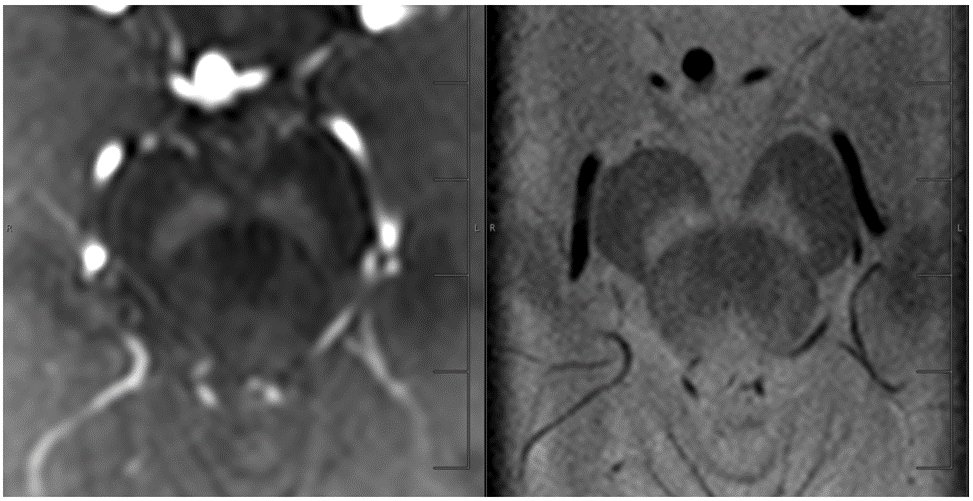
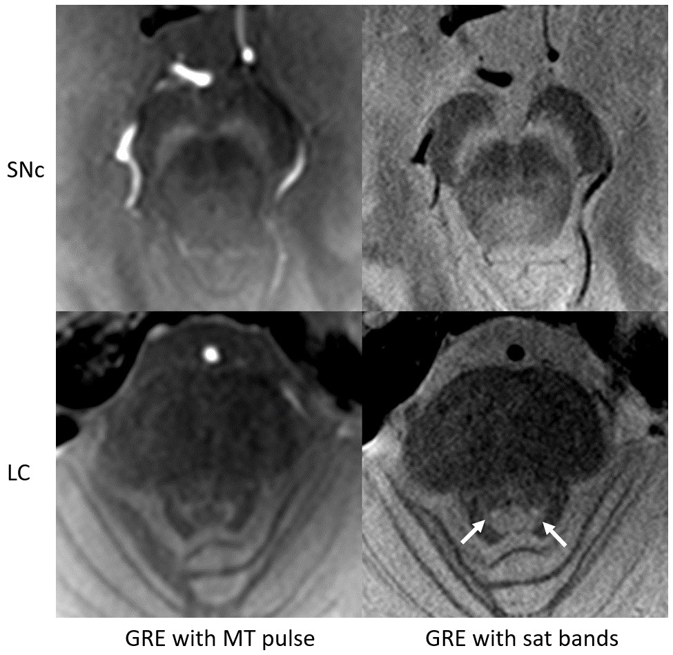
The contrast between NM-rich tissue and its surrounding tissues varies depending on the thickness of the saturation bands and their distance from the FOV center. Our results (not shown) reveal that a band thickness of 80mm and a distance of 60mm yield favorable SNc contrast against the surrounding tissues. This configuration also effectively reduces ghost artifacts and suppresses arterial blood signals. As depicted in Figure 2, the ghosting artifacts are notably mitigated with the reduced FOV method. Figure 3 presents a comparison between images acquired using the 3D GRE sequence with an MT pulse and images acquired using the 3D GRE sequence with three saturation bands. The comparison is made at two levels: SNc level and locus coeruleus (LC) level. It is evident that the reduced FOV method yields higher resolution, enhanced NM contrast, and a reduction in artifacts. Furthermore, the LC is identifiable in the lower right image acquired using the reduced FOV method.Discussion
GRE sequence family is the preferred candidate for NM imaging at 7T for its favorable low SAR characteristics. In GRE, each TR can accommodate MT pulse, ensuring equivalent MT effects on each k-space line. In our proposed method, three saturation bands were employed to replace the MT pulse, resulting in an improved NM image quality. This method could also be applied to turbo flash sequence, with saturation pulses played before multiple k-space line acquisition. However, this segmentation strategy may lead to diminished MT effects for later echoes. Similarly, other fast GRE sequences such as UTE and ZTE can also utilize the segmentation method for high resolution NM imaging, however, challenges, including addressing blurry artifacts and correcting gradient delays and waveform distortions, need to be addressed. In the product sequences, the three saturation bands are played sequentially, resulting in asymmetric MT and saturation effects for each saturation pulse. A potential solution is implementing simultaneous saturation bands to improve the saturation pulse efficiency and equalize the saturation effects. Please note that although the saturation band has the flexibility to adjust frequency offset (by changing the band distance to FOV center or band thickness), its pulse flip angle and bandwidth remain fixed.Conclusions
The reduced FOV method with three saturation bands offers potentials for higher resolution NM imaging at 7T with improved NM contrast and image quality.Acknowledgements
No acknowledgement found.References
[1] Sooyeon Ji et.al., Sandwich spatial saturation for neuromelanin-sensitive MRI: Development and multi-center trial, Neuroimage 264 (2022)Figures