4348
Evaluating the Prognostic Potential of High-Water Content Regions in Parkinson's Cognitive Impairment Progression1Department of Radiology, Ruijin Hospital, Shanghai Jiao Tong University School of Medicine, Shanghai, China, 2Department of Radiology, Wayne State University, Detroit, MI, United States, 3Department of Neurology, Ruijin Hospital, Shanghai Jiao Tong University School of Medicine, Shanghai, China, Shanghai, China, 4Philips Healthcare, Shanghai, China, 5Department of Neurology, Wayne State University, Detroit, MI, United States
Synopsis
Keywords: Parkinson's Disease, White Matter
Motivation: White matter damage has been known to accumulate prior to the onset of white matter hyperintensity, but there is currently no way to calculate the extent of damage.
Goal(s): Quantification of high-water content regions in Parkinson's patients based on proton spin density maps provides a new biomarker for mapping tissue damage and interpretation of WMH evolution.
Approach: Distribution of high-water content regions and WMH were mapped for Parkinson patients with different cognitive levels. Heatmaps were created for groups.
Results: High water content regions were mainly distributed along the ventricular wall. Water content and WMH correlated with CSF volumes with significant differences between groups.
Impact: Segmentation of the high-water content regions based on proton spin density maps provides the ability to interpret WMH evolution and provide a new potential biomarker to guide the early detection. These effects were dramatically seen in Parkinson’s patients with dementia.
Introduction
White matter damage is the result of ischemia, inflammation and degeneration of the lateral ventricular wall. The ventricular walls of the anterior and posterior horns of the lateral ventricles are subjected to increased mechanical tension, and this tension is further amplified with age-related ventricular enlargement, resulting in ventricular wall thinning, brain-fluid barrier dysfunction, and subsequent CSF leakage into the brain parenchyma [1-3]. Studies based on diffusion tensor imaging (DTI) fractional anisotropy (FA) have shown that normal-appearing white matter already begin to suffer from impaired fiber integrity [4-7]. Cerebral white matter hyperintensity (WMH) is characterized by the accumulation of pathologic changes in the white matter before the onset of WMH, but to date there is no method to assess the damage at any given time. Using STAGE (Strategic Acquisition of Gradient Echo) imaging[8], proton spin density maps can be generated. The proton spin density map can be used to identify the high-water content regions. Regions of abnormally high-water content can serve as new biomarkers of localized tissue damage and characterize Parkinson’s disease (PD) patients.Methods
A total of 23 healthy controls (HCs) and 138 PD patients were scanned at 3T using T1W, FLAIR and STAGE. The STAGE data were used to create proton spin density (water content) maps. Patients were divided into 3 groups based on their MoCA score, without dementia (NC), with dementia (PDD), and with mild cognitive impairment (MCI). The T2 FLAIR data was used to segment the WMH lesions and the proton spin density map was used to further segment high water content regions using a threshold of 0.85. The probability heat maps of WMH lesions and high-water content regions across for groups were generated by registering the subject data to the MNI atlas. The data were processed by SPM12, FSL, advanced normalization tool and Freesurfer. Tukey`s HSD were used for the statistical tests comparing the volumes of total WMH and high-water content regions in white matter for different groups.Results
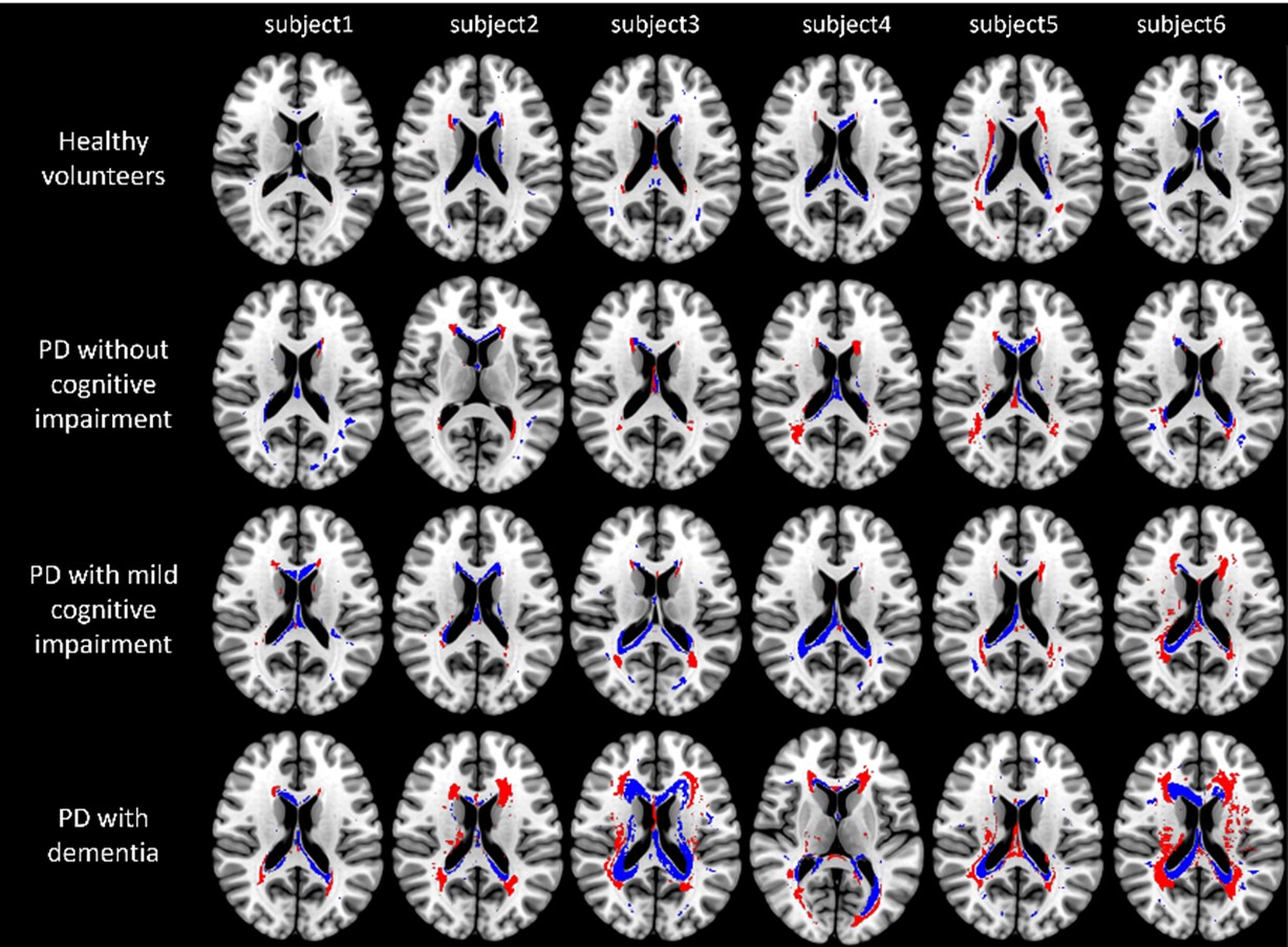
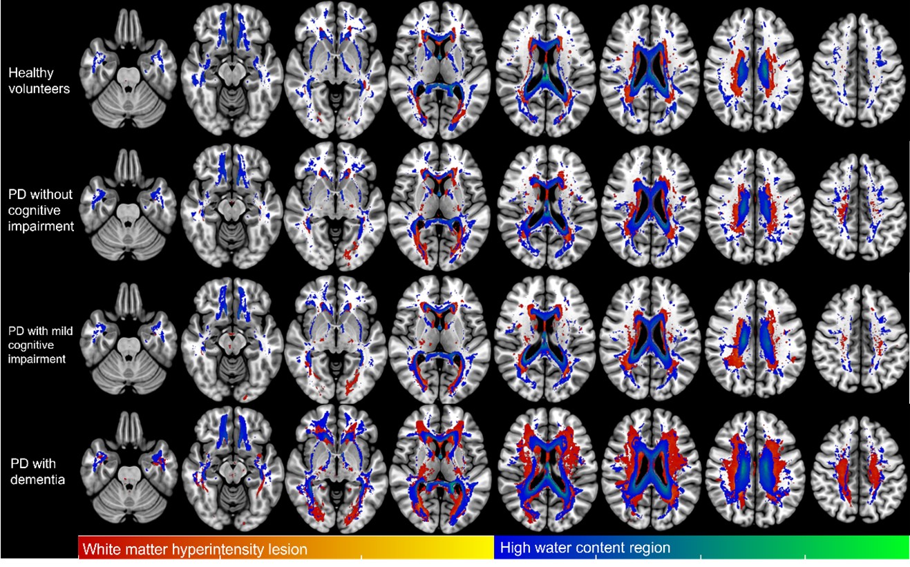
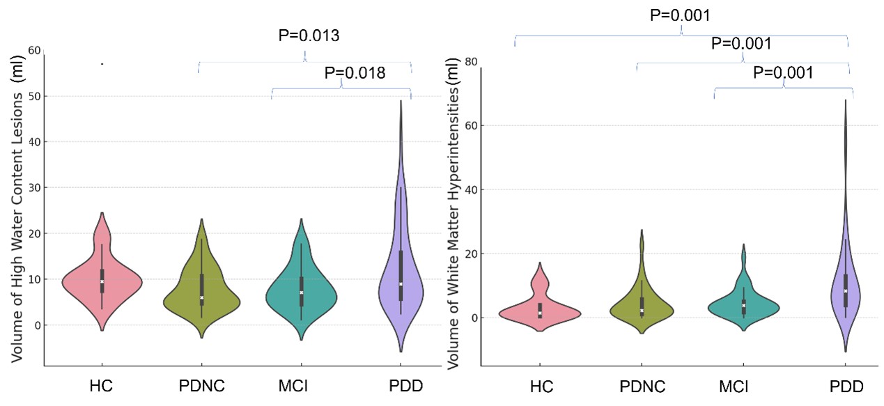
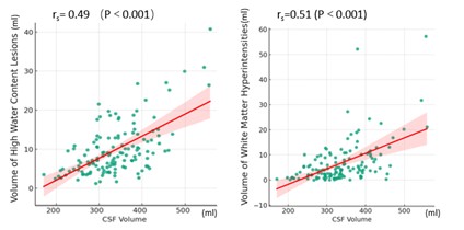
Figure 1 illustrates the segmentations of the total WMH and high-water content regions in six subjects. High water content regions were distributed along the ventricular wall, and the highest occurrence of high-water content lesions was in the anterior and posterior corners of the lateral ventricles and in the septum (Figure 2). Figure 3 displays that the difference in the volume of high-water regions between patients with PDD and HC, PDNC, and MCI, respectively, was statistically significant (P < 0.001). There was no difference between HC and PDD but there was a difference between PDD and PDNC (P=0.0139) and MCI (P=0.0182) and MCI (P=0.0182). Water content positively correlated with CSF volumes with significant differences between groups with different cognitive levels (rs= 0.49 with p < 0.001), as seen in Figure 4.Discussion
This study found that the distribution of high-water content regions around the white matter and the total CSF were significantly correlated. This suggests a relationship between the ventricles and the development of WMH. It may be caused by CSF gradually leaking into the adjacent brain parenchyma. The differences between various cognitive groups suggest that in healthy individuals, the self-protective mechanism protects adjacent tissue, while in PD patients, due to oxidative stress, mitochondrial dysfunction of ventricular cells and other pathologic alterations, the high-water content regions are unable to regress, and progresses gradually into WMHs[9, 10]. Therefore, volumetric quantification of the high-water content region holds significance for the study of WMH lesion evolution. In summary, segmentation of the high-water content regions based on proton spin density maps provides the ability to interpret WMH lesion evolution along the lateral ventricular wall. Mapping this information longitudinally could provide a new potential biomarker to guide the early detection and possible future interventions to prevent white matter damage. These effects were most dramatically seen in the Parkinson’s patients with severe cognitive impairment.Acknowledgements
This work was supported, in part, by the National Natural Science Foundation of China (grant number: 82271954, 81971576); Chinese National Science & Technology Pillar Program (grant number: 2022YFC2009900/2022YFC2009905) and the Innovative Research Team of High-level Local Universities in Shanghai.References
Figures