4346
SandwichNM Denoising with Deep Learning-Based Approach1Laboratory for Imaging Science and Technology, Department of Electrical and Computer Engineering, Seoul National University, Seoul, Korea, Republic of, 2Heuron Co.Ltd., Seoul, Korea, Republic of, 3Department of Neurology, Severance Hospital,, Yonsei University College of Medicine, Seoul, Korea, Republic of, 4Yongin Severance Hospital, Yonsei University College of Medicine, seoul, Korea, Republic of, 5Samsung Medical Center, Seoul, Korea, Republic of, 6Seoul National University Hospital, Seoul, Korea, Republic of, 7Department of Neurology, Inje University Sanggye Paik Hospital, Inje University College of Medicine, Seoul, Korea, Republic of
Synopsis
Keywords: Parkinson's Disease, Parkinson's Disease, Denoising
Motivation: SandwichNM is an advanced neuromelanin sensitive MRI method, but it use the same sequence twice and averages them to increase signal-to-noise ratio (SNR) requiring long scan time.
Goal(s): The objective is to preserve the SNR while reducing two scans into a single scan.
Approach: We proposed deep learning-based denoising method for sandwichNM image to reduce the number of scans.
Results: The proposed model achieved an increased PSNR and SSIM with utilizing single scan, which has reduced the scan time to half of the previous one.
Impact: SandwichNM is an advanced neuromelanin sensitive MRI technique but requires averaging two scans due to SNR issue. The proposed method enabled higher SNR from single scan which can be useful for scanning patients with Parkinson's disease with involuntary movements.
Introduction
Neuromelanin is a recognized biomarker for the diagnose of Parkinson’s disease (PD).1-3 SandwichNM is one of the neuromelanin sensitive MRI methods (NM-MRI), which provides homogeneous neuromelanin contrast across different vendors.4 However, sandwichNM requires long scan time, since it obtains two scans to raise SNR of the image. This makes it difficult to obtain high-quality images from PD patients with tremors or involuntary movements. Recently, there have been studies on using deep learning-based denoising for other NM-MRI methods.5,6 In this study, the deep learning-based sandwichNM denoising approach was proposed to obtain conventional image quality with a single scan (Fig. 1).Datasets
The data were collected using a 3T MRI scanner from two different vendors (Ingenia CX, Philips, Best, Netherlands; Skyra, Siemens, Erlangen, Germany). SandwichNM sequence was acquired twice (scan 1, scan 2) with the same scan parameters: TE = 4.1 ms for Philips and TE = 3.5 ms for Siemens, TR = 30 ms, flip angle = 14°, readout bandwidth = 168 Hz/pixel, matrix size = 320 × 220 × 32, resolution = 0.8 × 0.8 × 1.2 mm3, Each acquisition time was 2 min 45 s, Then the total scan time for the sandwichNM image was 5 min 30 s. The imaging slab was positioned perpendicular to the 4th ventricle, with the center slice aligned tangent to the upper aspect of the pons.A total of 287 subjects (166 from Philips scanner; 121 from Siemens scanner) were scanned. Following a quality assurance process, 26 subjects were excluded due to motion artifact. Consequently, for the purpose of training the network, the dataset was divided into 209 for training, 26 for validation, and 26 for testing (8:1:1 ratio). For training, each single scan was used as an input of the network, and the average of them was used as a label (Fig. 2). Patches were generated with the size of 64×64×16 overlapping 12.5% between neighboring patches. Patches that had over 50% of their area filled with missing data were not considered for training. A total of 66,096 patches were utilized in the training process.
Dataset Preprocessing
Rigid registration between scan 1 and scan 2 was proceeded using FLIRT7 before averaging. The brain masks were generated from averaged image by BET7 for removing background.Experiments
The architecture of our proposed model, 3D U-Net, is shown in (Fig. 3). It comprises a total of 36 convolutional layers, each followed by batch normalization and leaky ReLU activation function. The convolutional kernel size used is 3×3×3 with a stride size of 1×1×1.During training, we employed a learning rate of 1e-3 and applied a decay factor of 0.5 every 2 epochs. The optimization was done using the Adam optimizer, with a batch size of 64. Training was halted after 100 epochs. To improve generalization. We utilized L2 loss for training, and the entire network training was carried out using PyTorch8. The training process took approximately 19 hours, utilizing two NVIDIA GTX 1080 Ti GPUs.
We evaluated the denoising performance for two different vendors by comparing the PSNR and SSIM between input and reference with those between output and reference. Here we used averaged image as a reference.
Results
(Fig. 4) illustrates the result of the application of the proposed method to Siemens and Philips data. The input images (first column) show noisy images but our method successfully denoised them as shown in the output images (third column), demonstrating comparable noise level to label images (second column). Quantitative metrics (Fig. 4b) such as PSNR (33.4 ± 1.36 → 34.2 ± 1.31 dB in Siemens; 35.8 ± 1.39 → 36.2 ± 1.46 dB in Philips) and SSIM (0.905 ± 0.0134 → 0.917 ± 0.0109 in Siemens; 0.930 ± 0.0104 → 0.942 ± 0.0097 in Philips) also confirm the efficiency of our method.Discussion and Conclusion
In this study, we proposed deep learning-based denoising method for sandwichNM images. The conventional sandwichNM image requires averaging two scans to enhance SNR, which is time-consuming. To address this limitation, the method achieved an increase in SNR with using only a single scan. As a result, scan time was reduced from 5 min 30 s to 2 min 45 s, maintaining the image quality. Additionally, in (Fig. 4a), our method showed robust noise reduction capabilities across multi-vendor MRI scanners.Acknowledgements
This work is supported by the Korea Agency for Infrastructure Technology Advancement(KAIA) grant funded by the Ministry of Land, Infrastructure and Transport (Grant 21NPSS-C163415-01), Brain Korea 21 Plus Project in 0000, and Heuron Co., Ltd.References
1. Sulzer, D. et al. Neuromelanin detection by magnetic resonance imaging (MRI) and its promise as a biomarker for Parkinson’s disease. npj Park.’s Dis. 4, 11 (2018).
2. Ogisu, K. et al. 3D neuromelanin-sensitive magnetic resonance imaging with semi-automated volume measurement of the substantia nigra pars compacta for diagnosis of Parkinson’s disease. Neuroradiology 55, 719–724 (2013).
3. He, N., Chen, Y., LeWitt, P. A., Yan, F. & Haacke, E. M. Application of Neuromelanin MR Imaging in Parkinson Disease. J. Magn. Reson. Imaging 57, 337–352 (2023).
4. Ji, S. et al. Sandwich spatial saturation for neuromelanin-sensitive MRI: Development and multi-center trial. NeuroImage 264, 119706 (2022).
5. Oshima, Sonoko, et al. "Denoising approach with deep learning-based reconstruction for neuromelanin-sensitive MRI: image quality and diagnostic performance." Japanese Journal of Radiology (2023): 1-10.
6. Seada, S. A., Eerden, A. W. van der, Boon, A. J. W. & Hernandez-Tamames, J. A. Neuromelanin-MRI using 2D GRE and deep learning: considerations for improving the visualization of substantia nigra and locus coeruleus. arXiv (2023) doi:10.48550/arxiv.2306.16315.
7. Jenkinson, M., Beckmann, C. F., Behrens, T. E. J., Woolrich, M. W. & Smith, S. M. FSL. NeuroImage 62, 782–790 (2012).
8. Paszke, A. et al. PyTorch: An Imperative Style, High-Performance Deep Learning Library. arXiv (2019) doi:10.48550/arXiv.1912.01703.
Figures
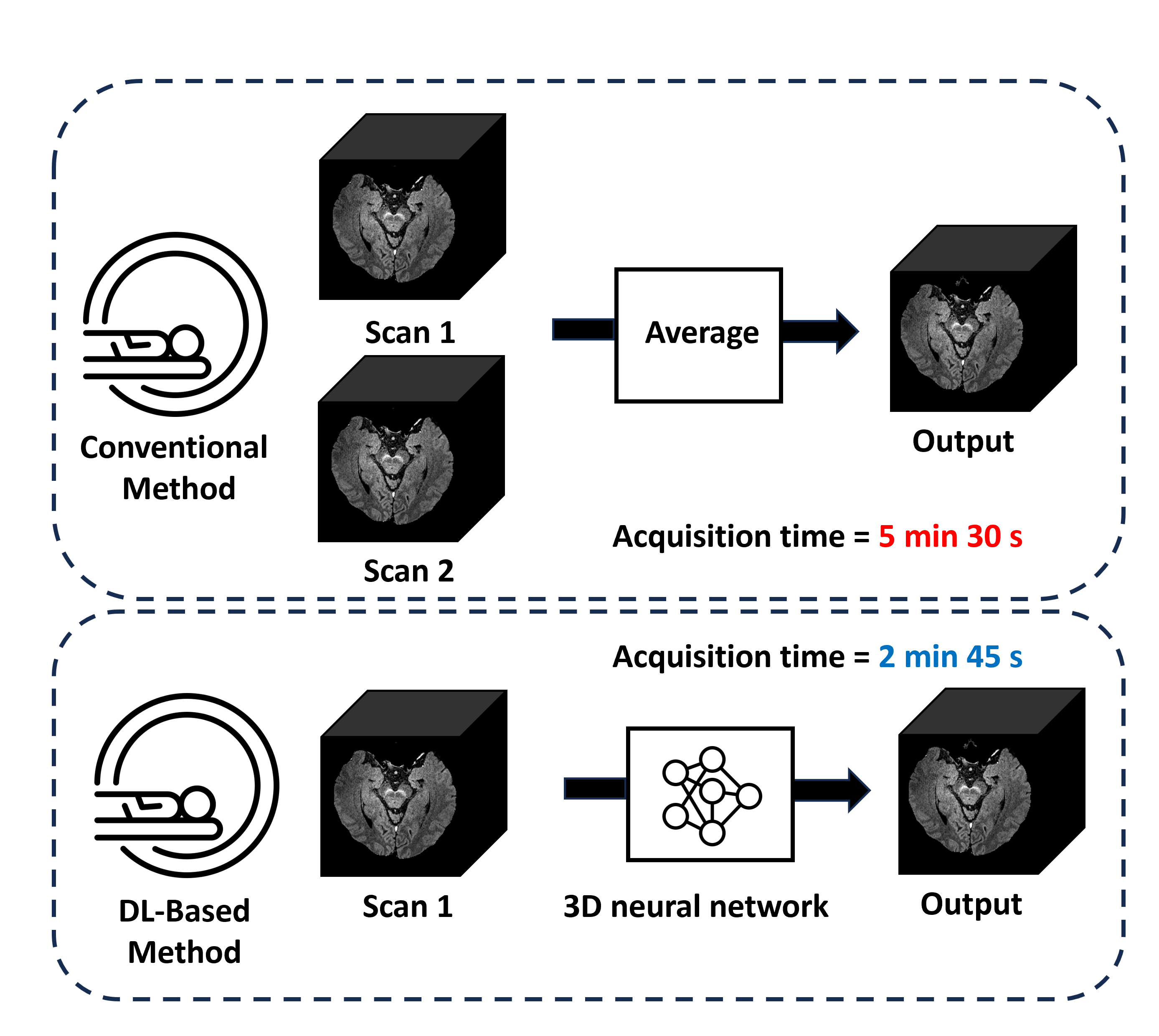
Fig. 1. Comparison of the pipeline of the conventional sandwichNM with that of the proposed method. Conventional sandwichNM imaging collects two scans in each subject and averages the images to obtain higher SNR. Our method only requires single scan and achieves comparable SNR with conventional sandwichNM acquisition by using denoising method.
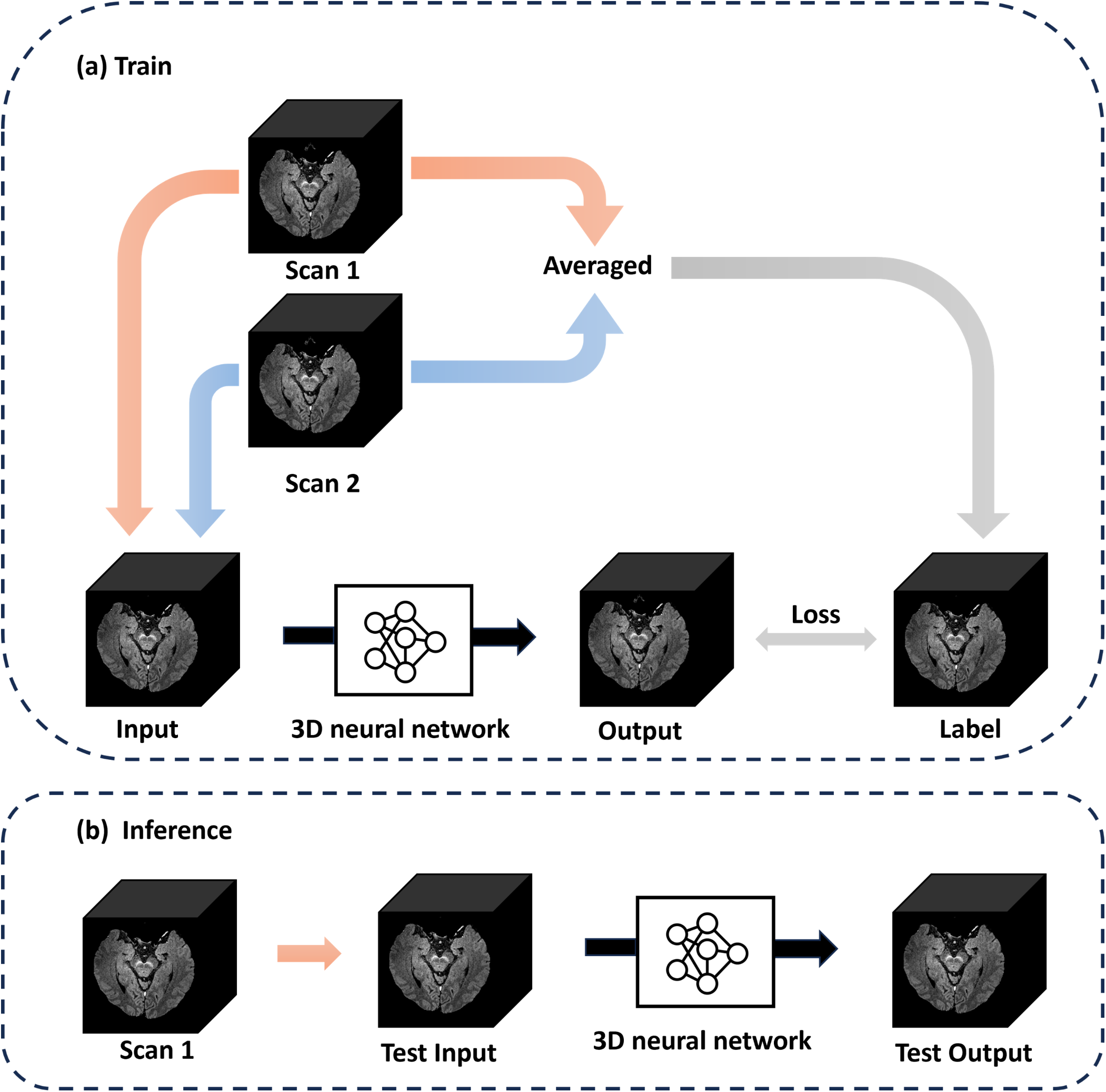
Fig. 2. Overview of sandwichNM denoising. Each single scan image was used as an input of the network, and averaged image as label. The images were divided into patches of size 64×64×16 voxels overlapping scheme of 12.5% between adjacent patches.
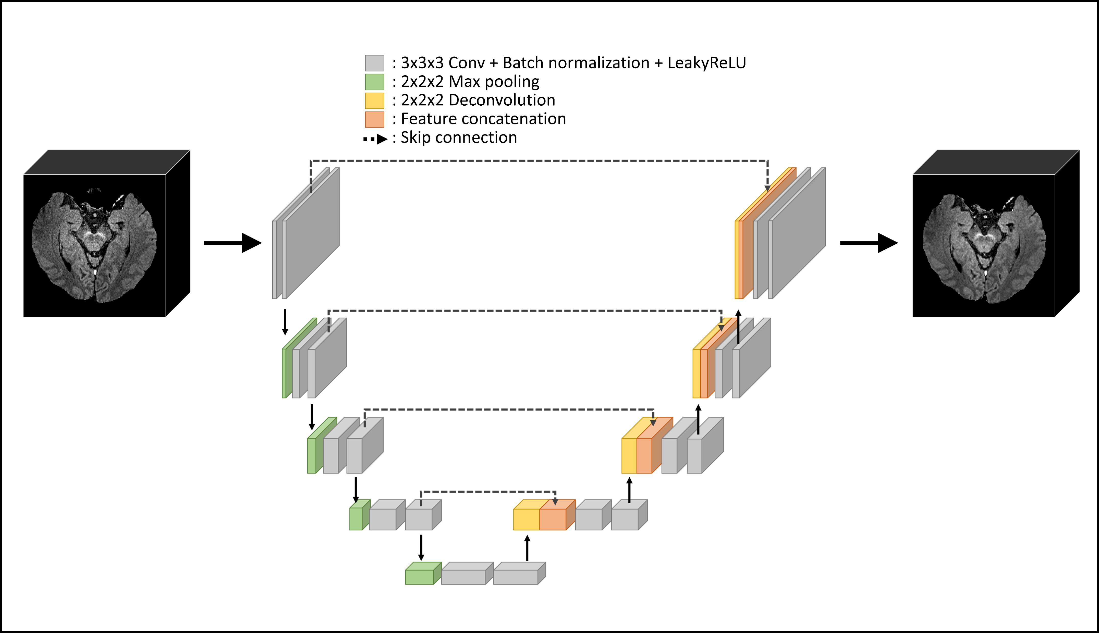
Fig. 3. Network structure of proposed model. By utilizing a 3D U-Net, inter-slice information is incorporated, thereby volumetric information of the brain can be well preserved.
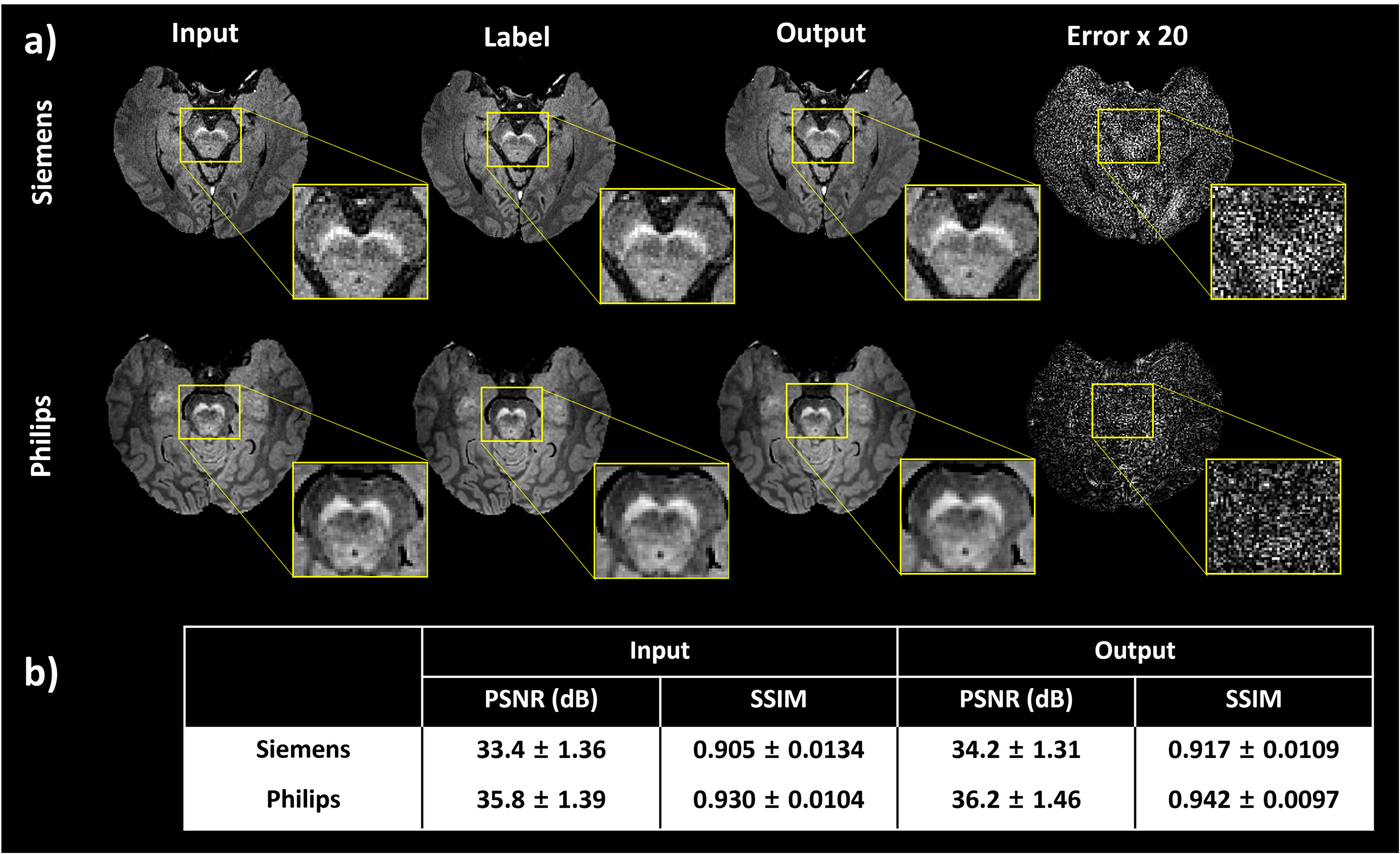
Fig. 4. Denoised results of real-world images. PSNR and SSIM values are presented in the table. Input refers to evaluation between input and label, while output refers to evaluation between label and output. PSNR and SSIM values both increased meaning a denoised output.