4344
4D flow in Parkinson’s disease: cognitive decline corresponds with impaired circle of Willis haemodynamics1New Zealand Brain Research Institute, Christchurch, New Zealand, 2Department of Medicine, University of Otago, Christchurch, New Zealand, 3Department of Electrical and Computer Engineering, University of Canterbury, Christchurch, New Zealand, 4Auckland Bioengineering Institute, University of Auckland, Auckland, New Zealand, 5Department of Neurology, Christchurch Hospital, Christchurch, New Zealand, 6School of Psychology, Speech and Hearing, University of Canterbury, Christchurch, New Zealand, 7School of Psychology and Centre for Brain Research, University of Auckland, Auckland, New Zealand, 8Centre for Advanced MRI, Auckland UniServices Limited, Auckland, New Zealand
Synopsis
Keywords: Parkinson's Disease, Parkinson's Disease
Motivation: The neurovascular changes associated with cognitive decline in Parkinson’s disease (PD) remain uncertain. Like Alzheimer’s disease, blood flow integrity may also play a significant role in maintaining cognitive function in PD.
Goal(s): To understand the relationship between cognitive impairment and circle of Willis anatomy and haemodynamics in PD.
Approach: 36 healthy controls and 81 individuals with PD completed 3T 4D flow MRI acquisition and an extensive neuropsychological battery.
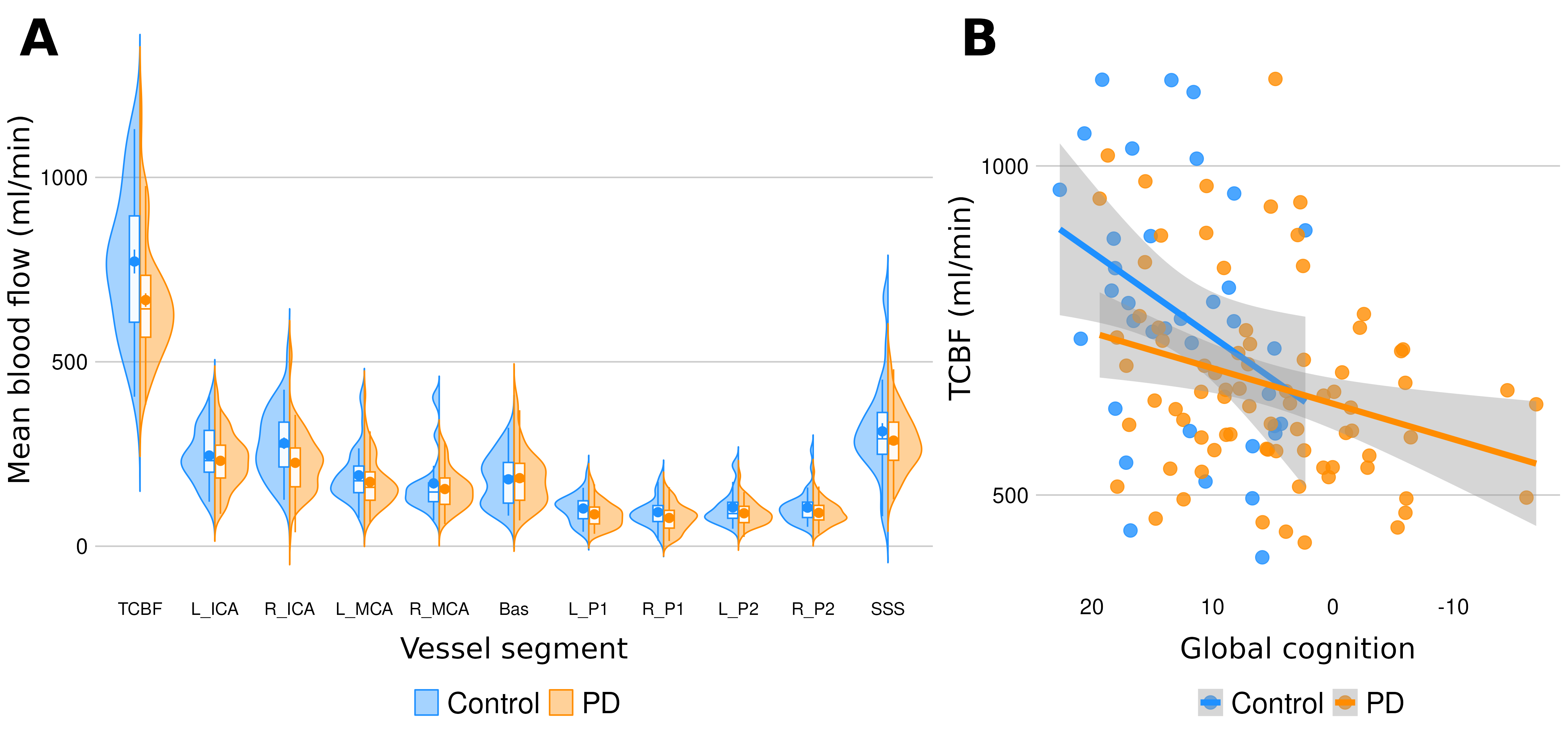
Results: The PD group showed 10% lower total cerebral blood flow (ml/min) and 7% lower flow velocity (cm/min) which were associated with poorer cognition.
Impact: 4D flow MRI revealed hypoperfusion of the circle of Willis in Parkinson’s disease which corresponded with poorer cognition. These results support future targeting of vascular mechanisms to alleviate cognitive impairment in Parkinson’s disease.
Introduction
Cognitive impairment is a core feature of Parkinson’s disease (PD), the fastest growing neurological disease worldwide1. The pathobiology driving cognitive impairment in PD is poorly understood, which prohibits effective treatment. Emerging evidence has implicated cerebral large vessel disease in cognitive impairment in healthy aging and Alzheimer’s disease2,3, supporting consideration of this mechanism in PD. To this end, this study applies 4D flow MRI as a novel approach to investigate the role of circle of Willis anatomy and haemodynamics in cognitive decline in PD.Methods
Data acquisition - A convenience sample of 81 individuals with PD and 36 healthy controls (HC) enrolled in the NZP3 longitudinal study4 (Christchurch, New Zealand) underwent comprehensive neuropsychological testing and 4D flow MRI acquisition. 4D flow scans scans were acquired using a prototype sequence on a 3T Siemens Skyra MRI (Erlangen, Germany), employing a 3D phase-contrast acquisition, with retrospective cardiac gating, velocity encoding (venc) = 100 cm/s, number of phases/bins per cardiac cycle = 20, field of view = 210 x 171 x 40 mm, centred on the circle of Willis, and acquired voxel size 1mm3.Image processing – Data were processed using QVT freeware5, where metrics were acquired from 10 vascular segments proximal and distal to the circle of Willis (see figure 1). Metrics evaluated at these points include mean blood flow (ml/min), mean blood velocity (cm/s), pulsatility index (PI), resistivity index (RI), and cross sectional vascular area (cm2). Internal carotid and posterior cerebral artery values were summed to provide a conservative estimate of total cerebral blood flow.
Analysis – Missing vascular values (18% across all vessels) were imputed via predictive means matching (18 imputations), and dimensionality reduction of the neuropsychological data was achieved via principal component analysis. Data were analysed using Bayesian regressions and model comparisons, controlling for age, sex, intracranial volume, and hypoplasia (anatomical variation of the circle of Willis, categorised as either normal, A1 type, P1 type, or A1-P1 combined).
Results
Relative to controls, individuals with PD exhibited lower total cerebral blood flow (HC = 844.6 ml/min [CI 755.3, 912.7], PD -9.8% [-18.0, -1.4%], figure 1), and mean blood flow velocity (HC = 1072.9 cm/min [CI 1021.3, 1124.0]; PD -6.9% [-9.7, -4.1%]) in the absence of evidence of changes to vascular anatomy (vessel area). Poorer global cognitive ability (principal component one representing 51% of variability in the data) was associated with reduced blood flow volume and velocity across all participants. An exploration of the relationship between flow metrics (flow volume and velocity, PI, RI and area) and vascular risk factors (pulse pressure, mean arterial pressure, orthostatic hypotension, neurogenic orthostatic hypotension, blood pressure, cholesterol) found that none of these indicators were useful predictors of 4D flow metrics in PD.Conclusion
These findings indicate that circle of Willis haemodynamics are substantially altered in PD, while additionally supporting previous findings in healthy aging and Alzheimer’s disease of an association between hypoperfusion and cognitive decline. The factors driving haemodynamic changes in PD require further elucidation. This work supports future investigations into cerebrovascular interventions to address cognitive decline in PD.Acknowledgements
This work is funded by Health Research Council, Canterbury Medical Research Foundation, New Zealand Neurological Foundation, Lottery Health Research, and University of Otago. We would like to acknowledge the support of Ayah Elsayed as well as staff at the New Zealand Brain Research Institute. We thank Siemens Healthcare for the provision of the 4D flow prototype sequence.References
[1] Dag A, Lucia B, Halliday GM, Geurtsen GJ, Ballard C, Ray CK, Weintraub D. Parkinson disease-associated cognitive impairment (primer). Nature Reviews: Disease Primers. 2021;7(1).
[2] Rivera-Rivera LA, Turski P, Johnson KM, Hoffman C, Berman SE, Kilgas P, et al. 4D flow MRI for intracranial hemodynamics assessment in Alzheimer’s disease. Journal of Cerebral Blood Flow & Metabolism. 2016 Oct;36(10):1718-30.
[3] Roberts GS, Peret A, Jonaitis EM, Koscik RL, Hoffman CA, Rivera-Rivera LA, et al. Normative cerebral hemodynamics in middle-aged and older adults using 4D flow MRI: initial analysis of vascular aging. Radiology. 2023 Mar 21;307(3):e222685.
[4] MacAskill MR, Pitcher TL, Melzer TR, Myall DJ, Horne KL, Shoorangiz R et al. The New Zealand Parkinson’s progression programme. Journal of the Royal Society of New Zealand. 2023 Aug 8;53(4):466-88.
[5] Roberts GS, Hoffman CA, Rivera-Rivera LA, Berman SE, Eisenmenger LB, Wieben O. Automated hemodynamic assessment for cranial 4D flow MRI. Magnetic Resonance Imaging. 2023 Apr 1;97:46-55.
Figures