4342
Changes in Elasticity and Microstructural Properties of the Brain due to Parkinson's Disease1Department of Biomedical Engineering and Health Systems, KTH Royal Institute of Technology, Stockholm, Sweden, 2Department of Molecular Medicine and Surgery, Karolinska Institute, Stockholm, Sweden, 3Department of Clinical Neuroscience, Karolinska Institute, Stockholm, Sweden
Synopsis
Keywords: Parkinson's Disease, Elastography
Motivation: MR Elastography (MRE) of the brain is a novel technique that elucidates the viscoelastic properties of the brain. Changes in e.g. stiffness of various brain regions often occur at an early stage in neuropathologies.
Goal(s): Assessing how viscoelastic properties of the brain changes with Parkinson’s disease, and study the microstructural mechanisms behind these changes.
Approach: We investigated how the viscoelasticity of the brain changes for diseased subjects compared to controls (21 subjects), and correlated these changes with microstructural properties (based on multidimensional diffusion imaging).
Results: We found a softening effect of the occipital and temporal lobes, possibly correlated with early neural atrophy.
Impact: The presented results show how PD affects the brain in a new combination of modalities which can help to better understand the pathology, which may, for example, lead to new methods for early PD diagnosis.
Introduction
Parkinson’s disease (PD) is the second most common neurological pathology, affecting about 1% of the population older than 60 years1. The disease predominantly affects the dopaminergic neurons in the brain, causing them to atrophy due to an increase of α-synuclein-aggregates. The mechanisms of this disease are however not fully understood, and more research is needed to understand its cause and ultimately to develop a cure. In this study, we used a combination of MR Elastography (MRE) in combination with multidimensional diffusion imaging (MUDI) to study the brain of PD subjects in comparison to healthy controls (HC), to understand which parts of the brain are predominately affected and how they are affected. With this combination, we were able to determine how the stiffness alters due to PD (as measured by MRE), and provide a tentative explanation for these alterations based on MUDI-derived properties. We found that the brain generally softens with PD, particularly in the occipital and the temporal lobes, most likely due to neural atrophy.Methods
| | HC | PD |
| Age mean (years) [min-max] | 56 [50-67] | 63 [47-78] |
| Gender (F:M) | 3:6 | 3:9 |
All MR images were acquired on a Philips Ingenia CX 3T scanner. MRE images were taken with an EPI sequence provided by Mayo clinic (see e.g. Ref 2 for details) in combination with a pneumatic driver from Resoundant (Rochester, MN, US) connected to a passive pillow which vibrated the heads of the subjects with a frequency of 60Hz. The resulting displacement images (3x3x3mm3 resolution) were inverted to viscoelastic parameters (given by the complex shear modulus, G*) with the use of a 3D inversion algorithm on the scanner using the AIDE algorithm3. Stiffness is defined by |G*|, and the viscosity related α parameter = 2/π arctan(G′′/G′ ), where G’ and G’’ are real and imaginary components of G*.
The MUDI images were acquired with an acquisition scheme explained in Ref 4 (2.5x2.5x2.5 mm3 resolution), using spherical and linear b-tensors with five b-values linearly spaced between 0 and 2000s/mm2. They were post-processed with the use of an open-source MD-dMRI software5. MUDI quantities include microscopic fractional anisotropy (μFA), mean diffusion (MD), and variance of MD (Var(MD)).
We registered and resampled all images to subject specific 1x1x1 skullstripped T1-maps, which had been parcellated into the Desikan-Killiany atlas6 with the use of FreeSurfer v7.2.
Results
Figure 2 shows the stiffness of the whole brain of HC and PD subjects plotted versus age. In figure 3a we show how the age of the HC and PD subjects correlate with the median value of various MR modalities (x-axis) in different segmented regions of interest (y-axis), where there is a general trend that age correlates negatively with stiffness and μFA, and positively with MD. Figure 3b shows how these values correlate with a PD diagnosis, after age-correction. There is a weak general trend for decreasing tissue stiffness for multiple regions of the brain, however this effect is most significant for the cerebrum, particularly the occipital and temporal lobes.Discussion and Conclusion
The results, as shown in Figures 2 and 3a indicate that the whole brain softens with age, which is in line with previous studies (see e.g. Hiscox et al7). This effect can be seen to correlate well with an increase in MD and a decrease in μFA. Such a correlation between MUDI-derived microstructural quantities and age has been previously reported and ascribed to indicate neural atrophy8, and is thus likely the mechanism in the present study as well. The viscoelastic properties generally decreased with PD compared to HC (in line with previous studies by Lipp et al9,10), however due to a slightly older PD cohort compared to the HC many of these correlations were not statistically significant after correcting for age. The main affected brain structures reside in the cerebrum, and we can see that, in particular, the occipital and temporal lobes have reduced stiffness due to the disease, consistent with previous studies (see e.g. Ref. 8 and 11). Just as for the aging effect, these structures exhibit changes in MUDI-quantities consistent with neural atrophy, however to a much lesser degree than for the age effects. It is noteworthy that the decrease in stiffness appears to be more significantly correlated with PD pathology than the MUDI-quantities, indicating that the stiffness is more sensitive to the pathology, and implies that the decrease in stiffness is not exclusively explained by atrophy.Although a larger cohort is necessary to increase statistical significance, the observed softening effects in combination with the MUDI-quantities presented here shows a promising method for investigating the underlying mechanisms of PD pathology.
Acknowledgements
We would like to acknowledge the support by prof. Richard Ehman and his co-workers at Mayo clinic (Rochester, MN, US) with providing hardware and software for the MRE pipeline, and for providing technical support and expertise on MRE. We would also like to acknowledge prof. Markus Nilsson and co-workers at Lund University for providing us with setting up MUDI at our scanner and providing help with the postprocessing of these images. This study was supported with funding from Digital Futures 2023, HMT-Hälsa, Medicin och Teknik 2022 and 2023 (FoUI-966774 and FoUI-992049), the Swedish Research Council (VR 2022-03389), and MedTechLabs 2023 (FoUI-991015). We would also like to thank the patients and healthy controls for volunteering, and the supporting staff from Philips.References
1. Tysnes, O.-B. & Storstein, A. Epidemiology of Parkinson’s disease. J Neural Transm (Vienna) 124, 901–905 (2017).
2. Kruse, S. A. et al. Magnetic resonance elastography of the brain. Neuroimage 39, 231–237 (2008).
3. Oliphant, T. E., Manduca, A., Ehman, R. L. & Greenleaf, J. F. Complex-valued stiffness reconstruction for magnetic resonance elastography by algebraic inversion of the differential equation. Magn Reson Med 45, 299–310 (2001).
4. Topgaard, D. Multidimensional diffusion MRI. Journal of Magnetic Resonance 275, 98–113 (2017).
5. Nilsson, M. et al. An open-source framework for analysis of multidimensional diffusion MRI data implemented in MATLAB. in Proc Intl Soc Mag Reson Med vol. 26 5355 (2018).
6. Desikan, R. S. et al. An automated labeling system for subdividing the human cerebral cortex on MRI scans into gyral based regions of interest. Neuroimage 31, 968–980 (2006).
7. Hiscox, L. V., Schwarb, H., McGarry, M. D. J. & Johnson, C. L. Aging brain mechanics: Progress and promise of magnetic resonance elastography. Neuroimage 232, (2021).
8. Kamiya, K. et al. Brain White-Matter Degeneration Due to Aging and Parkinson Disease as Revealed by Double Diffusion Encoding. Front Neurosci 14, 1091 (2020).
9. Lipp, A. et al. Cerebral magnetic resonance elastography in supranuclear palsy and idiopathic Parkinson’s disease. Neuroimage Clin 3, 381–387 (2013).
10. Lipp, A. et al. Progressive supranuclear palsy and idiopathic Parkinson’s disease are associated with local reduction of in vivo brain viscoelasticity. Eur Radiol 28, 3347–3354 (2018).
11. Pieperhoff, P. et al. Regional changes of brain structure during progression of idiopathic Parkinson’s disease – A longitudinal study using deformation based morphometry. Cortex 151, 188–210 (2022).
Figures
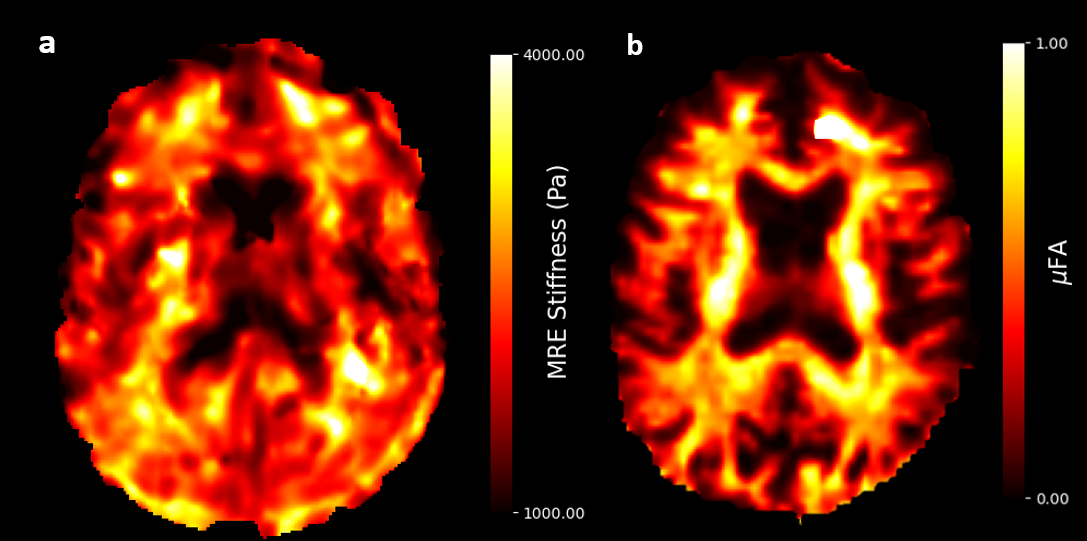
Figure 1. Example images of stiffness (1a) obtained from an MRE scan, and μFA (1b) as obtained from a multidimensional diffusion imaging (MUDI) scan.
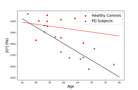
Figure 2. The average stiffness (|G*|) of the whole brain was 1915Pa (±87 Pa st.d.) for HC, and 1771Pa (±148Pa, st.d.) for PD patients. Assuming 0.5% decrease in stiffness per year7, and an age-difference between PD and HC of 7 years, the age-corrected stiffness for HC is 1848Pa. The Spearman correlation between average brain stiffness and age was -0.75 (p=0.0001), and the correlation between stiffness and PD diagnosis was -0.56 (p=0.012), after correcting for age.
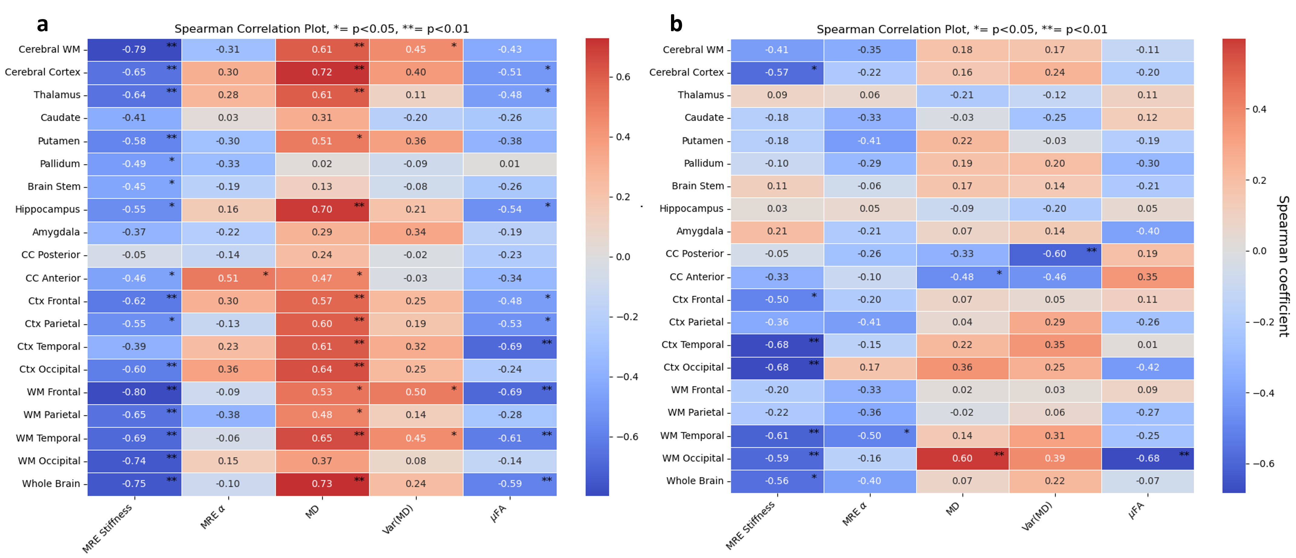
Figure 3. Spearman correlations between median values from different modalities (x-axis) at different regions (y-axis ) and age (3a, including both HC and PD) or PD diagnosis (3b) after age-correction. I.e. positive values indicate that the measured value by a specific modality in a specific region correlates positively with age (3a) or with a PD diagnosis (3b) . Ctx=cortex, WM=white matter. * indicates that p<0.05 and ** indicates that p<0.01