4340
Beyond the dipole: unveiling the hidden impact of nondipolar shifts on MRI phase contrast anisotropy in the brain1Department of Computer Science and Automation, Technische Universität Ilmenau, Ilmenau, Germany, 2Buffalo Neuroimaging Analysis Center, Department of Neurology at the Jacobs School of Medicine and Biomedical Sciences, University at Buffalo, The State University of New York, Buffalo, NY, United States, 3Center for Biomedical Imaging, Clinical and Translational Science Institute, University at Buffalo, The State University of New York, Buffalo, NY, United States
Synopsis
Keywords: Quantitative Imaging, Quantitative Susceptibility mapping
Motivation: To elucidate the contribution of nondipolar frequency shifts to the anisotropy of phase contrast in brain MRI and decode the underpinning biophysical mechanisms of phase contrast.
Goal(s): To differentiate the roles of magnetic susceptibility and nondipolar frequency shifts across various head orientations in MRI scans.
Approach: Utilizing DEEPOLE QUASAR, the study compares susceptibility estimates and nondipolar frequency shifts in anisotropic brain regions across multiple head orientations.
Results: Nondipolar frequency shifts played a substantial role in the anisotropy of frequency shifts, with DEEPOLE QUASAR providing more stable susceptibility estimates than conventional QSM, regardless of head orientation.
Impact: The study establishes the substantial influence of nondipolar frequency shifts on MRI phase contrast anisotropy, questioning established assumptions from susceptibility tensor imaging and quantitative susceptibility mapping, thereby paving the way for more accurate brain tissue characterization.
Introduction
Quantitative susceptibility mapping (QSM) uses the phase of the magnetic resonance imaging (MRI) signal to quantify the magnetic susceptibility of tissues, which, in the brain, can be used to deduct contents of paramagnetic iron and diamagnetic myelin.1Experimental evidence and theoretical considerations suggest that, in brain tissue, the formation of phase shifts depends not only on bulk magnetic susceptibility but also on tissue orientation, microstructure, and chemical exchange.2 We recently presented DEEPOLE QUASAR, a method that maps these nondipolar frequency shifts that are not accounted for in the conventional QSM model (Fig. 1).3
In this work, we further investigated the underpinnings of phase contrast in the brain by studying the magnetic susceptibility and nondipolar frequency shifts under different orientations relative to the main magnetic field.
Methods
Solution techniques: We used the deep learning-based method DEEPOLE QUASAR to estimate magnetic susceptibility and nondipolar frequency shifts from frequency maps.3 For comparison with conventional QSM, we used a deep learning based QSM algorithm (in-house improved version4 of DeepQSM5).Methods validation: We first confirmed that in the absence of orientation dependency in the sources, the methods do not hallucinate any orientation dependency. We used a realistic brain susceptibility map and simulated phase data under different magnetic field orientations.
Real phase data: We studied the orientation dependence in the liquid-filled lateral ventricles (isotropic) and regions of the brain that are known to be structurally anisotropic. We evaluated the voxel values of highly directional white matter fiber bundles (corpus callosum, optic radiations, and internal capsule) with regards to the underlying fiber orientation relative to the main magnetic field. We used MRI scans from eight patients, measured under 11-29 orientations to the main magnetic field.6
Results
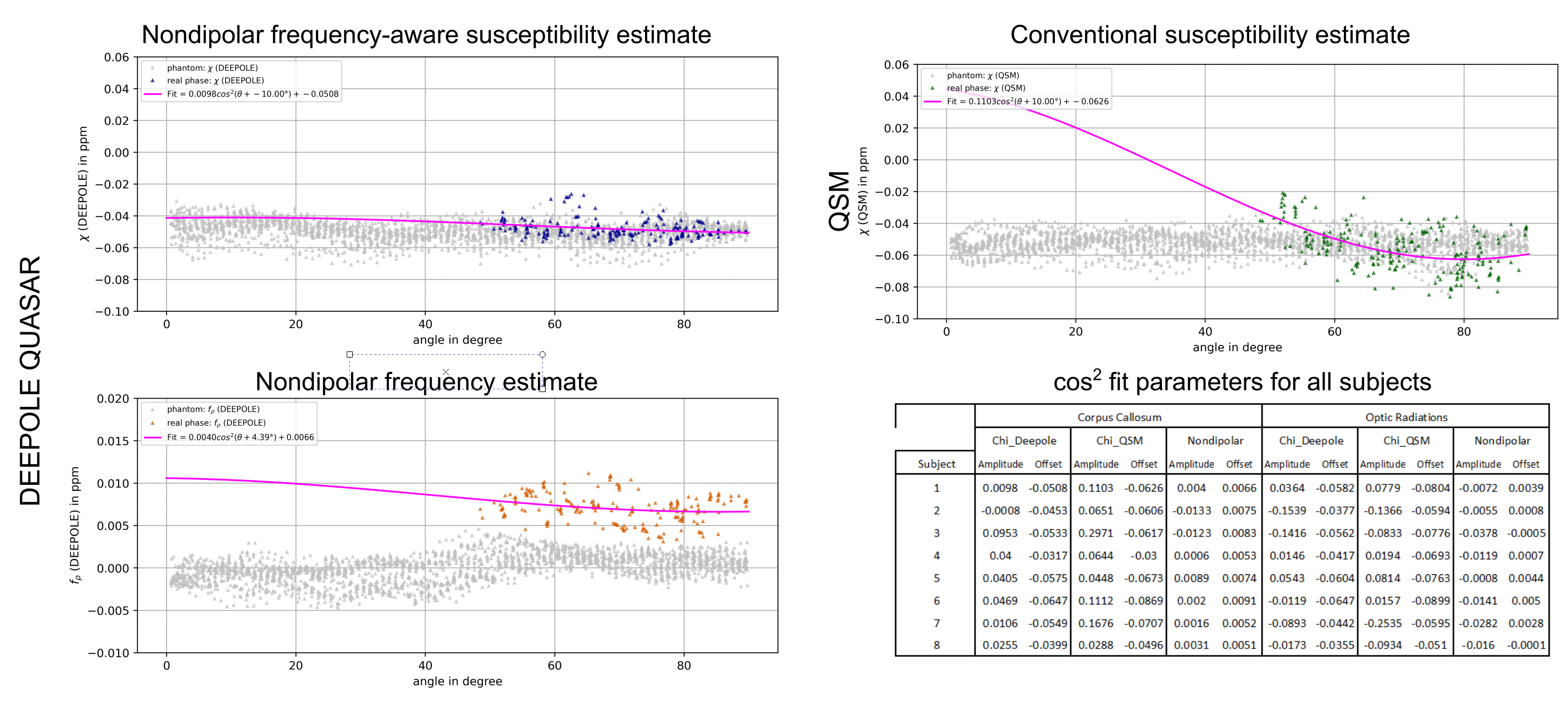
In our isotropic phantom, neither DEEPOLE QUASAR nor QSM hallucinated orientation dependency (Fig. 3, light gray markers in the background).In real data, orientation dependency with QSM was substantially stronger than with DEEPOLE QUASAR (Fig. 3). Neither DEEPOLE QUASAR’s susceptibility estimate nor its nondipolar frequency shift maps showed substantial orientation dependence.
Fig. 2 shows solutions of both methods from a representative subject under 8 different head orientations, as well as the standard deviations across the solutions under different oblique head orientations (right-most column). Fig. 3 shows the orientation dependence of the susceptibility and nondipolar frequency shift in the corpus callosum region of the same subject. We made comparable observations in the two studied corpus callosum regions, two optic radiation regions, and two internal capsule regions.
Discussion
Our findings with DEEPOLE QUASAR suggest that anisotropy in frequency shifts is not solely attributable to magnetic susceptibility but potentially a misinterpretation of nondipolar frequency shifts, which are neglected by the conventional QSM model. Our findings suggest that QSM and STI may require to incorporate nondipolar contributions into their physical models.The limited range of head tilt angles achievable in typical MRI scans, however, constrains our observations of the full angular spectrum, indicating the need for dedicated MRI protocols that can capture a wider range of orientations for a more comprehensive analysis.
Conclusion
The study presents a reassessment of quantitative susceptibility mapping in light of the previously ignored, yet theoretically and experimentally shown nondipolar frequency shifts. Solutions from DEEPOLE QUASAR, a novel technique for mapping susceptibility and nondipolar shifts suggest that susceptibility anisotropy in white matter is smaller than previously though and might instead be the result of a misinterpretation of nondipolar shifts, which could stem from chemical exchange or tissue microstructure.Acknowledgements
The research was supported by the Free State of Thuringia grant ThiMEDOP (2018 IZN 0004) with funds of the European Union (EFRE), the German Federal Ministry of Education and Research (BMBF) grant AVATAR (16KISA024, funded by the European Union - NextGenerationEU), the German Academic Exchange Service (DAAD PPP 57599925), and an ISMRM Research Exchange Grant awarded to T.J. Research reported in this publication was partially supported by the National Institute of Neurological Disorders and Stroke of the National Institutes of Health under Award Number R01NS114227 (F.S.) and the National Center for Advancing Translational Sciences of the National Institutes of Health under Award Number UL1TR001412 (F.S.). The content is solely the responsibility of the authors and does not necessarily represent the official views of the National Institutes of Health.References
1. Schweser, F., Deistung, A., Lehr, B. W. & Reichenbach, J. R. Quantitative imaging of intrinsic magnetic tissue properties using MRI signal phase: An approach to in vivo brain iron metabolism? NeuroImage 54, 2789–2807 (2011).
2. Schweser, F. & Zivadinov, R. Quantitative susceptibility mapping (QSM) with an extended physical model for MRI frequency contrast in the brain: a proof-of-concept of quantitative susceptibility and residual (QUASAR) mapping. NMR in Biomedicine 31, e3999 (2018).
3. Jochmann, T., Haueisen, J., Zivadinov, R. & Schweser, F. U2-Net for DEEPOLE QUASAR–A Physics-Informed Deep Convolutional Neural Network that Disentangles MRI Phase Contrast Mechanisms. (2019).
4. Jochmann, T., Haueisen, J. & Schweser, F. How to train a Deep Convolutional Neural Network for Quantitative Susceptibility Mapping (QSM). in International Society of Magnetic Resonance in Medicine (ISMRM) 28th Annual Meeting, Virtual Conference (2020).
5. Bollmann, S. et al. DeepQSM - using deep learning to solve the dipole inversion for quantitative susceptibility mapping. NeuroImage 195, 373–383 (2019).
6. Shi, Y. et al. Towards in vivo ground truth susceptibility for single-orientation deep learning QSM: A multi-orientation gradient-echo MRI dataset. NeuroImage 261, 119522 (2022).
Figures
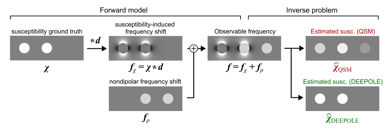
Fig. 1. Physical model of DEEPOLE QUASAR in comparison to QSM. d: magnetic dipole kernel, * : 3D convolution, χ: magnetic susceptibility, fρ: nondipolar frequency
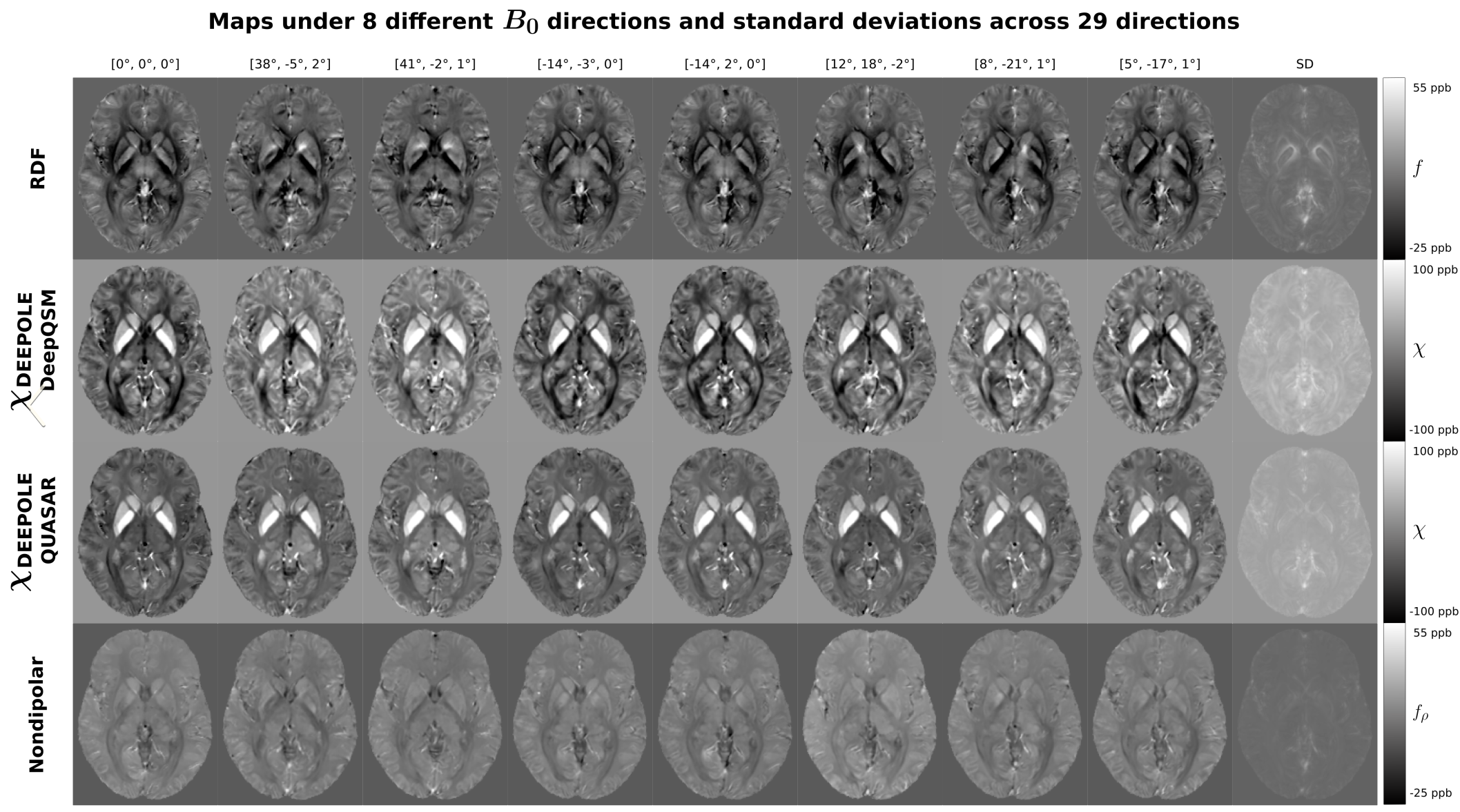
Fig. 2. Comparison of a RDFs and their estimated sources under different head orientations. While QSM yields just a susceptibility map χ, DEEPOLE QUASAR derives a susceptibility estimate and a separate nondipolar contrast. In the bottom row, the standard deviations over the susceptibility maps of all 26 head orientations of the subject are shown. The standard deviation map acquired from the DEEPOLE QUASAR estimates shows less variation between the head orientations.