4330
Physics-Driven Learned Deconvolution of Multi-Spectral Fluorine-19 MRI with Multiple Agents Using Radial Sampling1Department of Electrical and Computer Engineering, University of California San Diego, La Jolla, CA, United States, 2Department of Radiology, University of California San Diego, La Jolla, CA, United States
Synopsis
Keywords: Signal Modeling, Cell Tracking & Reporter Genes, Machine Learning/Artificial Intelligence, Contrast Agents, Data Acquisition, Data Processing, Modelling, Multi-Contrast, Non-Proton
Motivation: Detection of multiple cell targets separately labeled with different chemically-shifted, paramagnetic 19F tracers can benefit from radial k-space sampling pulse sequences; however, radial sampling can lead to non-linear smearing chemical shift artifacts.
Goal(s): Our goal is to develop suitable modeling of the radial chemical shifts and a physics-informed deconvolution scheme to unmix multi-spectral components.
Approach: We proposed a novel Radon transform modeling of forward operator for radial chemical shifts and introduced machine learning based method for multi-spectral deconvolution.
Results: Radial chemically-shifted artifacts are significantly reduced via the deep unrolling learned deconvolution algorithm, especially for low signal-to-noise-ratio (SNR) and highly undersampled acquisitions.
Impact: Unlike Cartesian chemical shifts that result in image displacements, radial chemical shifts produce more complex artifacts. To effectively unmix the multi-spectral 19F components, we developed an analytical model using Radon transform and a data-driven deconvolution method based on deep unrolling.
Introduction
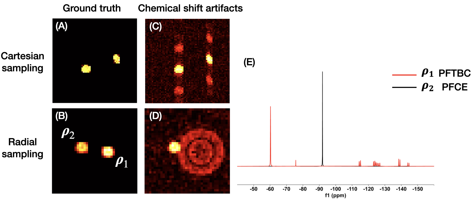
Fluorine-19 (19F) MRI uses perfluorocarbon (PFC) tracers to enable background-free, “hot-spot” in vivo cell tracking1. Detection of multiple cell targets requires separating images acquired using different PFC molecules (Fig. 1A-B) with different chemical shifts. The resulting shift artifacts using Cartesian k-space sampling appear as displacements (Fig. 1C) along the readout direction2. Moreover, highly sensitive imaging can be achieved using newer, metallo-PFC (MPFC) nanoemulsion probes which have an order of magnitude T1 reduction3; these tracers are optimally imaged using radially sampled ultrashort echo-time (UTE) pulse sequences4 to overcome paramagnetic shortening of T2, which can also be combined with k-space undersampling and compressed sensing reconstruction for further sensitivity enhancement4. A key challenge with radially sampled data is that the frequency offsets lead to non-linear smearing chemical artifacts throughout the image (Fig. 1D).In this work, we develop a model of the chemical shift effects with radial k-space sampling. Furthermore, we exploit a physics-informed primal-dual unrolling strategy that enables simultaneous artifact removal, as well as low SNR detection.
Theory and Methods
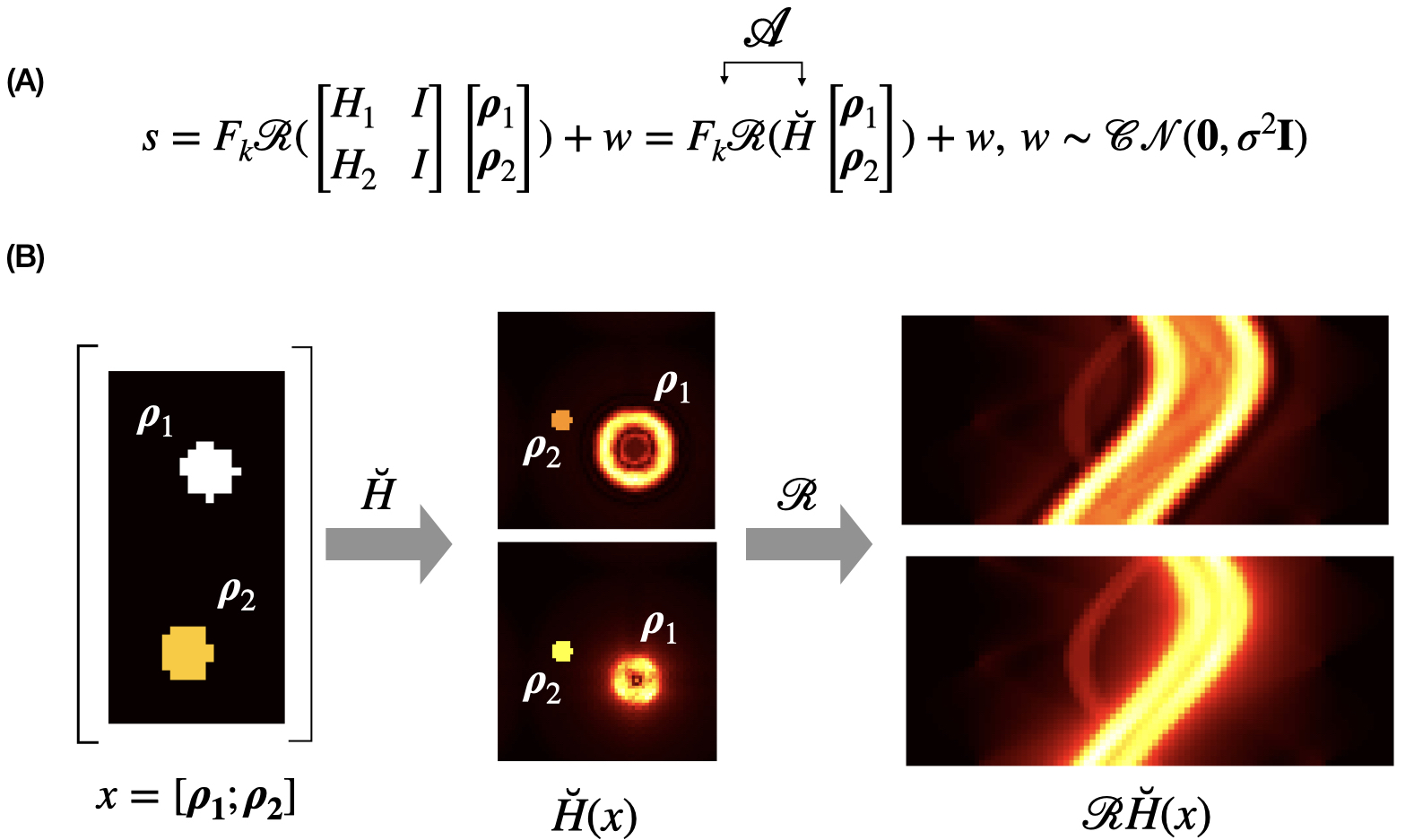
From Fourier slice theorem, radial k-space data $$$s(k, \Phi)$$$ can be obtained by taking 1D Fourier transform $$$F_k$$$ along the $$$t$$$ direction of the projection data $$$R_{\Phi}(t)$$$, where $$$R$$$ denotes Radon transform5. We consider two PFC imaging tracers with multiple chemical frequency shifts (Fig. 1E), representing perfluorotert-butyl cyclohexane (PFTBC as $$$\rho_1$$$) and perfluoro-15-crown-5-ether (PFCE as $$$\rho_2$$$). The reference frequency is set at -92.5 ppm for PFCE. By acquiring $$$P\geq2$$$ sets of measurements $$$s=\{s_j\}_{j=1}^P$$$ with varying readout bandwidths and hence varying effective sampling time6, distinct harmonic structure can be produced. We model the radial chemical shift as described in Fig. 2. $$$\breve{H}$$$ represents the spectral mixing matrix, where the corresponding convolution kernels can be analytically derived7. Given the overall forward sensing operator $$$\mathscr{A}=F_kR\breve{H}$$$ (Fig.2A), the corresponding inverse problem is$$\min_{x=[\textrm{ρ}_1;\textrm{ρ}_2]}\|s-\mathscr{A}x\|_2+\lambda\mathscr{D}\left(x\right)\quad(1)$$
where the regularizer $$$\mathscr{D}(x)$$$ encodes the structural prior of $$$x$$$. Here we employ the total-variation norm $$$\|\nabla x\|_1$$$, which naturally promotes sparsity. Eq.(1) is solved by a primal-dual hybrid gradient (PDHG) scheme using the explicit proximal operators8. To further improve the performance under low SNR and undersampling scenarios, we consider learning an end-to-end data-driven prior from training sample pairs $$$(s^{(1)},x^{(1)}),\dots,(s^{(N)},x^{(N)})$$$. The goal is to learn a non-linear mapping $$$\mathscr{A}_{\theta}^{\dagger}$$$ based on the model in Fig.2A such that $$$\mathscr{A}_{\theta}^{\dagger}(s)\approx x$$$. The objective is to minimize the empirical loss$$\hat{L}(\theta)=\frac{1}{N}\sum_{i=1}^{N}\|\mathscr{A}_{\theta}^{\dagger}(s^{(i)})-x^{(i)}\|_2\quad(2)$$Inspired by the learned CT reconstruction8, we replace the proximal operators with convolutional neural networks (CNNs) for learned proximals to unroll the primal-dual (UPD) algorithm for radial multi-spectral deconvolution. We trained UPD with 250 synthetic PFC image pairs (each consisting of two component images of 642 size) to simulate19F phantoms4, along with synthetic UTE data generated using the equation in Fig.2A. This yields input-SNRs of 20 and 5 dB, which corresponds to moderate and high noise, respectively. The number of radial projections were chosen to be 10 and 20 (i.e., 10, 5-fold undersampling). $$$\breve{H}$$$ was derived from the chemical shift spectra displayed on Fig.1E with readout bandwidth (BW) =100 kHz–200 kHz. Test data were generated using a separate set of phantom images with the same $$$\mathscr{A}$$$ and noise levels. We examined the PDHG reconstruction with optimally tuned parameters as a baseline. The network was trained by minimizing Eq.2 using Adam optimizer. All the implementations are based on Python using Operator Discretization Library (ODL)9 and Pytorch.
Results
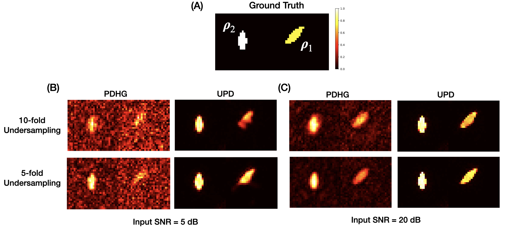
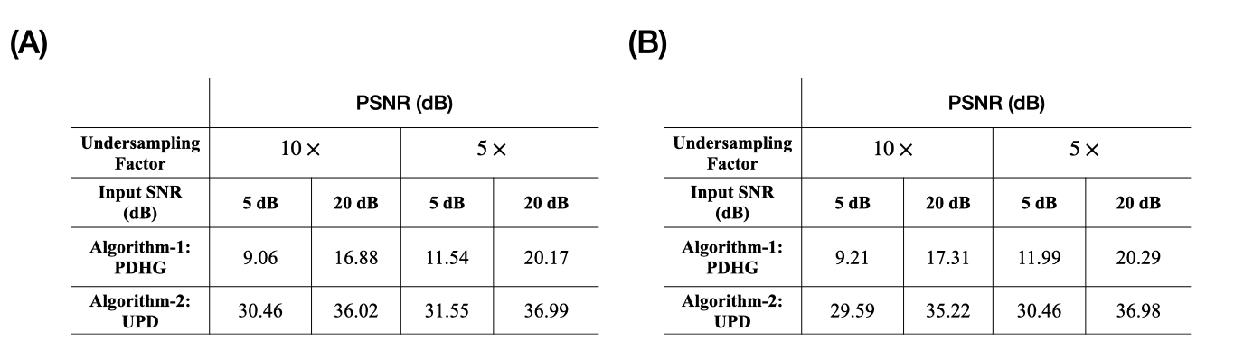
Proper modeling of $$$\mathscr{A}$$$ enabled successful chemical shift deconvolution across all the test cases. For input SNR = 5 dB, some of the weak 19F signals are not detected using PDHG (Fig. 3B) due to the limitations of structural prior under highly noisy scenarios. In contrast, learned UPD is capable of enhancing the signal detection with much more pronounced noise removal effects, which yields > 10 dB improvement in peak-signal-to-noise-ratio (PSNR,$$$20\log_{10}(M/\|\mathscr{A}_{\theta}^{\dagger}(s^{(i)})-x^{(i)}\|_2)$$$, where $$$M$$$ denotes the maximum possible pixel value) compared to PDHG (Fig. 4). Approximately ~1 dB improvement is achieved using $$$\breve{H}$$$ with BW = 200 kHz, 100 kHz (Fig.4A) against BW = 150 kHz, 100 kHz, respectively (Fig.4B). This implies that joint design of $$$R$$$ and $$$\breve{H}$$$ in the overall sensing operator $$$\mathscr{A}$$$ is necessary to guarantee recovery.Discussion and Conclusion
The modeling of radial MRI chemical shifts as convolutional effects provides a new avenue to unmix the component images. By leveraging the connection between radial sampling and Radon transform, learned primal-dual through unrolling significantly improves the performance of radial multi-spectral deconvolution of highly noisy and compressed data. In addition to the detection of multiple 19F tracers in vivo, these methods provide a new avenue to unmix chemical shifts for other X-nuclei applications with short T2 components.Acknowledgements
Funding for ETA was provided by National Institutes of Health (NIH) grant R01-CA269860. Funding for PP was provided by by Office of Naval Research (ONR) grant ONRN00014-19-1-2227 and Department of Energy (DE) grant DE-SC0022165.References
1. Ahrens ET, Helfer BM, O'Hanlon CF, et al. Clinical cell therapy imaging using a perfluorocarbon tracer and fluorine‐19 MRI. Magnetic resonance in medicine. 2014;72(6):1696-701.
2. Schoormans J, Calcagno C, Daal MR, et al. An iterative sparse deconvolution method for simultaneous multicolor 19F‐MRI of multiple contrast agents. Magnetic resonance in medicine. 2020;83(1):228-39.
3. Kislukhin AA, Xu H, Adams SR, Narsinh KH, Tsien RY, Ahrens ET. Paramagnetic fluorinated nanoemulsions for sensitive cellular fluorine-19 magnetic resonance imaging. Nature materials. 2016;15(6):662-8.
4. Chen J, Pal P, Ahrens ET. Enhanced detection of paramagnetic fluorine‐19 magnetic resonance imaging agents using zero echo time sequence and compressed sensing. NMR in Biomedicine. 2022;35(8):e4725.
5. Han Y, Yoo J, Kim HH, et al. Deep learning with domain adaptation for accelerated projection‐reconstruction MR. Magnetic resonance in medicine. 2018;80(3):1189-205.
6. Engström M, McKinnon G, Cozzini C, et al. In‐phase zero TE musculoskeletal imaging. Magnetic resonance in medicine. 2020;83(1):195-202.
7. Doganay O, Wade T, Hegarty E, et al. Hyperpolarized 129Xe imaging of the rat lung using spiral IDEAL. Magnetic Resonance in Medicine. 2016;76(2):566-76.
8. Adler J, Öktem O. Learned primal-dual reconstruction. IEEE transactions on medical imaging. 2018;37(6):1322-32.
9. J. Adler, H. Kohr, and O. Öktem. (2017). Operator Discretization Library (ODL). [Online]. Available: https://github.com/odlgroup/odl
Figures