4328
Restriction-induced time-varying transcytolemmal exchange rate: Revisiting diffusion MRI-based Kӓrger exchange model1Center for Nano and Micro Mechanics, Department of Engineering Mechanics, Tsinghua University, Beijing, China, 2Center for Biomedical Imaging Research, Department of Biomedical Engineering, School of Medicine, Tsinghua University, Beijing, China, 3Institute of Imaging Science, Vanderbilt University Medical Center, Nashville, TN, United States, 4Department of Biomedical Engineering, Vanderbilt University, Nashville, TN, United States, 5Department of Radiology and Radiological Sciences, Vanderbilt University Medical Center, Nashville, TN, United States, 6Department of Physics and Astronomy, Vanderbilt University, Nashville, TN, United States
Synopsis
Keywords: Signal Modeling, Microstructure, water exchange, quantitative microstructure imaging
Motivation: The diffusion MRI (dMRI) based two-compartmental Kӓrger exchange model is widely used to characterize transcytolemmal water exchange, but the influence of diffusion restriction remains unclear.
Goal(s): To investigate how diffusion restriction and other acquisition and microstructural parameters impact the estimation of transcytolemmal water exchange rate constants in the Kӓrger model.
Approach: Finite-difference-based numerical simulations were performed to quantitatively investigate time-varying transcytolemmal exchange rates of magnetization.
Results: When the compartmental size is large, e.g. 15 μm (close to typical cancer cell sizes), Kӓrger-model-derived exchange rate constants will be significantly dependent on time, diffusion gradient waveforms and microstructural features, resulting in overestimation of water exchange.
Impact: This
work elucidates the influence of diffusion restriction on the estimation of
transcytolemmal water exchange rate constants in the two-compartmental Kӓrger
model, revealing that water exchange is overestimated in large compartmental
sizes such as in tumors.
Introduction
Diffusion MRI (dMRI) provides a non-invasive approach to probe biological microstructures. Quantitative information is extracted through biophysical modeling and data fitting1,2. Typically, the current models ignore transcytolemmal water exchange3-5. Recent studies suggest that this simplification is inappropriate and the parameter reflecting water exchange is an important clinical biomarker6,7. Incorporating water exchange into modeling can provide more accurate and rich microstructural information.A general two-compartment (intra- and extracellular) model describes the exchange of magnetizations (Min and Mex) as8,9:
$$\begin{aligned}& \frac{d M_{i n}}{d t}=-q^2 D_{i n} M_{i n}-k_{i n}^m M_{i n}+k_{e x}^m M_{e x} \\& \frac{d M_{e x}}{d t}=-q^2 D_{e x} M_{e x}-k_{e x}^m M_{e x}+k_{i n}^m M_{i n}\end{aligned}\tag{1}$$
where q2 is the diffusion-weighted acquisition parameter, Din and Dex are compartmental diffusivities, $$$k_{i n}^m$$$ and $$$k_{e x}^m$$$ are time-varying intra-to-extra and extra-to-intra exchange rates of magnetizations.
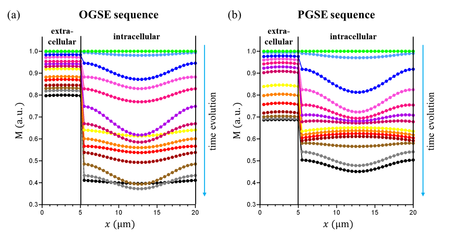
The Kӓrger model assumes $$$k_{i n}^m$$$ and $$$k_{e x}^m$$$ are time-independent and equal to exchange constants $$$k_{i n}$$$ and $$$k_{e x}$$$ of water molecules. It is based on two assumptions10: sufficiently long diffusion time and slow transcytolemmal water exchange. However, these two conditions are invalid in e.g., tumors with large cell sizes and high membrane permeability. Membrane-induced restriction leads to inhomogeneous magnetization distribution inside cells, with high magnetization near boundaries i.e., “edge-enhancement”11 (Fig.1).
Here, we quantitatively investigate how acquisition and microstructural factors impact the derivation of actual $$$k_{i n (e x)}^m$$$ from $$$k_{i n (e x)}$$$. The results will inspire us to revise the Kӓrger model to obtain more accurate information about tissue microstructure.
Method
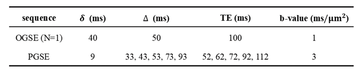
We use a finite difference method12 to simulate the time evolution of the magnetization distribution in a one-dimensional, two-compartmental, exchanging model system. This model is periodic along the x-axis, i.e., each compartment is exchanging with other compartments at both ends. The basic microstructural parameters are: intra-/extracellular compartment size=15/5μm, permeability Pm=0.01 μm/ms, and Din=Dex=1.56 μm2/ms. Both OGSE and PGSE sequences are performed, as shown in Table 1.$$$k_{i n ( e x )}^m(t)$$$ and $$$k_{i n ( e x )}(t)$$$ are obtained by calculating the ratio of the net outflow of magnetizations or molecules to the total amount in the compartment at time-t. Note that $$$k_{i n ( e x )}(t)$$$ is constant in each simulation. We introduce a dimensionless parameter to describe the deviation of $$$k_{i n ( e x )}^m(t)$$$ from $$$k_{i n ( e x )}(t)$$$:
$$\alpha_{i n(e x)}(t)=\frac{k_{i n(e x)}^m(t)}{k_{i n(e x)}(t)}\tag{2}$$
For each simulation, we also calculate an effective $$$\alpha_{i n ( e x )}$$$ by integral over time:
$$\alpha_{i n(e x)}=\frac{1}{\mathrm{TE}} \int_0^{\mathrm{TE}} \alpha_{i n(e x)}(t) d t\tag{3}$$
Furthermore, we investigate the effects of eight microstructural and acquisition factors on $$$\alpha_{i n ( e x )}$$$ by controlling other variables. They are: b-value, intracellular compartment size, effective diffusion time, permeability, Din, Dex, TE, and (Δ-δ)-value. The range of the above-mentioned parameters is set to meet experimental studies and clinical conditions5,13-16.
Results and Discussion
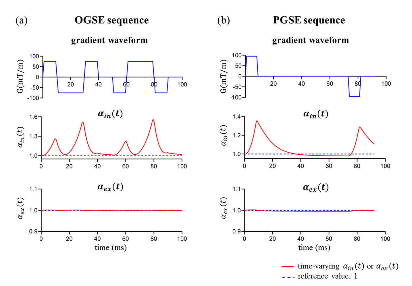
Fig 1 shows the magnetization distribution for (a) OGSE and (b) PGSE sequences. For the intracellular compartment, the edges hinder water diffusion, resulting in weaker signal attenuations near the boundaries. For the extracellular space, the magnetization distribution is almost uniform because this compartment is very narrow.Fig 2 shows the time-dependent $$$\alpha_{i n ( e x )}(t)$$$ during diffusion encoding for (a) OGSE and (b) PGSE sequences. The value of $$$\alpha_{i n}(t)$$$ is typically larger than 1 while $$$\alpha_{e x}(t)$$$ is always near 1.
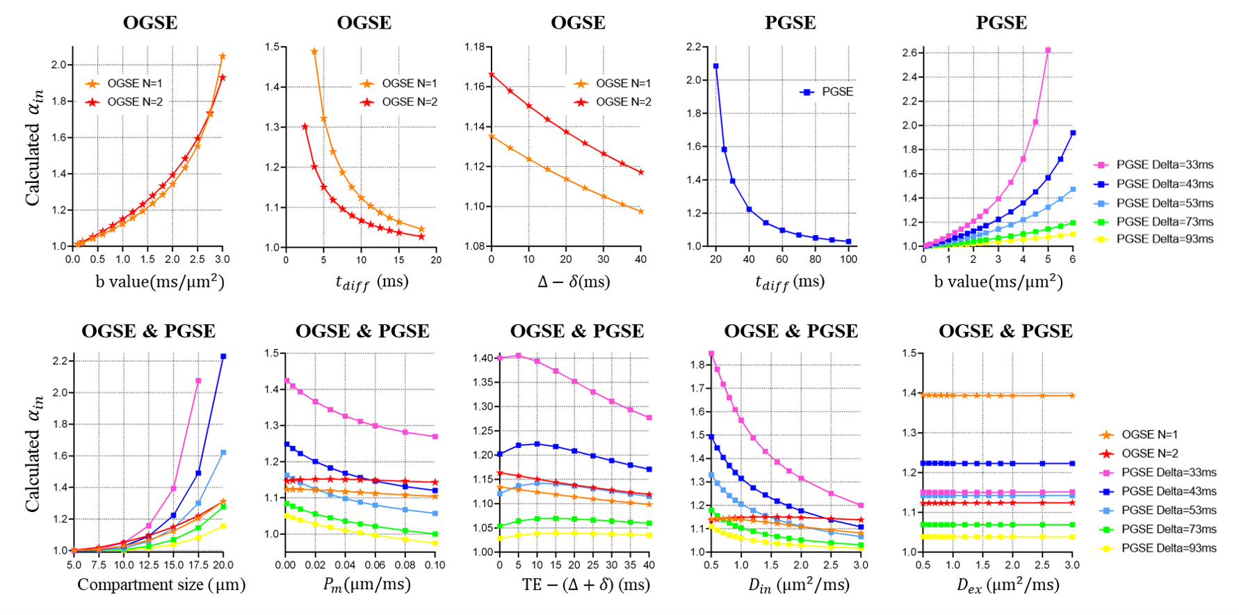
Fig. 3 shows the calculated effective $$$\alpha_{i n}$$$ for OGSE and PGSE sequences, as functions of different factors, respectively. The $$$\alpha_{i n}$$$ value is larger than 1 in most cases, and its variations are obvious within the predefined range of b-values, intracellular compartment sizes, and effective diffusion times tdiff. These factors play dominant roles for $$$\alpha_{i n}$$$>1 (i.e., $$$k_{i n}^m>k_{i n}$$$). In contrast, the effects of other factors are limited. These results demonstrate the necessity for $$$k_{i n}^m$$$-correction in the Kӓrger-model-based methods.
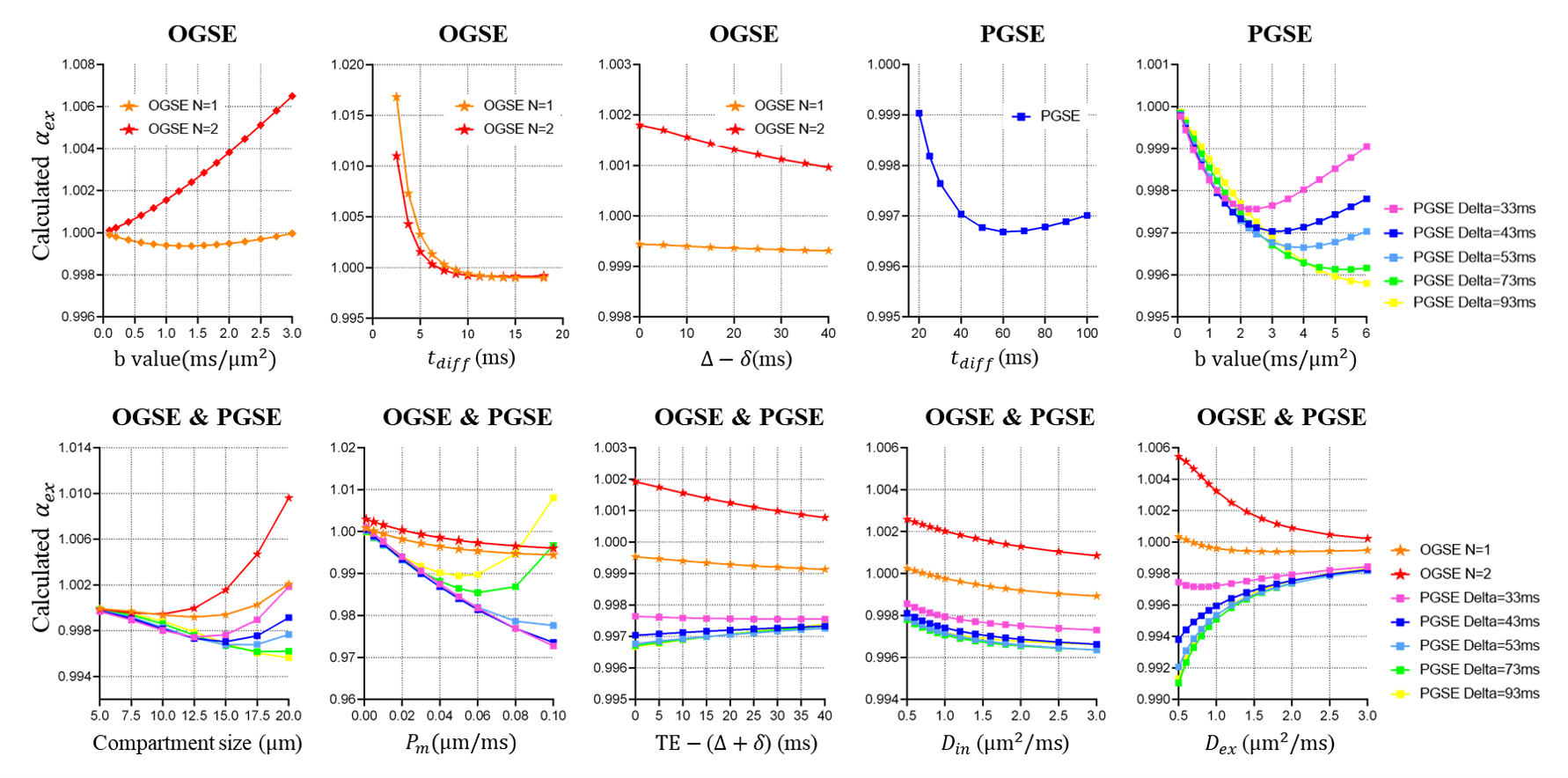
Fig. 4 shows the calculated effective $$$\alpha_{e x}$$$ for OGSE and PGSE sequences. The $$$\alpha_{e x}$$$ values are always near 1, and the deviation is negligible. These results indicate that there is no need to correct for $$$\alpha_{e x}$$$ because $$$k_{e x}^m=k_{e x}$$$ always holds.
Conclusion
In this work, we performed finite-difference-simulations to investigate time-varying magnetization exchange rates. When compartmental size is large e.g., 15 μm, the restriction-induced edge-enhancement leads to enhanced magnetizations involved in water exchange, then the percentage of exchanged magnetizations becomes higher than that of exchanged molecules. Furthermore, we determine the dominant factors in the edge-enhancement effect, which can inspire us to modify the Kӓrger-model-based methods to correct for overestimated exchange constants and provide more accurate microstructural information.Acknowledgements
No acknowledgement found.References
1. Gore JC, Xu J, Colvin DC, Yankeelov TE, Parsons EC, Does MD. Characterization of tissue structure at varying length scales using temporal diffusion spectroscopy. Nmr Biomed 2010;23(7):745-756.
2. Novikov DS, Fieremans E, Jespersen SN, Kiselev VG. Quantifying brain microstructure with diffusion MRI: Theory and parameter estimation. Nmr Biomed 2019;32:e3998.
3. Panagiotaki E, Walker-Samuel S, Siow B, et al. Noninvasive quantification of solid tumor microstructure using VERDICT MRI. Cancer Research 2014;74(7):1902.
4. Jiang X, Li H, Xie J, Zhao P, Gore JC, Xu J. Quantification of cell size using temporal diffusion spectroscopy. Magn Reson Med 2016;75(3):1076-1085.
5. Xu J, Jiang X, Li H, Arlinghaus LR, Gore JC. Magnetic resonance imaging of mean cell size in human breast tumors. Magn Reson Med 2019;83(6):2002-2014.
6. Li H, Jiang X, Xie J, Gore JC, Xu J. Impact of transcytolemmal water exchange on estimates of tissue microstructural properties derived from diffusion MRI. Magn Reson Med 2017;77(6):2239-2249.
7. Jiang X, Devan SP, Xie J, Gore JC, Xu J. Improving MR cell size imaging by inclusion of transcytolemmal water exchange. Nmr Biomed 2022;35(12):e4799.
8. Karger J, Pfeifer H, Heink W. Principles and Application of Self-Diffusion Measurements by Nuclear Magnetic Resonance. Advances in Magnetic and Optical Resonance 1988;12:1-89.
9. Stanisz GJ, Li JG, Wright GA, Henkelman RM. Water dynamics in human blood via combined measurements of T2 relaxation and diffusion in the presence of gadolinium. Magnetic Resonance in Medicine Official Journal of the Society of Magnetic Resonance in Medicine 1998;39(2):223-233.
10. Fieremans E, Novikov DS, Jensen JH, Helpern JA. Monte Carlo study of a two-compartment exchange model of diffusion. Nmr Biomed 2010;23(7):711-724.
11. Sukstanskii AL, Ackerman JJH, Yablonskiy DA. Effects of barrier-induced nuclear spin magnetization inhomogeneities on diffusion-attenuated MR signal. Magn Reson Med 2003;50(4):735-742.
12. Xu J, Does MD, Gore JC. Numerical study of water diffusion in biological tissues using an improved finite difference method. Physics in Medicine & Biology 2007;52(7):111-126.
13. Åslund I, Nowacka A, Nilsson M, Topgaard D. Filter-exchange PGSE NMR determination of cell membrane permeability. Journal of Magnetic Resonance 2009;200(2):291-295.
14. Meier C, Dreher W, Leibfritz D. Diffusion in compartmental systems. II. Diffusion-weighted measurements of rat brain tissue in vivo and postmortem at very large b-values. Magn Reson Med 2003;50(3):510-514.
15. Jelescu IO, de Skowronski A, Geffroy F, Palombo M, Novikov DS. Neurite Exchange Imaging (NEXI): A minimal model of diffusion in gray matter with inter-compartment water exchange. Neuroimage 2022;256:119277.
16. Zhang J, Lemberskiy G, Moy L, Fieremans E, Novikov DS, Kim SG. Measurement of cellular-interstitial water exchange time in tumors based on diffusion-time-dependent diffusional kurtosis imaging. Nmr Biomed 2021;34(6):e4496.
Figures