4327
Monitoring treatment response of nasopharyngeal carcinoma to vascular normalization with Amide Proton Transfer Imaging1Department of Radiology, Affiliated Hospital of Guizhou Medical University, Guiyang, China, 2Philips Healthcare, Shanghai, China
Synopsis
Keywords: Signal Modeling, Cancer
Motivation: The effectiveness of anti-angiogenesis therapy in patients with advanced nasopharyngeal carcinoma is not optimal, and early monitoring of treatment effectiveness can reduce unnecessary treatment for patients.
Goal(s): The focus of this study was to ascertain whether Amide Proton Transfer- Magnetic Resonance Imaging (APT-MRI) could be effectively used to evaluate tumor response at an early stage.
Approach: The research involved an examination of the correlation between APTw values and tumor cell proliferation, achieved by subjecting preclinical models to diverse antiangiogenic therapies.
Results: The results of our study demonstrated a marked positive correlation between APTw and the expression of Ki67, a tumor proliferation marker.
Impact: This study is useful for the clinical application of anti-angiogenesis therapy. The study provides an in-depth exploration of the mechanisms and efficacy of this therapeutic approach, offering a comprehensive understanding that is crucial for its successful implementation in clinical practice.
Introduction
The clinical efficacy of chemo-radiotherapy alone in patients with locally advanced nasopharyngeal carcinoma is suboptimal. Several researchers have discovered that anti-angiogenic therapy shows promise as an anti-tumor treatment. This therapy not only inhibits tumor angiogenesis but also promotes tumor vascular normalization [1]. The human recombinant vascular endothelial growth factor (VEGF) monoclonal antibody Bevacizumab (BEV) has been widely used in clinical practice. However, BEV often develops drug resistance, and its long-term effectiveness is not ideal [2]. TGF-β strongly influences tumor proliferation, angiogenesis, invasion, and therapy resistance. It is an important target for tumor treatment [3]. This study hypothesized that TGF-β may be a key target for overcoming resistance to anti-angiogenesis therapy in nasopharyngeal carcinoma. Due to the high cost of antiangiogenic therapy, early monitoring of tumor response can reduce unnecessary treatment for patients. Chemical exchange saturation transfer (CEST) is an MRI contrast mechanism that utilizes the chemical exchange of unstable protons in biomolecules with solvent hydrants [4]. APT is a variant of CEST-MRI that is sensitive to amide proton exchange on the main and side chains of proteins. The APT signal depends on the exchange rate of the amide proton with the solvent, and this exchange rate is base-catalyzed [5], making APT sensitive to tissue pH [6]. Previous studies combining in vivo APT-MRI measurements and in vitro histological protein concentration measurements of rat brain metastases showed that approximately 66% of APT-MRI signals originated from changes in cytoplasmic protein concentration, while the remaining 34% resulted from changes in tumor pH value [7]. Therefore, this study discusses the application value of APT-MRI in monitoring tumor treatment response, which is beneficial for implementing personalized anti-tumor treatment in clinical practice.Materials and Methods
40 subcutaneous xenograft tumor models of nasopharyngeal carcinoma were randomly divided into 4 groups (10 in each group): the control group, galunisertib group, BEV group, and galunisertib + BEV group. MRI and histopathological analysis were performed at different time points (day 0 and 25). The MRI experiment was conducted using a 3.0T scanner (Elition X, Philips Healthcare, Best, the Netherlands) equipped with an eight-channel mouse coil (HC607P, Hezi Medical Technology Co., Ltd., Wuxi, China). The scanning sequence included T1-weighted imaging (T1WI), T2-weighted imaging (T2WI), and APT. The IVIM parameters were evaluated using the bi-exponential model, and the corresponding parameter maps were generated. The APTw images were reconstructed using the built-in APT image reconstruction algorithm. Region of interest (ROI) was plotted based on the T1WI and T2WI images, including the entire tumor region, and the APT weighted values of each ROI were recorded. The correlation between MRI parameters and pathological parameters was determined using Pearson’s correlation analysis.Results
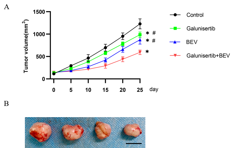
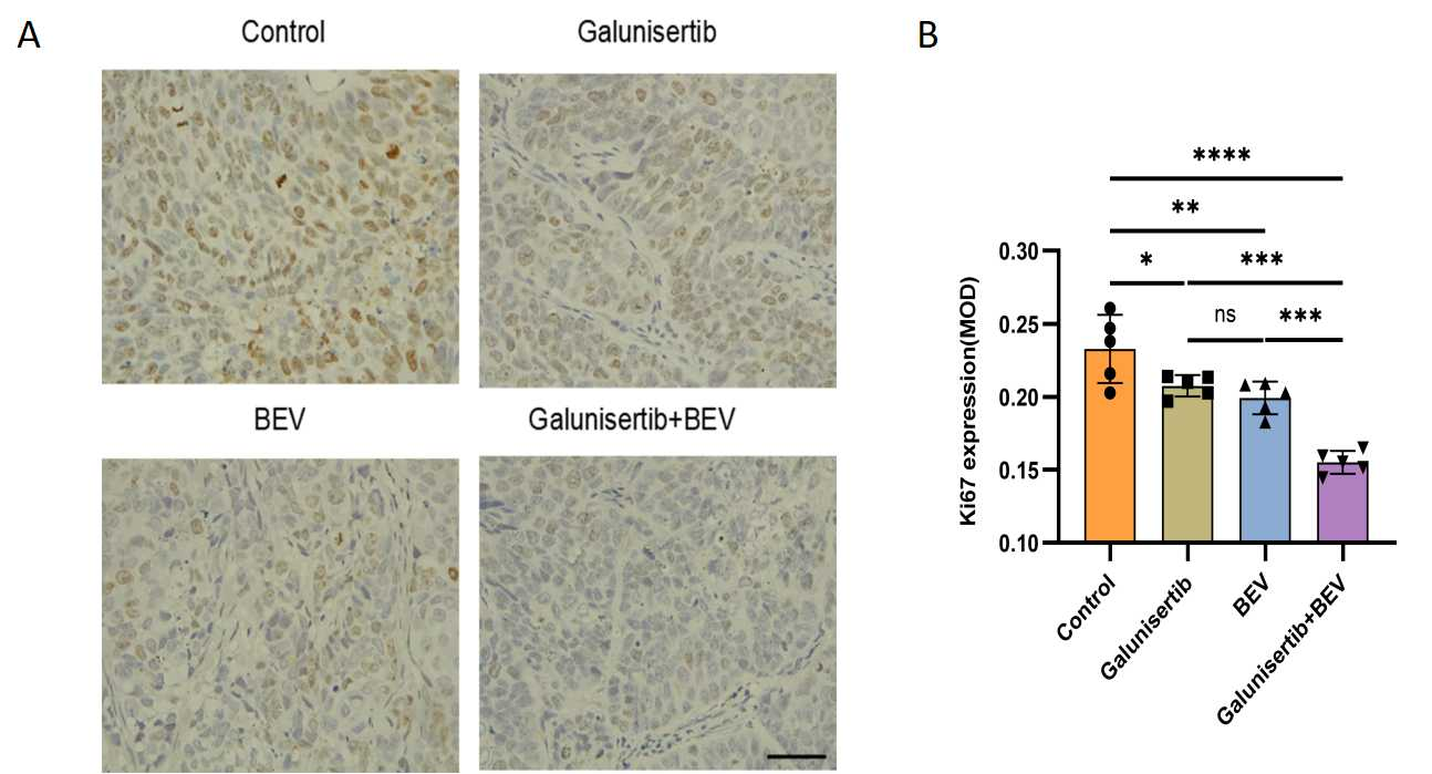
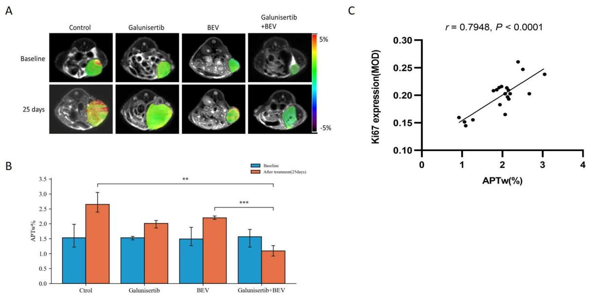
Galunisertib, BEV, and the combination of Galunisertib+BEV effectively suppressed tumor growth. After 25 days of treatment, the mean tumor size in the Galunisertib+BEV combination group was significantly smaller than that in the Galunisertib and BEV monotherapy groups, as well as the control group (P < 0.05) (Fig. 1). Immunohistochemical staining revealed that the combined treatment group significantly inhibited tumor cell proliferation compared to the BEV monotherapy group (P < 0.05) (Fig. 2). The initial APTw levels did not differ significantly among all groups before treatment. However, after 25 days of intervention, the APTw levels increased in the galunisertib monotherapy, BEV monotherapy, and control groups, with the control group showing the greatest increase. Only the galunisertib + BEV combination treatment group exhibited a decrease in APTw. On day 25 after treatment, the APTw levels in the combined treatment group decreased considerably compared to those in the control and BEV monotherapy groups (P < 0.05) (Fig. 3). Correlation analysis revealed a significant positive correlation between APTw and the expression of the tumor proliferation marker Ki67 (Fig. 3).Discussion
This study demonstrated that Galunisertib can enhance the antitumor effect of BEV. APT imaging is a novel MRI technology used to evaluate the levels of mobile proteins and peptides in tissues by saturating peptide bonds with amide proton. APT can be used to assess efficacy in glioma, rectal, and breast cancers [8]. The intensity of the APT signal is influenced by the protein level and cell density of tumor tissue [9]. The APTw value exhibited a significantly positive correlation with the tumor proliferation marker Ki67, consistent with previous studies on breast cancer and rectal cancer [10].Conclusion
Our study demonstrates that the combination of Galunisertib and BEV is more effective than any single treatment in slowing down the growth of tumors. APTw can indicate the level of proliferation in tumor cells. APT-MRI can be utilized to identify the response of antitumor therapy.Acknowledgements
No acknowledgement found.References
[1] Gong X, et al. The Treatment Combining Antiangiogenesis with Chemoradiotherapy Impinges on the Peripheral Circulation Vascular Endothelial Cells and Therapeutic Effect in the Patients with Locally Advanced Nasopharyngeal Carcinoma. Biomed Res Int. 2022 Jul;2022:1787854.
[2] Chelvanambi M, et al. STING agonist-based treatment promotes vascular normalization and tertiary lymphoid structure formation in the therapeutic melanoma microenvironment. J Immunother Cancer. 2021 Feb;9(2):e001906.
[3] Peng D, et al. Targeting TGFB signal transduction for fibrosis and cancer therapy. Mol Cancer. 2022 Apr;21(1):104.
[4] van Zijl PCM, et al. Magnetization Transfer Contrast and Chemical Exchange Saturation Transfer MRI. Features and analysis of the field-dependent saturation spectrum. Neuroimage. 2018 Mar; 168:222–41.
[5] Zhou J, et al. Using the amide proton signals of intracellular proteins and peptides to detect pH effects in MRI. Nat Med. 2003 Aug; 9:1085–90.
[6] Tee YK, et al. Comparing different analysis methods for quantifying the MRI amide proton transfer (APT) effect in hyperacute stroke patients. NMR Biomed. 2014 Sep; 27:1019–29.
[7] Ray KJ, et al. Tumor pH and Protein Concentration Contribute to the Signal of Amide Proton Transfer Magnetic Resonance Imaging. Cancer Res. 2019 Apr;79(7):1343-1352.
[8] Park JE, et al. Identification of Early Response to Anti-Angiogenic Therapy in Recurrent Glioblastoma: Amide Proton Transfer-weighted and Perfusion-weighted MRI compared with Diffusion-weighted MRI. Radiology. 2020 May;295(2):397-406.
[9] Zhou J, et al. Three-dimensional amide proton transfer MR imaging of gliomas: Initial experience and comparison with gadolinium enhancement. J Magn Reson Imaging. 2013 Nov;38(5):1119-28.
Figures