4326
Virtual brain modelling of the cerebro-cerebellar loop dynamics with region-specific mean field formalism1Department of Brain and Behavioral Sciences, Università di Pavia, Pavia, Italy, 2Digital Neuroscience Centre, IRCCS Mondino Foundation, Pavia, Italy, 3NMR Research Unit, Queen Square MS Centre, Department of Neuroinflammation, UCL Queen Square Institute of Neurology, Faculty of Brain Sciences, University College London, London, United Kingdom
Synopsis
Keywords: Signal Modeling, fMRI (resting state), Neuro, brain modeling, The Virtual Brain
Motivation: Brain dynamic simulators rely on the same model for each grey matter region.
Goal(s): We embed region-specific mean field models (MFs) into virtual brains by designing a flexible framework linking input/output signals from different MFs.
Approach: We integrate a recently-developed cerebellar MF into “The Virtual Brain” platform. We simulate brain dynamics with cerebellar MF for cerebellar nodes and connect cerebellar MF input/output with MFs previously used for other regions.
Results: The multi-modEl framework is ready to be used with any number of different MFs; moreover, in the cerebellum, using its realistic MF improves 7-folds the correlation between simulated and empirical functional connectivity.
Impact: Simulations of brain dynamics rely on assigning the same model to all brain regions, not capturing cortical microcircuits diversities. We developed a framework to connect region-specific mean field models and demonstrated an improved performance in the cerebellum, towards personalized simulations.
Introduction
The Virtual Brain (TVB) is a data-driven brain dynamic framework that enables simulating brain activity starting from subject-specific structural connectivity (SC) and functional connectivity (FC), extracted from diffusion weighted imaging and functional MRI (fMRI) respectively1. TVB computes a simulated Blood Oxygen Level Dependent (BOLD) signal by solving mathematical models (i.e., mean field models (MFs)) of the functional activity of each node (i.e., grey matter region), structurally connected through the SC matrix2. Simulated BOLD signals are optimized by maximizing the Pearson Correlation Coefficient (PCC) between the simulated FC and the empirical FC. TVB applications in clinic and research are growing, e.g. to improve pre-operative planning in epilepsy or to study neurodegenerative diseases3-5. However, TVB simulations employ the same model for all nodes, not accounting for intrinsic region-specific brain activities1. We provide a framework to differentiate node dynamics, by associating different MFs to different regions, known to be characterised by different microcircuits. Specifically, we focus on the cerebro-cerebellar loop by (i) integrating a validated cerebellar MF6 and (ii) connecting its input/output with MFs available within the pool of TVB models that we used for other regions7–9. This pipeline can be used to integrate any number of region-specific MF, improving the specificity of TVB predictions and its personalisation.Methods
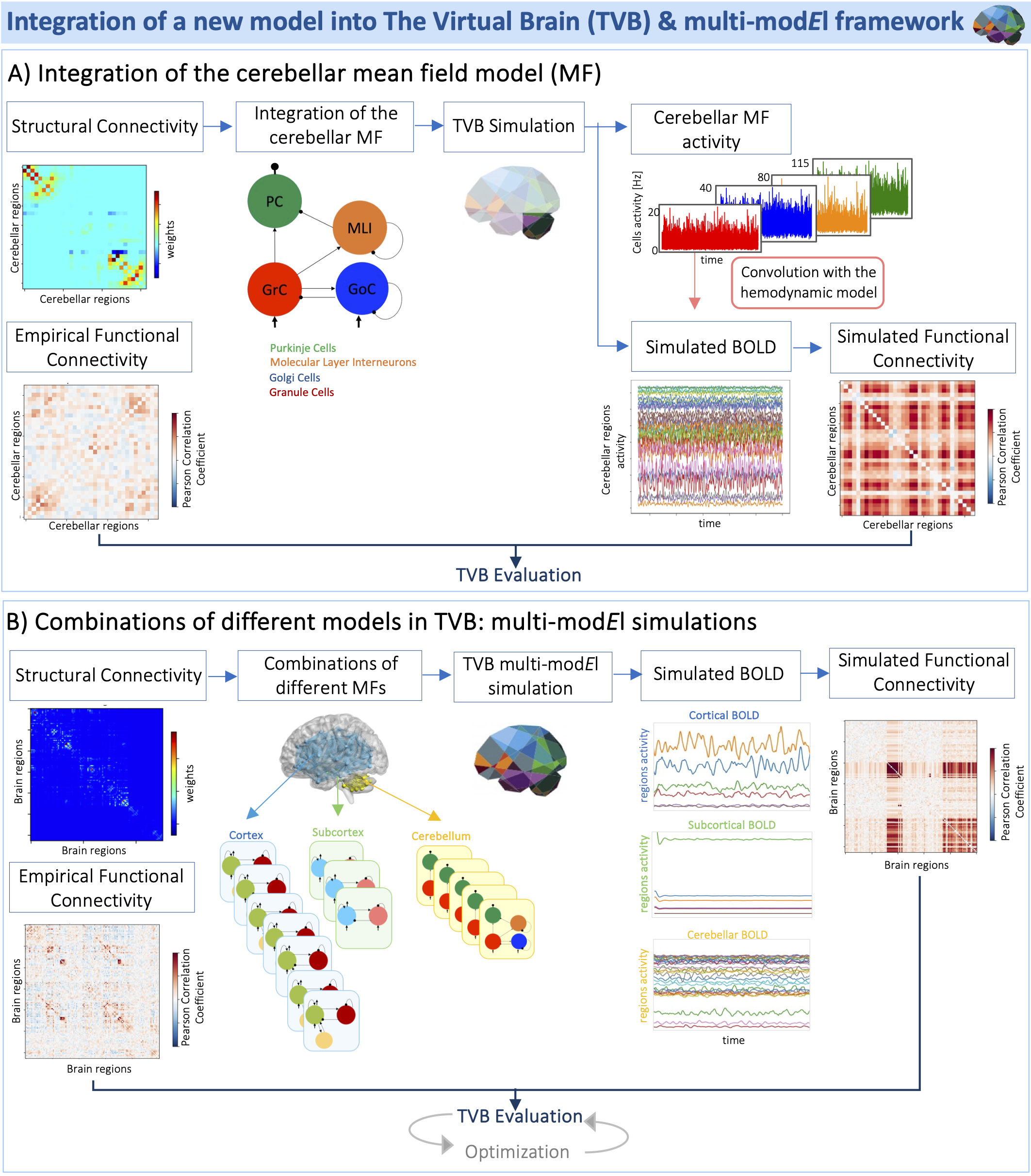
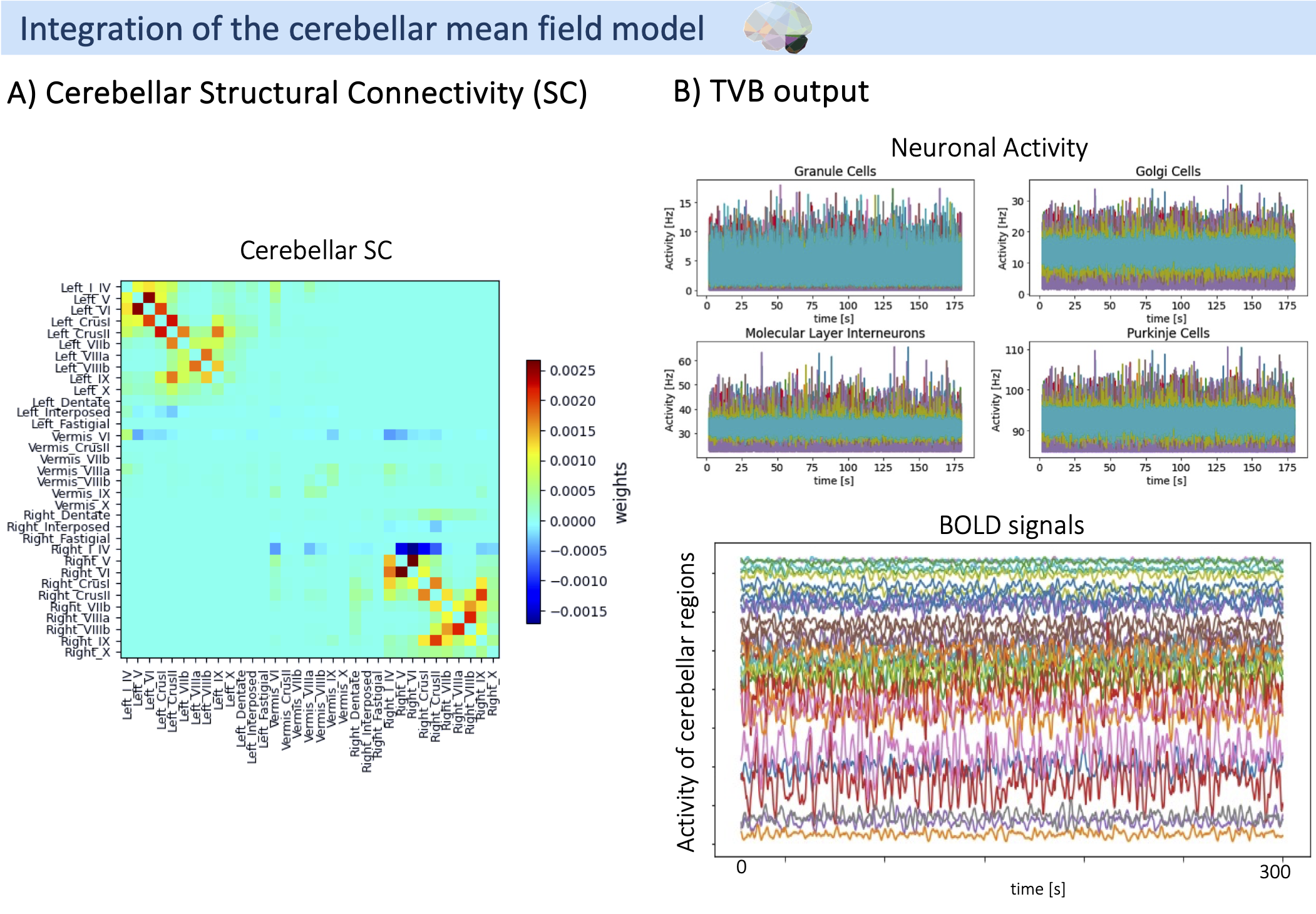
Our pipeline is reported in Figure 1. SC and FC were computed from a subject of the Human Connectome Project data (http://db.humanconnectome.org). We addressed how to: (i) integrate the cerebellar MF into the TVB platform, and (ii) how to connect its input/output with other MFs for cerebral cortical and subcortical regions. A TVB simulation using the same generic MF in all nodes (i.e., Wong-Wang model10 for cerebral cortex, subcortex and cerebellum) was implemented as the standard reference to compare (i) and (ii)11. In our developed framework, it was important to take care of the fact that excitatory long-range connections, weighted by SC, linked all MF models of cerebral regions7, in contrast to the inhibitory output of the specific cerebellar MF, connected to deep cerebellar nuclei12. To capture this excitatory/inhibitory signals, the SC was multiplied by a matrix of value -1 for all connections from the cerebellar cortex to the deep cerebellar nuclei and +1 everywhere else.- (i) Cerebellar MF integration into TVB (Figure 2). SC and empirical FC of the cerebellum were used as input for TVB simulation. Quantitative scores (PCC and Spearman correlation coefficients, cosine similarity, Mean Absolute Error (MAE), and Root Mean Square Error (RMSE)) were computed to evaluate the similarity between empirical and simulated FC. PCC was compared with the PCC of the standard TVB simulation.
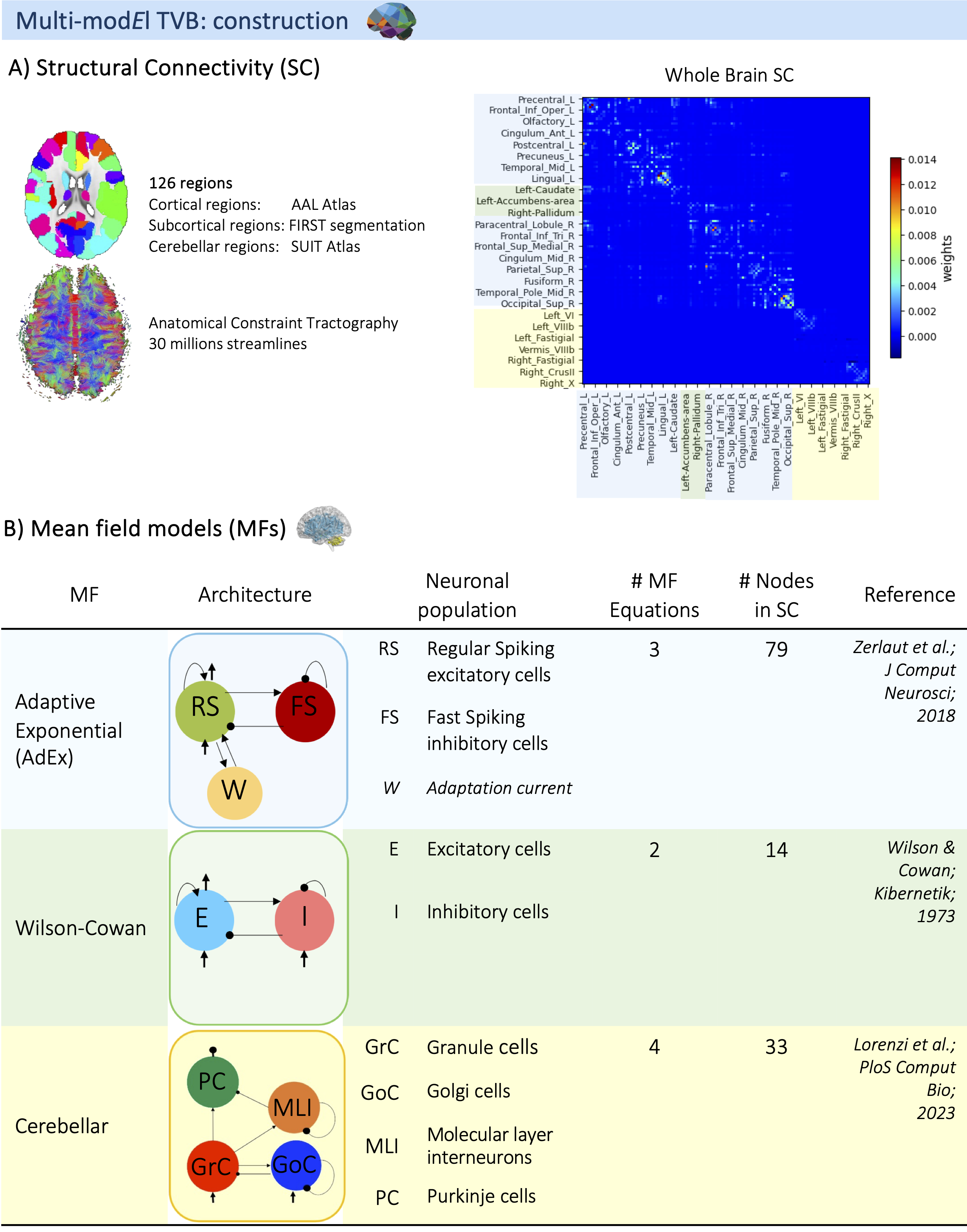
- (ii) Connection of different MFs: Multi-modEl framework (Figure 3). Whole-brain SC and empirical FC were used as input. The cerebellar MF13 was associated to cerebellar nodes, Wilson-Cowan MF14 to subcortical structures and Adaptive Exponential (AdEx) MF9 to cortical regions. Simulated and empirical FC were evaluated as in (i). PCC of the cerebellar subnetwork and the cerebral subnetwork (cortical and subcortical regions) were compared with the PCC of the standard TVB simulation11.
Results
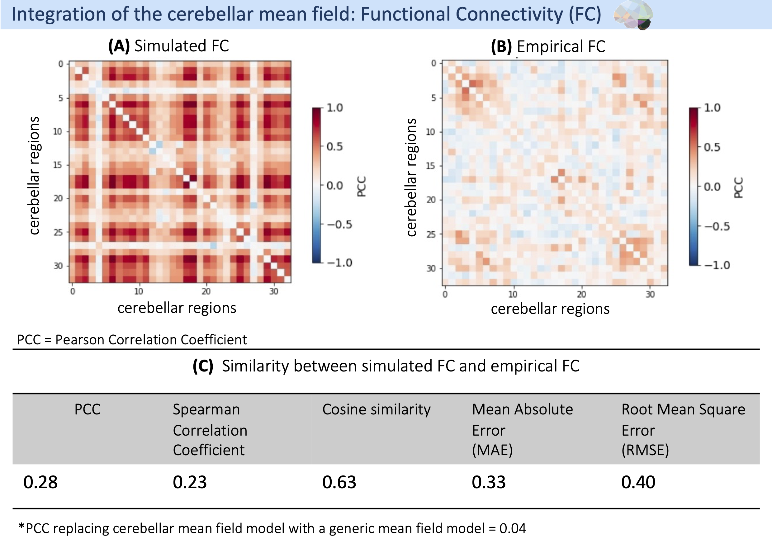
- (i) Cerebellar simulated FC is reported in Figure 4. PCC computed between simulated and empirical FC using the standard TVB was 0.04, while PCC increased 7-fold up to 0.28 by integrating the cerebellar MF.
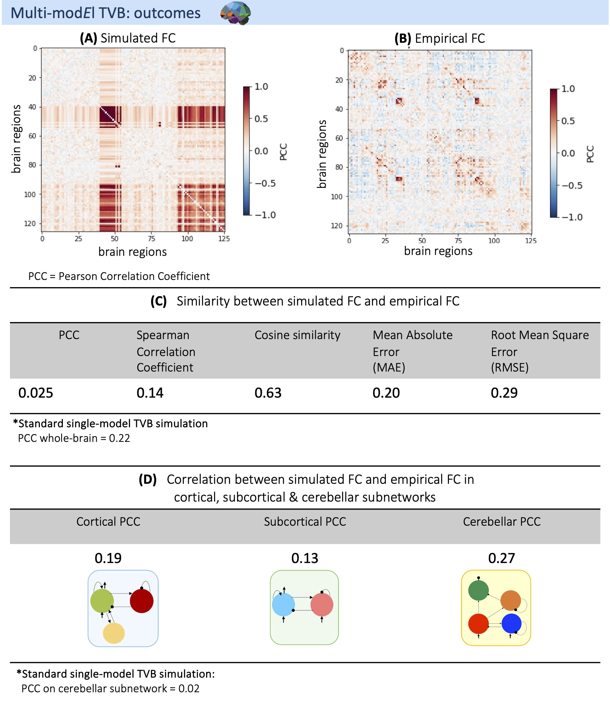
- (ii) Multi-modEl simulated FC is reported in Figure 5. MAE and RMSE show an error less than 30% between simulated and empirical FC. Compared to the PCC computed with the standard framework, whole-brain and cerebral PCC decreased, while the cerebellar PCC increased as in (i).
Discussion
The integration of a cerebellar MF model into TVB was successful and demonstrated the possibility to connect signals from MF models specific to different grey matter regions. Indeed, the cerebellar PCC increased 7-fold both in the cerebellar network (i) and in the whole-brain multi-modEl simulation (ii). The cerebellar MF, though, was recently developed starting from realistic models of cellular microcircuits (bottom-up), while the Wilson-Cowan and AdEx MF are simplified top-down models of cerebral function; therefore, our cerebellar result emphasizes the impact of incorporating biologically grounded models into BOLD simulators that need to be extended to all cortical regions. Moreover, further work should include strategies for parameter optimisation specific to this multi-modEl framework. In conclusion, TVB subject-specific functional parameters can now be extended to regional levels, opening up new opportunities for region-specific investigations and potentially advancing diagnostic and treatment applications.Conclusion
By integrating cerebellar MF and establishing a multi-modEl framework in TVB we set the foundation for personalized, region-specific simulations.Acknowledgements
This research has received funding from the European Union’s Horizon 2020 Framework Program for Research and Innovation under the Specific Grant Agreement No. 945539 (Human Brain Project SGA3) to ED, CGWK, FP. CGWK received funding from BRC (#BRC704/CAP/CGW), MRC (#MR/S026088/1), Ataxia UK, Rosetree trust (#PGL22/100041 and #PGL21/10079). CGWK is a shareholder in Queen Square Analytics Ltd. This research has also received funding from Centro Fermi project “Local Neuronal Microcircuits” to ED. Special acknowledgement to EBRAINS and FENIX for informatic support and infrastructure. RL have been supported by Human Brain Project SGA3. This work was also supported by #NEXTGENERATIONEU (NGEU) and funded by the Ministry of University and Research (MUR), National Recovery and Resilience Plan (NRRP), project MNESYS (PE0000006) – A Multiscale integrated approach to the study of the nervous system in health and disease (DN. 1553 11.10.2022) to ED, CGWK and CC; and Project EBRAINS-Italy (IR00011) - (M4C2 Line 3.1 of the PNRR, Action 3.1.1 - CUP B51E22000150006) to ED and CC.References
[1] Sanzleon P, Knock SA, Woodman MM, et al. The virtual brain: A simulator of primate brain network dynamics. Front Neuroinform. 2013;7(MAY). doi:10.3389/fninf.2013.0001
[2] Ritter P, Schirner M, Mcintosh AR, Jirsa VK. The Virtual Brain Integrates Computational Modeling and Multimodal Neuroimaging. Brain Connect. 2013;3(2):121-145. doi:10.1089/brain.2012.0120
[3] Jirsa VK, Proix T, Perdikis D, et al. The Virtual Epileptic Patient: Individualized whole-brain models of epilepsy spread. Neuroimage. 2017;145:377-388. doi:10.1016/j.neuroimage.2016.04.049
[4] Monteverdi A, Palesi F, Costa A, et al. Subject-specific features of excitation / inhibition profiles in neurodegenerative diseases. 2022;(August):1-17. doi:10.3389/fnagi.2022.868342
[5] Zimmermann J, Perry A, Breakspear M, et al. Differentiation of Alzheimer’s disease based on local and global parameters in personalized Virtual Brain models. NeuroImage Clin. 2018;19(April):240-251. doi:10.1016/j.nicl.2018.04.017
[6] Mazzoni A, Lindén H, Cuntz H, Lansner A, Panzeri S, Einevoll GT. Computing the Local Field Potential (LFP) from Integrate-and-Fire Network Models. PLoS Comput Biol. 2015;11(12):1-38. doi:10.1371/journal.pcbi.1004584
[7] Goldman JS, Kusch L, Aquilue D, et al. A comprehensive neural simulation of slow-wave sleep and highly responsive wakefulness dynamics. Front Comput Neurosci. 2023;16. doi:10.3389/fncom.2022.1058957
[8] Wilson HR, Cowan JD. Excitatory and Inhibitory Interactions in Localized Populations of Model Neurons. Biophys J. 1972;12(1):1-24. doi:https://doi.org/10.1016/S0006-3495(72)86068-5
[9] Zerlaut Y, Chemla S, Chavane F, Destexhe A. Modeling mesoscopic cortical dynamics using a mean-field model of conductance-based networks of adaptive exponential integrate-and-fire neurons. J Comput Neurosci. 2018;44(1):45-61. doi:10.1007/s10827-017-0668-2
[10] Wong KF, Wang XJ. A recurrent network mechanism of time integration in perceptual decisions. J Neurosci. 2006;26(4):1314-1328. doi:10.1523/JNEUROSCI.3733-05.2006
[11] Palesi F, Lorenzi RM, Casellato C, et al. The Importance of Cerebellar Connectivity on Simulated Brain Dynamics. Front Cell Neurosci. 2020;14(July):1-11. doi:10.3389/fncel.2020.00240
[12] D’Angelo E. Physiology of the Cerebellum. Vol 154. 1st ed. Elsevier B.V.; 2018. doi:10.1016/B978-0-444-63956-1.00006-0
[13] Lorenzi RM, Geminiani A, Zerlaut Y, et al. A multi-layer mean-field model of the cerebellum embedding microstructure and population-specific dynamics. PLoS Comput Biol. 2023:1-30. doi:10.1371/journal.pcbi.1011434
[14] Wilson HR, Cowan JD. A mathematical theory of the functional dynamics of cortical and thalamic nervous tissue. Kybernetik. 1973;13(2):55-80. doi:10.1007/BF00288786
Figures