4325
IVIM diffusion parameter highlights placental microstructural damage in patients with previous SARS-CoV-2 pregnancy infection1Physics Dpt Sapienza University of Rome, National Research Council, Institute for Complex Systems, Rome, Italy, 2Physics, Sapienza University of Rome, Rome, Italy, 3Radiological, Oncological and Pathological Sciences, Policlinico Umberto I, Sapienza University of Rome, Rome, Italy, 4Maternal and Child Health and Urological Sciences, Policlinico Umberto I, Sapienza University of Rome, Rome, Italy
Synopsis
Keywords: Placenta, Placenta, Perfusion, Diffusion, COVID-19, Microstructural damage
Motivation: Currently, no study has investigated the role of diffusion-MRI to assess the placenta of women getting SARS-CoV-2 infection.
Goal(s): To study the placental tissues abnormalities due to the infection.
Approach: Pregnant women COVID-19 group (n=14) and pre-pandemic healthy women (n=19) were investigated using IVIM protocol at 1.5T.
Results: D was significantly higher in the COVID-19 compared to that of the age-matched healthy group. No-significant difference between f values was found in the two groups suggesting no-specific microstructural damage with no-perfusion alteration. A significant negative correlation was found between D and GA only in the COVID-19 reflecting a possible senescence process due to COVID-19.
Impact: Diffusion MRI underline higher D value in SARS-Cov-2 compared to healthy placentas, which can be explained as a microstructural deterioration of the placental tissue. Further investigations will allow us better to understand the effects of SARS-Cov-2 infection on human tissues.
INTRODUCTION
Severe acute respiratory syndrome coronavirus-2 (SARS-CoV-2) has been a major global health problem since December 2019. To our knowledge, no study has investigated the role of diffusion MRI to assess the placenta of women getting SARS-CoV-2 infection during the COVID-19 pandemic in COVID-19 negative women. This work aimed to investigate whether pregnant women's mild and moderate SARS-CoV-2 infection was associated with microstructural and vascular changes in the placenta observable in vivo by Intravoxel Incoherent Motion (IVIM) at different gestational ages (GA).METHODS
Pregnant women during the SARS-CoV-2 pandemic (COVID-19 group, n=14) and pre-pandemic healthy pregnant women (n=19) were investigated. MRI IVIM protocol at 1.5T was constituted of diffusion-weighted (DW) images with TR/TE=3100/76ms and 10 b-values (0,10,30,50,75,100,200,400,700,1000s/mm2). f and D IVIM maps were obtained1,2. Due to the heterogeneity of placental tissue3,4 maternal and fetal placentas were analyzed separately. Differences between IVIM parameters D (diffusion), and f (fractional perfusion) quantified in the two groups were evaluated using the ANOVA test with Bonferroni correction and linear correlation between IVIM metrics and GA, COVID-19 duration, the delay time between a positive SARS-CoV-2 test and MRI examination (delay-time exam+) was studied by Pearson-test.RESULTS
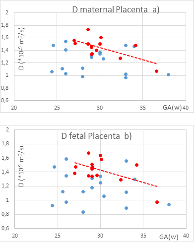
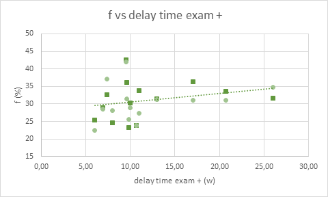
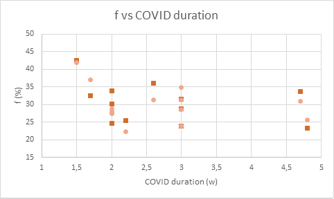
D was significantly higher in the COVID-19 placentas (in fetal placenta, D=(1.43±0.17)*10-3mm2/s, in maternal placenta D=(1.45±0.16)*10-3mm2/s ) compared to that of the age-matched healthy group (in fetal placenta D=(1.18±0.23)*10-3mm2/s, in maternal placenta D=(1.24±0.20)*10-3mm2/s ) with p<0.04 in fetal and p<0.007 in maternal site, Fig. 1. No significant difference between f values was found in the two groups suggesting no-specific microstructural damage with no perfusion alteration (potentially quantified by f) in mild/moderate SARS-Cov-2 placentas. A significant negative correlation was found between D and GA in the COVID-19 placentas whereas no significant correlation was found in the control placentas reflecting a possible accelerated senescence process due to COVID-19. A trend of increasing f as a function of delay time exam + was found considering all fetal and maternal placental sites of COVID-19 group (Fig. 2). Finally, a significant negative correlation (r=-0.92, p<0.0002) was found between f and COVID duration for SARS-CoV-2 infection duration less than 2.2 months (Fig. 3).DISCUSSION
We report impaired microstructural placental development during pregnancy and the absence of perfusion-IVIM parameter changes that may indicate no perfusion changing through microvessels and microvilli in the placentas of pregnancies with mild/moderate SARS-Cov-2 after reaching negativity. Although the increase in D is a nonspecific marker, it is well known that D increases in case of damage to the tissue microstructure. Therefore, in vivo diffusion MRI investigation of previous SARS-CoV-2 positive placentas suggests microstructural placenta damage. Although the results do not show an alteration of placental perfusion in COVID-19 group, it seems that f tends to increase with increasing delay time between a positive SARS-CoV-2 test and MRI examination (Fig. 3), and f increases with decreasing the time interval between positive and negative SARS-CoV-2 test (Fig. 3). Therefore, timing and duration of infection could affect placental perfusion.CONCLUSION
Since the subjects analyzed were affected by mild or moderate SARS-Cov-2 symptoms, we did not expect to find differences in IVIM parameters between the pre-pandemic control and COVID-19 groups, or, at most, we expected to find, in agreement with the literature5,6, alteration of perfusion parameters. Contrary to expectations, diffusion MRI results underline a clear alteration of D parameter (higher value in SARS-Cov-2 compared to healthy placentas), which can be explained as a microstructural deterioration of the placental tissue. These results, therefore, deserve further investigations that will allow us better to understand the effects of SARS-Cov-2 infection on human tissues.Acknowledgements
No acknowledgement found.References
1. A Maiuro, A Antonelli, L Manganaro, S Capuani. Molecular diffusion and perfusion of biological water quantified by MRI for the diagnosis of pathological human placentas. IL NUOVO CIMENTO C (2020) 43, 124
2. Maiuro A, Ercolani G, Di Stadio F, Antonelli A, Catalano C, Manganaro L, Capuani S, Two-Compartment Perfusion MR IVIM Model to Investigate Normal and Pathological Placental Tissue. J Magn Reson Imaging 2023.
3. Capuani S, Guerreri M, Antonelli A, et al. Diffusion and perfusion quantified by Magnetic Resonance Imaging are markers of human placenta development in normal pregnancy. Placenta. 2017;58:33-39.
4. Slator PJ, Hutter J, McCabe L, et al. Placenta microstructure and microcirculation imaging with diffusion MRI: Placenta Microstructure and Microcirculation Imaging. Magn Reson Med. 2018;80(2):756-766.
5. Vivanti AJ, Vauloup-Fellous C, Prevot S, et al. Transplacental transmission of SARS-CoV-2 infection. Nat Commun. 2020;11(1):3572.
6. Antonelli A, Capuani S, Ercolani G, et al. Human placental microperfusion and microstructural assessment by intra-voxel incoherent motion MRI for discriminating intrauterine growth restriction: a pilot study. J Matern Fetal Neonatal Med. Published online March 15, 2022:1-8.
Figures