4323
Comparative Evaluation of Diffusion Weighted Imaging Sequences for Predicting Muscle Invasion in Bladder Cancer1Chongqing University Cancer Hospital, Chongqing, China, 2GE HealthCare MR Research, Beijing, China
Synopsis
Keywords: Urogenital, Bladder, cancer
Motivation: The limitations of current diffusion-weighted imaging (DWI) techniques in predicting muscle invasion in bladder cancer necessitate a comprehensive comparison of different DWI sequences to improve diagnostic accuracy.
Goal(s): Evaluate the image quality of four diffusion sequences(ssDWI/FOCUS/MUSE/FOCUS-MUSE) and assess their effectiveness in predicting muscle invasion in bladder cancer.
Approach: 44 primary bladder cancer patients underwent imaging using clinical and four diffusion weighted imaging (DWI) sequences
Results: Among the four diffusion sequences, field-of-view optimized and constrained undistorted single-shot (FOCUS) combined with multiplexed sensitivityencoding (MUSE) has the best image quality, and the highest accuracy in predicting muscle invasion of bladder cancer.
Impact: FOCUS-MUSE diffusion weighted imaging can accurately predict muscle invasion of bladder cancer.
Introduction
Bladder cancer is a common malignancy that requires accurate diagnosis and staging for appropriate treatment planning. Diffusion-weighted imaging (DWI) is commonly used in bladder imaging to assess tissue microstructure and cellular organization. However, current DWI techniques have limitations, including bladder motion, susceptibility artifacts, and low signal-to-noise ratio (SNR), which can affect image quality and accuracy in predicting muscle invasion in bladder cancer.[1]To overcome these limitations, different DWI sequences, including ss-DWI, MUSE, FOCUS, and FOCUS-MUSE, have been developed[2]. Each sequence offers unique advantages and potential improvements in image quality and diagnostic accuracy. However, a comprehensive comparison of these sequences in terms of image quality and their ability to predict muscle invasion is lacking.This study aims to compare the image quality of these four DWI sequences and evaluate their performance in predicting muscle invasion in bladder cancer patients. By identifying the most effective DWI sequence, we can enhance the accuracy of muscle invasion prediction and improve clinical decision-making for bladder cancerMaterials and Methods
Patients:This study was approved by the Institutional Review Board. Forty-four primary bladder cancer patients were finally enrolled , consisting of 13 women and 32 men.
MRI Acquisition:All patients underwent standard preoperative MRI examination on a 3.0T MR scanner (Premier, GE Healthcare, USA) with 32-channels flexible coil. The imaging parameters of the four DWI sequences are presented in Table 1.
Data Analysis:Image quality was assessed by calculating the signal-to-noise ratio (SNR) and contrast-to-noise ratio (CNR) for tumor in all four diffusion sequences. Two experienced radiology diagnostic physicians provided subjective evaluations of clarity, distortion, and artifacts using the Likert score.Statistical tests, including the Friedman test with post hoc Wilcoxon signed-rank test for SNR and CNR comparisons and the Kruskal Wallis H-test for score distribution differences, were conducted. The performance of muscle invasion prediction in bladder cancer was assessed using receiver operating characteristics curve analysis. P<0.05 was considered significant. The consistency of the subjective rating results reported by the two raters was evaluated by kappa consistency test (kappa value >0.75 indicated high consistency, 0.4–0.75 indicated moderate consistency, and <0.4 indicated low consistency). Intraclass correlation coefficient was calculated based on a two-way mixed model to test the consistency between the two readers. ICC values of <0.50, 0.50–0.75, 0.75–0.90, and >0.90 represent poor, moderate, good, and excellent degree of agreement between the readers, respectively.
Results
The measurement values between the two technicians showed average to good consistency. The consistency between the two readers regarding the SNR (b = 50), CNR (b = 50), and SNR (b = 1400) of the three DWI sequences were high or moderate. The subjective scores for clarity, distortion, and artifacts showed statistically significant differences among the four diffusion weighted image (p<0.05)(Table2). The FOCUS-MUSEsequence exhibited the highestclarity, minimal distortion, and minimal artifacts. Significant differences in SNR (b=50) and SNR (b=1000) values were observed among thefour diffusion weighted sequences (p<0.05), while the difference in CNR (b=50) values among the four sequences was not statistically significant (p>0.05)(Table3). By comparing ROC curves, we found that the area under FOCUS-MUSE curve was the highest, which was the best predictor of muscle invasion of bladder cancer (Figure1).Discussion and Conclusion
In this study, we compared four DWI sequences, namely ss-DWI, MUSE, FOCUS, and FOCUS-MUSE, to assess their image quality and predictive capability for muscle invasion in bladder cancer. Our findings highlight the importance of selecting an optimal DWI sequence to overcome the limitations associated with bladder motion, susceptibility artifacts, and low SNR in current imaging techniques.Among the evaluated sequences, FOCUS-MUSE demonstrated superior image quality, characterized by minimal distortion and artifacts, and higher SNR. FOCUS-MUSE DWI with a slight rotation of the field of excitation and applied with a slight rotation of the field of excitation and applied motion compensation using an in-plane registration algorithm and complex averaging, which can be used to improve image sharpness and the estimation accuracy of the ADC maps, was in agreement with the results of the subjective rating. The CNR did not significantly differ among the four DWI sequences.Distortion of bladder images is a common issue during pelvic scanning, which could be mainly caused by the adjacent bones and gas in the rectum. However, the FOCUS-MUSE DWI technique has a higher spatial resolution[3], and therefore, the distortion of bladder images was not substantial. FOCUS-MUSE emerged as the most effective sequence, offering improved image quality and enhanced predictive capability for muscle invasion. The application of FOCUS-MUSE in clinical practice can lead to more accurate staging of bladder cancer and facilitate better treatment planning.Acknowledgements
The authors would like to thank all study participants who were enrolled in this study.References
[1] Lin W-C, Chen J-H. Pitfalls and Limitations of Diffusion-Weighted Magnetic Resonance Imaging in the Diagnosis of Urinary Bladder Cancer. Translational Oncology. 2015;8(3):217-30.
[2] Chen H, Chen L, Liu F, Lu J, Xu C, Wang L. Diffusion-weighted magnetic resonance imaging in bladder cancer: comparison of readout-segmented and single-shot EPI techniques. Cancer Imaging. 2019;19(1).
[3] Bai Y, Pei Y, Liu WV, Liu W, Xie S, Wang X, et al. MRI: Evaluating the Application of FOCUS‐MUSE Diffusion‐Weighted Imaging in the Pancreas in Comparison With FOCUS, MUSE, and Single‐Shot DWIs. Journal of Magnetic Resonance Imaging. 2022;57(4):1156-71.
Figures
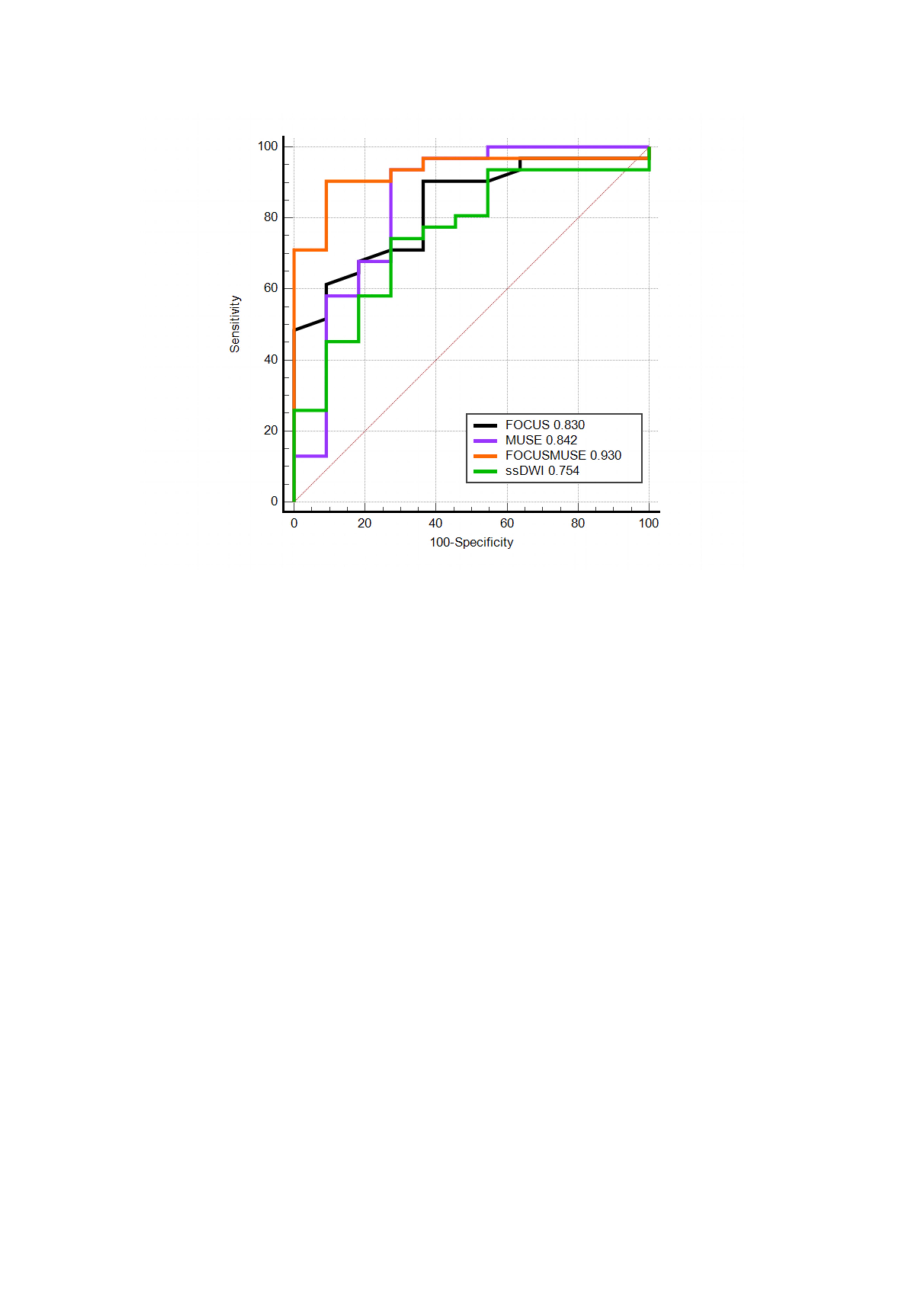
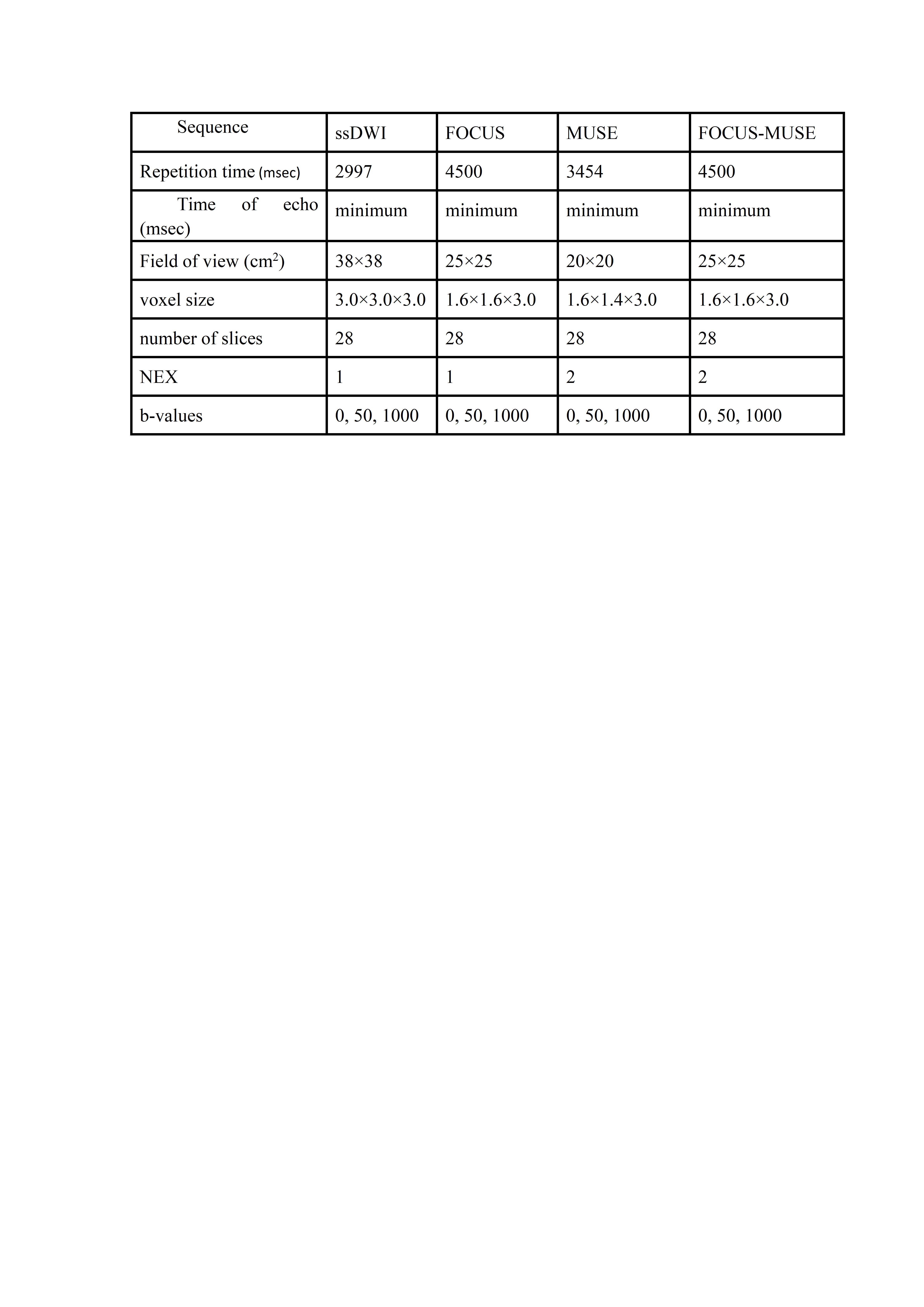
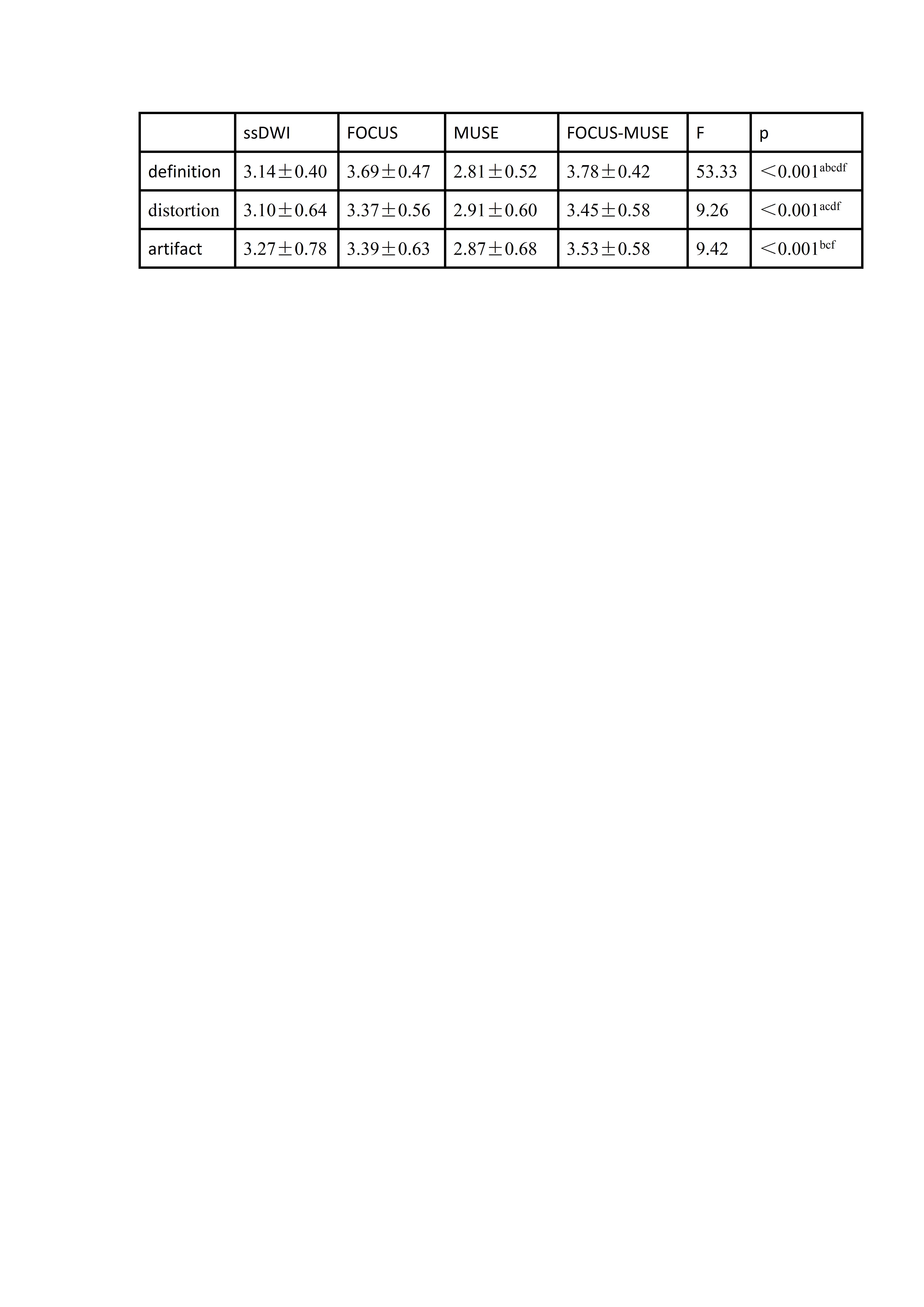
Table2 Comparison of subjective scores for four diffusion sequences
a p< 0.05, post-hoc comparison between ssDWI and FOCUS sequences; b p< 0.05, post-hoc comparison between ssDWI and MUSE sequences; c p< 0.05, post-hoc comparison between FOCUS and MUSE sequences; d p< 0.05, post-hoc comparison between ssDWI and FOCUS-MUSE sequences; e p< 0.05, post-hoc comparison between FOCUS and FOCUS-MUSE sequences; f p< 0.05, post-hoc comparison between MUSE and FOCUS-MUSE sequences.
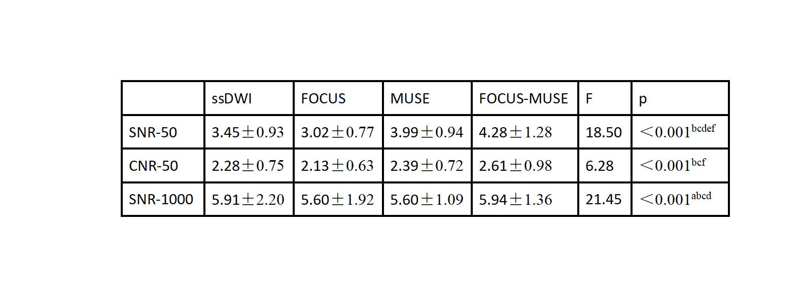
Table3 Objective Evaluation Comparison of Four Dispersion Sequences
a p< 0.05, post-hoc comparison between ssDWI and FOCUS sequences; b p< 0.05, post-hoc comparison between ssDWI and MUSE sequences; c p< 0.05, post-hoc comparison between FOCUS and MUSE sequences; d p< 0.05, post-hoc comparison between ssDWI and FOCUS-MUSE sequences; e p< 0.05, post-hoc comparison between FOCUS and FOCUS-MUSE sequences; f p< 0.05, post-hoc comparison between MUSE and FOCUS-MUSE sequences.