4320
Automatic Quantification of Image Distortion in Prostate Diffusion-Weighted Imaging1Biomedical Imaging Research Institute, Cedars-Sinai Medical Center, Los Angeles, CA, United States, 2Bioengineering, University of California, Los Angeles, Los Angeles, CA, United States, 3Siemens Healthineers, Austin, TX, United States
Synopsis
Keywords: Prostate, Prostate, DWI distortion
Motivation: DWI is crucial for prostate cancer imaging, but its susceptibility to image distortion poses challenges to reading and leads to a substantial rate of nondiagnostic scans.
Goal(s): This study aimed to develop and compare two algorithms for automatic distortion assessment for prostate DWI.
Approach: Two automatic distortion assessment methods were developed based on image segmentation, and deformable registration. Both were validated and compared using radiology grading as the reference.
Results: Both distortion assessment methods quantified the levels of image distortion in prostate DWI consistent with visual assessments and correlated well with expert ratings. The deformable registration-based approach appeared to outperform its counterpart.
Impact: The developed methods for automatic assessment of image distortion may assist in acquiring high-quality prostate DWI and reducing patient recalls.
1. INTRODUCTION:
Diffusion-Weighted Imaging (DWI) is the core sequence of the prostate MRI exams and plays a crucial role in evaluating prostate cancer1,2. However, the single-shot echo-planar (SS-EPI) technique utilized in most prostate exams is prone to image distortion as it is sensitive to static magnetic field (B0) inhomogeneities3,4. Severe image distortions present substantial challenges in accurate diagnostic interpretations and lead to nondiagnostic scans, as recognized in the standardized reporting and interpretation system (PI-RADS)5. Automatic quantification of image distortion may provide immediate feedback to the scan operator thus avoiding possible patient recalls.We aim to develop two tailored methods for quantifying prostate DWI distortion. One is based on improving on a previously proposed segmentation-based method6 with slice normalization. The other is based on deformable-registration to quantify the distortion. Expert radiologist’s ratings are used as the reference to validate and compare the two proposed methods.
2. METHODS:
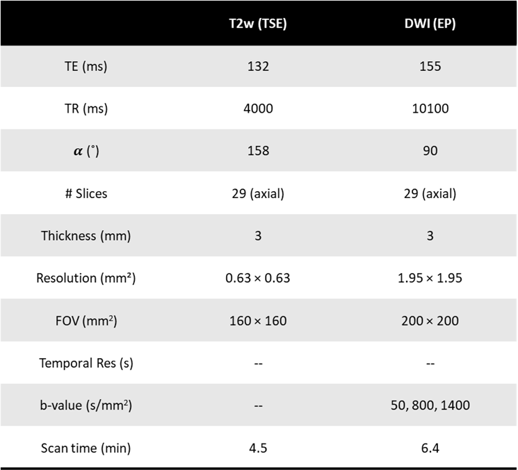
2.1 Study datasetThis study consists of 24 cases of mpMRI clinical prostate MRI. All patients were scanned on a 3 T clinical scanner (Biograph mMR; Siemens Healthineers, Erlangen, Germany) with detailed T2wighed and DWI protocol parameters listed in Figure 1. In this work, only low-b value DWI scans (b = 50) were used.
2.2 Data preprocessing
The low b-value DWIs were first resampled to have the same space resolution as T2WIs, then both images were cropped to have same field of view for further analysis. Whole gland prostate outlining of DWI and T2WI were drawn slice by slice using 3D-slicer7 separately.
2.3 Computational methods for distortion assessment
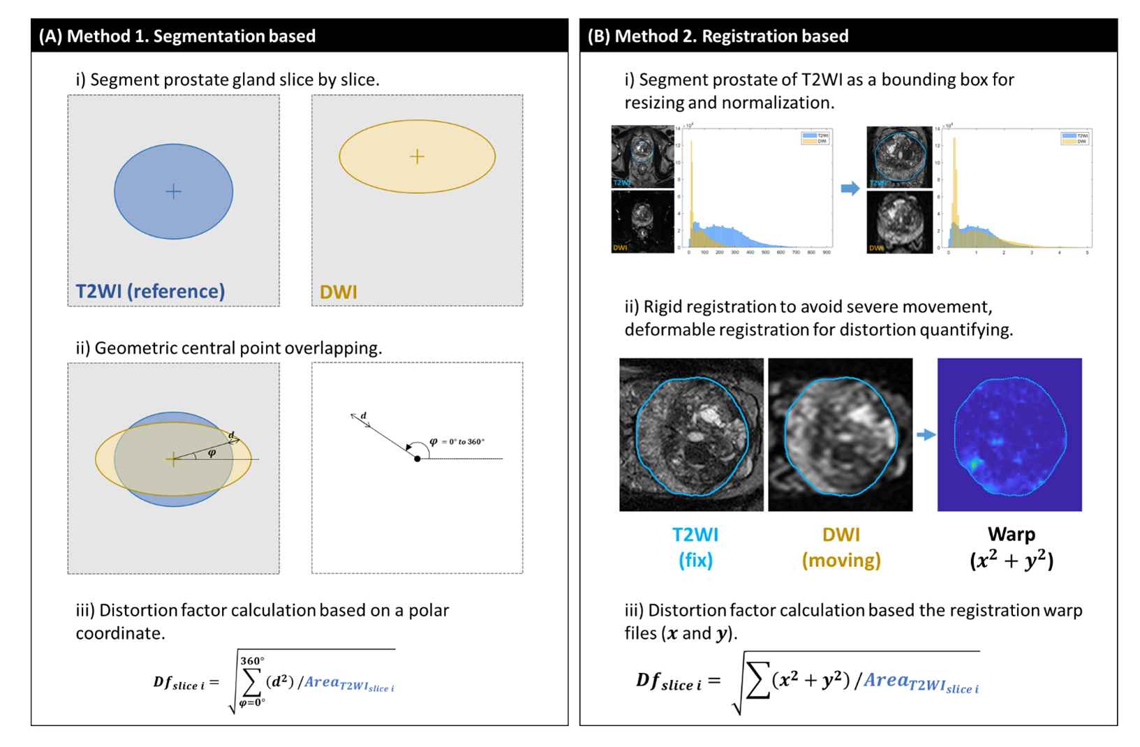
Method 1 segmentation-based method. Using the whole prostate gland of T2WI as a non-distorted volume reference, based on polar coordinates calculate the distortion factor (DF) of each slice with the detailed algorithm shown in Figure 2(A).
Method 2 intensity-registration-based method. As shown in Figure 2(B), the intensity-registration-based method was conducted by MATLAB 2023a. Preprocessed T2WI and DWI were resized and normalized on the T2WI prostate gland segmentation as a bounding box. Rigid registration was first performed to avoid severe patient moving. Then DF was calculated from the warp information generated after the deformable registration.
2.4 Evaluation
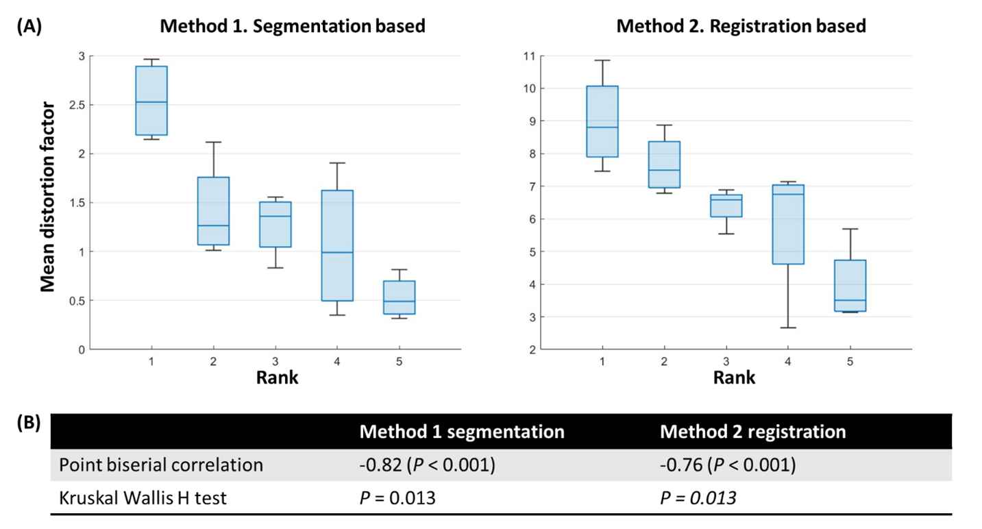
For each case, the mean DF of the two methods will be calculated by eliminating the top 2 and the last 2 slices, to reduce the segmentation error. An experienced radiologist provided a 5-point ranking8 for the low-b value DWI based on the level of image distortion (1: poor to 5: excellent). Point biserial correlation and Kruskal Wallis H test were performed on the graded scores and the DF of both methods.
3. RESULTS:
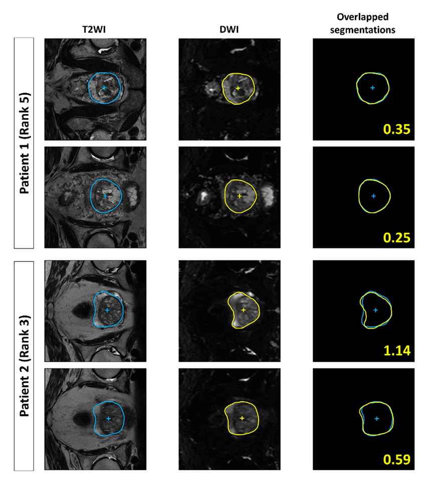
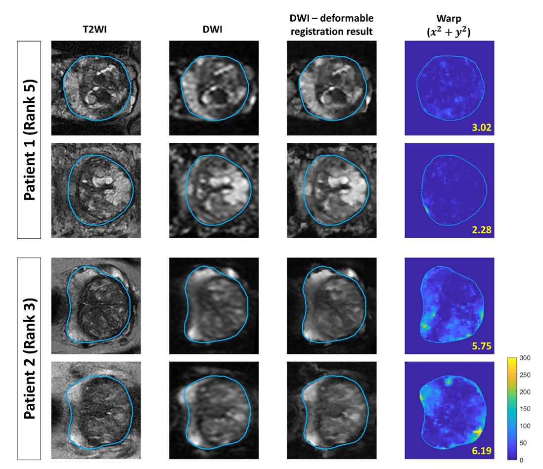
The segmentation-based method (Method 1) and intensity-registration-based method (Method 2) were applied to the DWI images of all 24 patients. Figure 3 displays the DF calculated by Method 1 for two example patients, while Figure 4 shows the DF conducted by Method 2 of the exact same slices for these patients as in Figure 3. The radiologist separately ranked the distortion of these two patients as 3 (intermediate distortion) and 5 (no distortion).Both methods accurately calculated DF consistent with the radiologists' assessment. Examining the mean DF at a patient level (excluding the initial and final two factors to mitigate segmentation errors) revealed a noticeable negative correlation with the assigned rankings, as depicted in Figure 5.
Statistical analyses, including point biserial correlation and the Kruskal Wallis H test, were conducted between the mean DF and the discrete ranks, providing further evidence supporting the methods' reliability.
4. DISCUSSION AND CONCLUSION:
We developed two automatic distortion quantification methods for assessing prostate DWI. In the testing on real-world clinical prostate MRI data, both methods demonstrated a statistically significant correlation with expert radiologist grading. The segmentation-based method provided plausible distortion quantification factors that matched visual assessments in the testing cases; however, its accuracy was contingent upon the precision of the prostate gland outline. Conversely, the registration method only required an approximate whole gland segmentation as a bounding box for resizing and intensity normalization, thus providing a more robust quantification of image distortion.Future work will focus on improving the accuracy of prostate gland segmentation, refining the registration algorithms, and potentially developing a hybrid method. Additionally, expanding the training and testing dataset and incorporating lesion ROI-based analysis may aid in exploring the potential clinical applications.
The study examined two methods for quantifying DWI distortion. Both the segmentation-based method and the registration-based method revealed a robust correlation between quantitative DF and qualitative radiologist grading, which showed the reliability and potential of these techniques to enhance diagnostic accuracy and treatment planning for prostate cancer by aiding in the acquisition of high-quality DWI images.
Acknowledgements
This work is supported by Siemens Healthineers, USA.References
1. Baliyan V, Das CJ, Sharma R, Gupta AK. Diffusion weighted imaging: Technique and applications. World J Radiol. 2016;8(9):785-798. doi:10.4329/wjr.v8.i9.785
2. Koh DM, Collins DJ. Diffusion-Weighted MRI in the Body: Applications and Challenges in Oncology. American Journal of Roentgenology. 2007;188(6):1622-1635. doi:10.2214/AJR.06.1403
3. Le Bihan D, Poupon C, Amadon A, Lethimonnier F. Artifacts and pitfalls in diffusion MRI. Journal of Magnetic Resonance Imaging. 2006;24(3):478-488. doi:10.1002/jmri.20683
4. Poustchi-Amin M, Mirowitz SA, Brown JJ, McKinstry RC, Li T. Principles and Applications of Echo-planar Imaging: A Review for the General Radiologist. RadioGraphics. 2001;21(3):767-779. doi:10.1148/radiographics.21.3.g01ma23767
5. Weinreb JC, Barentsz JO, Choyke PL, et al. PI-RADS Prostate Imaging - Reporting and Data System: 2015, Version 2. Eur Urol. 2016;69(1):16-40. doi:10.1016/j.eururo.2015.08.052
6. Gill AB, Czarniecki M, Gallagher FA, Barrett T. A method for mapping and quantifying whole organ diffusion-weighted image distortion in MR imaging of the prostate. Sci Rep. 2017;7:12727. doi:10.1038/s41598-017-13097-6
7. Fedorov A, Beichel R, Kalpathy-Cramer J, et al. 3D Slicer as an image computing platform for the Quantitative Imaging Network. Magn Reson Imaging. 2012;30(9):1323-1341. doi:10.1016/j.mri.2012.05.001
8. Lawrence EM, Zhang Y, Starekova J, et al. Reduced field-of-view and multi-shot DWI acquisition techniques: prospective evaluation of image quality and distortion reduction in prostate cancer imaging. Magn Reson Imaging. 2022;93:108-114. doi:10.1016/j.mri.2022.08.008
Figures