4319
The value of diffusion-weighted magnetic resonance imaging (DW-MRI) in the evaluation of fetal kidney development1Sichuan Provincial Maternity and Child Health Care Hospital, Chengdu, China, 2GE Healthcare, Beijing, China
Synopsis
Keywords: Fetal, Kidney, Fetal kidney, diffusion-weighted imaging, kidney development
Motivation: Fetal magnetic resonance technology provides morphological data of the fetal kidneys, as well as preliminary exploration of kidney function through DW-MRI.
Goal(s): This project aims to investigate the correlation between gestational age and multi-quantitative MR parameter values of the fetal kidney.
Approach: The kidney size, thickness, T2 signal and ADC quantitative parameters of normal fetuses from 24 to 40 weeks of gestation were collected, analyzing the correlation between each parameter value and gestational age.
Results: In conclusion,there was a positive correlation between fetal kidney size, parenchymal thickness and gestational age, and a negative correlation between renal ADC, T2 signal values and gestational age.
Impact: This project achieved a preliminary assessment of fetal kidney development and provide essential quantitative data references for different developmental stages of the fetal kidney.
introduction
Prenatal ultrasound (US) is commonly used to detect fetal structural urinary tract abnormalities, but it is limited by factors such as fetal or maternal conditions, which can affect the accuracy of diagnosis [1]. However, fetal magnetic resonance technology has rapidly advanced and now provides morphological data of the fetal kidneys, as well as preliminary exploration of kidney function through DW-MRI [2-3].DW-MRI is a widely utilized and well-established MRI technique. This technique provides valuable information on biological and physical characteristics, such as tissue cells and density, microstructure, and microcirculation [4,6]. The evaluation of fetal kidney development using DW-MRI is primarily done in two ways: qualitative assessment of signal intensity and quantitative measurement of the apparent diffusion coefficient (ADC). The aim of our study is to investigate the correlation between gestational age and multi-quantitative MR parameter values of the fetal kidney [5].Methods
Study population: 313 normal fetal kidneys that have undergone fetal abdominal MRI examination at our hospital between October 2018 and October 2022. Written informed consents were obtained from all patients.MRI scanning: All MRI scan were performed on a 1.5T MRI scanner (Signa HDXt 1.5T, GE Healthcare, Milwaukee, USA) Scanning sequence is crown, vector, axis T2WI; crown, axis T1WI and DWI (b-value 600).
Data measurement: Measure the size of both kidneys (Anteroposterior Diameter (AD), Transverse Diameter (TD), Major diameter (MD), renal parenchyma thickness (RPT), kidney T2 signal value, kidney apparent diffusion coefficient (ADC).
Statistics: The Pearson Correlation Coefficient was used to analyze the correlation between AD, TD, MD, RPT, T2 signal value, ADC for both kidneys and gestational age.
Results
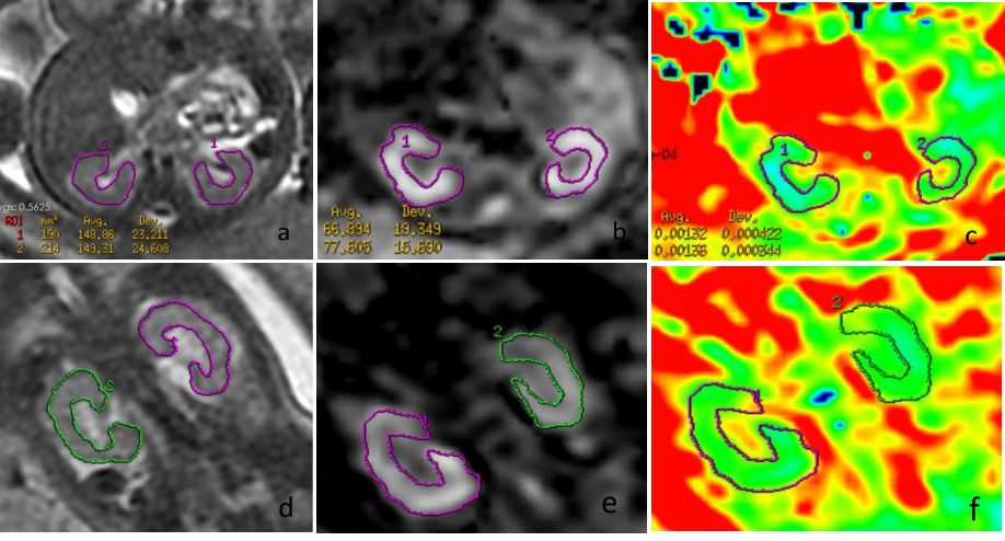
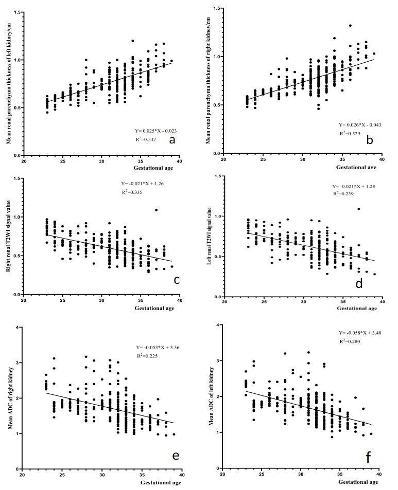
All measured values of kidney size and kidney parenchyma thickness at different gestational ages are shown in Table 1. The value of fetal kidney size and kidney parenchyma thickness were positively correlated with gestational age, p<0.0001(Figure 2). Right renal parenchyma Pearson correlation = 0.724. Left renal parenchyma Pearson correlation = 0.733. Kidney size and parenchymal thickness help predict kidney function-kidney size correlates with the number of nephrons [7].Renal T2WI signal value was inversely correlated with gestational age, p<0.0001(Figure 2). Right renal T2 signal Pearson correlation = -0.583, left renal T2 signal Pearson correlation = -5.88.DW-MRI fetal kidneys have high signal and ADC low signal (Figure 1), and renal ADC values for different gestational age are shown in Table 2. Renal ADC values were inversely correlated with gestational age, and ADC values decreased with increasing gestational age (Figure 2). The best result was expressed with the regression equation: right renal ADC (mm2/s) =3.356–0.0527*(GA) (p<0.0001, Pearson correlation = -0.566),Left renal ADC (mm2/s) = 3.481–0.05795*(GA) (p<0.0001,Pearson correlation = -0.61). Quantification of ADC values associated with gestational age is a true functional parameter that can help define normal renal function [8].Discussion and Conclusion
In conclusion, MRI examination holds significant clinical value in the assessment of fetal kidney structure and development. Fetal MR can also provide crucial functional information regarding normal kidney development, enhance awareness of fetal kidney development, and ultimately aid in pregnancy management, as well as the diagnosis and treatment of newborns.Acknowledgements
No acknowledgement found.References
[1] Hill BJ, Joe BN, Qayyum A, et al. Supplemental value of MRI in fetal abdominal disease detected on prenatal sonography: preliminary experience. AJR Am J Roentgenol. 2005 Mar;184(3):993-8.
[2] Cassart M, Massez A, Metens T, et al. Complementary role of MRI after sonography in assessing bilateral urinary tract anomalies in the fetus. AJR Am J Roentgenol. 2004 Mar;182(3):689-95.
[3] Vermoolen MA, Kwee TC, Nievelstein RA. Apparent diffusion coefficient measurements in the differentiation between benign and malignant lesions: a systematic review. Insights Imaging. 2012 Aug;3(4):395-409.
[4] Colagrande S, Carbone SF, Carusi LM, Cova M, Villari N. Magnetic resonance diffusion-weighted imaging: extraneurological applications. Radiol Med. 2006 Apr;111(3):392-419. English, Italian.
[5] Prasad PV, Priatna A. Functional imaging of the kidneys with fast MRI techniques. Eur J Radiol. 1999 Feb;29(2):133-48.
[6] Thoeny HC, De Keyzer F. Extracranial applications of diffusion-weighted magnetic resonance imaging. Eur Radiol. 2007 Jun;17(6):1385-93.
[7] Al Salmi I, Al Hajriy M, Hannawi S. Ultrasound Measurement and Kidney Development: a Mini-Review for Nephrologists. Saudi J Kidney Dis Transpl. 2021 Jan-Feb;32(1):174-182.
[8] Manganaro L, Francioso A, Savelli S, Tomei A, Fierro F, Di Maurizio M, Coratella F, Perrone A, Ballesio L, Giancotti A, Porfiri L, Marini M. Fetal MRI with diffusion-weighted imaging (DWI) and apparent diffusion coefficient (ADC) assessment in the evaluation of renal development: preliminary experience in normal kidneys. Radiol Med. 2009 Apr;114(3):403-13.
Figures