Sheng Zhang1, Xia Wang1, Xiaofan Liu1, Chanjuan Yu1, Gang Tian1, Xiuzheng Yue2, and Yuedong Han1
1Xi'an GaoXin Hospital, Xi'an, China, 2Philips Healthcare, Xi'an, China
Synopsis
Keywords: Kidney, Kidney, normal right kidney; amide proton transfer; characteristic
Motivation: The changes in renal protein levels are closely related to physiological and pathological functions. Amide proton transfer weighted (APTw) imaging has the function of non-invasive and targeted detection of tissue-free proteins or peptides and is of great significance in the preliminary study of acute and chronic kidney diseases.
Goal(s): The study of APT imaging in normal kidneys still needs to be completed
Approach: The APT images study of 49 volunteers in this study.
Results: The APT values of the renal cortex and medulla were consistent, and the medulla was significantly higher than the cortex.
Impact: APT imaging is initially applied to the urinary system, such as chronic kidney disease, kidney cancer, etc. In this study, the application value of APTw imaging in renal dermal medulla was investigated
Introduction:
APTw imaging has the function of non-invasive and targeted detection of tissue-free
proteins and peptides [1]. It has initially obtained good clinical value in the
study of acute and chronic kidney diseases [2-3]. The reabsorption of amino
acids and other protein substances is one of the kidney's primary functions,
which lays a theoretical foundation for applying APTw imaging in the kidney.
Targeted understanding of the characteristics of normal renal cortical and
medullary protein levels not only can help to recognize the change
characteristics of free proteins in normal kidneys but also lays a foundation
for the early study of disease.
Method:
58 healthy
volunteers from September 2021 to February 2022 were enrolled for intermittent
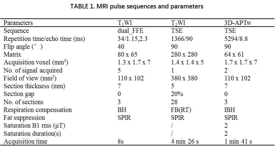
breath-hold right kidney axial 3D-APT imaging using 3.0T (Philips Ingenia CX,
Best, Netherland). The specific imaging parameters are shown in Table 1. The
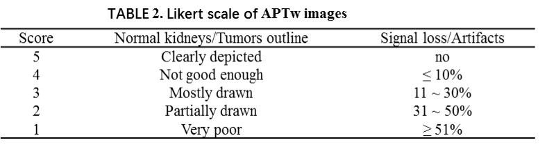
scores of APTw images were more than 2 points on the 5-Likert scale [4]. (Table 2) They were considered successful images and
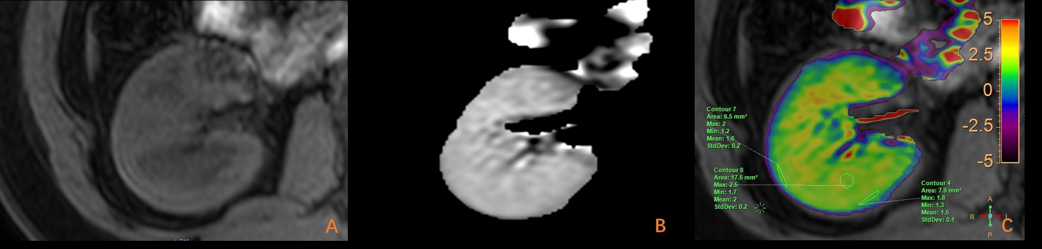
were used to measure APT values by two radiologists. Three circular regions of interest
(ROIs) with an area of 0.2-0.4 cm2 were used to measure the APT values
of the cortex and medulla, respectively (Fig. 1), avoiding imaging defects on
the pseudo-colored maps, artifacts, kidney sinuses, and visible vessels. ROI delineation was then reviewed and
approved by a radiologist with more than 10 years of experience in urinary
system imaging. The intraclass correlation coefficient
(ICC) was used to evaluate the consistency of APT values between two
radiologists, and an ICC of > 0.74
was considered excellent. The t-tests were used to analyze the
difference in APT values between cortex and medulla in all data and different
genders. All statistical tests were 2 sided. A P value less than 0.05 indicated
a statistically significant difference.
Results:
9 healthy volunteers were
excluded from the study due to claustrophobia (2 cases) and breathing training
failure before (3 cases) or during the 3D-APTw imaging scan (4 cases). Finally,
49
volunteers (aged 33.10 ± 1.54 years, 13 male) were enrolled. The inter-observer agreement was excellent
for the APT values of the cortex (ICC = 0.951) and medulla (ICC
= 0.940). The APT values of
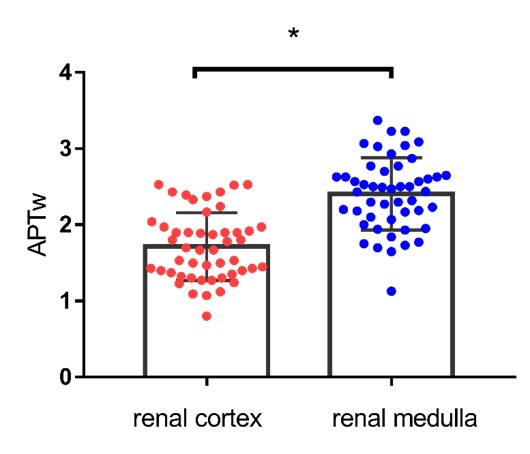
the medulla (2.407% ± 0.474%) were
higher than the cortex (1.714% ± 0.448%). The difference was statistically significant (P < 0.001) (Fig. 2). There was no significant difference in the
renal cortex (P = 0.804) and medulla (P = 0.465) between
different genders.
Discussion:
This study's high consistency of APT values in the normal renal cortex and
renal medulla reflected the feasibility of accurate renal APT imaging. The
differences in APT values between the renal medulla and cortex suggested that
the difference in renal tissue microstructure is the main reason, such as the
skeleton protein, blood flow, and other factors. This study also reflected the
ability of APTw imaging to detect trace protein changes in tissues, which will
have significant potential for future research.
Conclusion:
APT imaging of the normal kidney medulla and cortex was
highly consistent, and APT values of the kidney medulla were significantly
higher than the cortex. This study provides a theoretical basis for the study
of noninvasive kidney proteins.Acknowledgements
NoReferences
[1]
Zhou J, Heo HY, Knutsson L, et al. APT‐weighted MRI: techniques, current neuro
applications, and challenging issues[J]. J Magn Reson Imaging 2019, 50(2):
347‐364. DOI: 10.1002/jmri.26645.
[2]
Liu J, Han Z, Chen G, et al. CEST MRI of sepsis-induced acute kidney injury.
NMR Biomed 2018; 31: e3942.
[3]
Ju Y, Liu AL, Wang Y, et al. Amide proton transfer magnetic resonance imaging
to evaluate kidney impairment in patients with chronic kidney disease. Magn
Reson Imaging 2022; 87:177-182.
[4] Tu W, Alzahrani A, Currin S, Walsh C, Narayanasamy S,
McInnes MDF, Schieda N. Evaluation of a free-breathing respiratory-triggered
(Navigator) 3-D T1-weighted (T1W) gradient recalled echo sequence (LAVA) for
detection of enhancement in cystic and solid renal masses. Eur Radiol 2019;
29:2507-2517.
[5]
Togao O, Keupp J, Hiwatashi A, et al. Amide proton transfer imaging of brain
tumors using a self-corrected 3D fast spin-echo Dixon method: Comparison With
separate B0 correction. Magn Reson Med 2017; 77:2272-2279.