4310
Quantitative Apparent Diffusion Coefficient Metrics for MRI-only Suspicious Breast Lesions: Any Added Clinical Value?1Beijing hospital, Beijing, China, 2bejing hospital, beijing, China
Synopsis
Keywords: Breast, Breast, diffusion-weighted imaging; apparent diffusion coefficient; MRI; breast lesions.
Motivation: A simple, objective, and noninvasive approach is required to improve the diagnostic accuracy of MRI-only suspicious lesions.
Goal(s): We aimed to assess whether using standard DWI sequences could increase the diagnostic performance of MRI for MRI-only breast lesions and reduce unnecessary MRI-guided biopsies.
Approach: In our current work, we intend to use the quantitative assessment of the heterogeneity of the ADC value to explore the diagnostic value of conventional DWI sequences for MRI-only lesions.
Results: Our results showed that quantitative ADC metrics based on large 2D-ROI measurements, particularly mean ADC values, exhibited superior performance over BI-RADS features in distinguishing MRI-only suspicious lesions.
Impact: DWI quantitative parameters may significantly improve diagnostic ability of BI-RADS-based features of breast MRI and provide additional value for the discrimination of MRI-only suspicious lesions.
Background
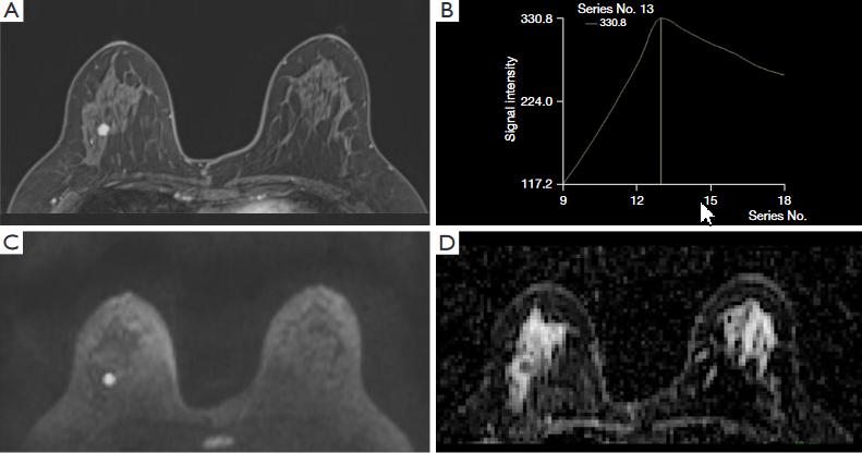
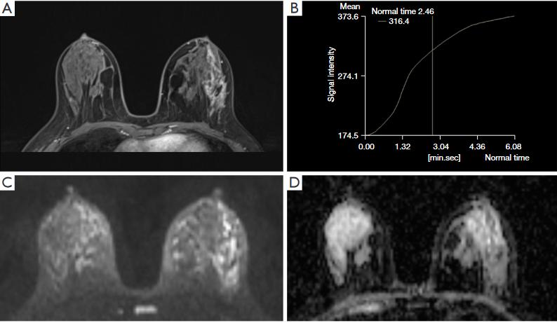
Suspicious breast lesions (Breast Imaging Reporting and Data System, BI-RADS 4 or 5) detected only by magnetic resonance imaging (MRI) and invisible on other initial imaging modalities (MRI-only lesions) are usually small and poorly characterized in previous literature(1, 2), thus making diagnosis and management difficult. This study aimed to investigate the clinical significance of quantitative apparent diffusion coefficient (ADC) metrics derived from conventional diffusion-weighted imaging (DWI) on evaluating MRI-only lesions.Methods
A total of 90 suspicious MRI-only lesions were evaluated, including 51 malignant and 39 benign lesions. Morphological and kinetic characteristics of all lesions (termed BI-RADS parameters) were described according to the BI-RADS lexicon on dynamic contrast enhancing imaging. Minimum, maximum, and mean ADC values (ADCmin, ADCmax, ADCmean) were obtained by measuring the ADC map of DWI using large 2D-region of interest (ROI). ADC heterogeneity was then obtained by the following formula: ADCheterogeneity = (ADCmax - ADCmin) / ADCmean. Diagnostic performance of these parameters was assessed and compared using the receiver operating characteristic curve (ROC).Results
Of the 90 MRI-only lesions, there were 45 masses and 45 non-mass lesions. Among BI-RADS parameters, only the two different kinetic patterns were significantly different between benign and malignant group (P = 0.005 and P < 0.001, respectively). The area under ROC (AUC) of combined significant ADC parameters (ADCmin, ADCmean, and ADCmax, all P ≤ 0.001) was significantly higher than that of the two different kinetic patterns (P = 0.006 for both). For MRI-only masses, only ADCmean and ADCmax, among all BI-RADS and ADC parameters, had diagnostic value (combined AUC = 0.833). For non-mass lesions, size, distribution, ADCmin, and ADCmean were significantly different between benign and malignant group (P = 0.004, P < 0.001, P = 0.001, and P < 0.001, respectively). In addition, ADCmean had the highest diagnostic performance among all ADC parameters, regardless of mass or non-mass (AUC = 0.825 and 0.812, respectively). ADCheterogeneity showed no significant differences, no matter in mass or non-mass group (P = 0.62 and 0.43, respectively).Discussion
Our findings showed that AUC of ADC parameters for diagnosing MRI-only lesions was higher than that of BI-RADS features. In the subgroup analysis, MRI-only masses had no pathognomonic BI-RADS features, and we noticed that these masses were small, almost none exceeding one centimeter. For non-mass lesions, we showed that the distribution (e.g., segmental and linear) and size of non-mass lesions were proper BI-RADS features for differentiating MRI-only lesions; However, when combining the two parameters, the AUC was only 0.745. Simultaneously, our study showed that ADCmean had the highest diagnostic performance among all the significant ADC parameters, regardless of tumor type (mass or non-mass). This result is inconsistent with the finding of Hirano et al.(3). The discrepant results may be related to the small lesion size and less necrosis of masses in our study. In addition, it is related to the different ROI delineation methods(4). Our current study fails to reflect the value of ADCheterogeneity in the differential diagnosis of MRI-only lesions. This may be due to the small size of MRI-only masses and their limited voxels. Meanwhile, non-mass lesions are mixed with adipose and glandular tissues which probably affect the evaluation of tumor heterogeneity. Additionally, the delineation of single-slice ROIs may not accurately reflect internal tumor heterogeneity.Conclusions
In differentiating MRI-only suspicious lesions, quantitative ADC metrics generally performed better than BI-RADS parameters, and ADCmean is still the best ADC parameter to distinguish MRI-only lesions.Acknowledgements
No acknowledgement found.References
1. Arponen O, Masarwah A, Sutela A, et al.Incidentally detected enhancing lesions found in breast MRI: analysis of apparent diffusion coefficient and T2 signal intensity significantly improves specificity.Eur Radiol 2016,26:4361-4370.
2. Teifke A, Lehr H A, Vomweg T W, Hlawatsch A and Thelen M.Outcome analysis and rational management of enhancing lesions incidentally detected on contrast-enhanced MRI of the breast.AJR. American Journal of Roentgenology 2003,181:655-662.
3. Hirano M, Satake H, Ishigaki S, Ikeda M, Kawai H and Naganawa S.Diffusion-weighted imaging of breast masses: comparison of diagnostic performance using various apparent diffusion coefficient parameters.AJR. American Journal of Roentgenology 2012,198:717-722.
4. Kim J J, Kim J Y, Suh H B, et al.Characterization of breast cancer subtypes based on quantitative assessment of intratumoral heterogeneity using dynamic contrast-enhanced and diffusion-weighted magnetic resonance imaging.Eur Radiol 2022,32:822-833.
Figures