4307
Radiomic Features of Placental Magnetic Resonance Imaging Identify Adverse Pregnancy Outcomes: A Pilot Study1Radiology, UT Southwestern Medical Center, Dallas, TX, United States, 2Bioengineering, The University of Texas at Dallas, Richardson, TX, United States, 3Center for Imaging and Surgical Innovation, The University of Texas at Dallas, Richardson, TX, United States, 4Obstetrics & Gynecology, UT Southwestern Medical Center, Dallas, TX, United States, 5Parkland Health, Dallas, TX, United States
Synopsis
Keywords: Placenta, MR Value, chronic hypertension, radiomic analysis, preeclampsia, small for gestational age
Motivation: Chronic hypertension (cHTN) is a significant obstetric condition with increased risk of adverse outcomes. Radiomic analysis of MR images may be able to predict outcomes before development of clinical findings.
Goal(s): Our goal was to compare MR T2W radiomic features among 4 clinical outcomes: normal, cHTN, preeclampsia with severe features, and small for gestational age (SGA).
Approach: We applied ROI-based radiomics analysis on prospectively collected T2W MR images in normal and cHTN pregnancies.
Results: We found that MR T2W radiomic features as early as 16 weeks can identify those patients who develop preeclampsia with severe features and whose infants are born SGA.
Impact: We demonstrated that MR T2W radiomic features as early as 16 weeks can identify those patients who develop preeclampsia with severe features and whose infants are born small for gestational age.
Introduction
The human placenta is a complex organ with unique functionality, undergoing extensive growth and remodeling through trophotrophism and maturation of the villous trees within the intervillous spaces of cotyledons. Lack of this remodeling can result in placental-mediated disease which can result from maternal disease (e.g. chronic hypertension). Despite its complexity the human placenta remains poorly understood due to limited research access in utero.Chronic hypertension (cHTN) accounts for 14% of mortality worldwide,1 affects 3-5% of pregnant women,2 and has known adverse pregnancy outcomes. Up to 30% of pregnant women with cHTN deliver preterm from placental-mediated complications of superimposed preeclampsia or small for gestational age (SGA).2
The objective of this work is to evaluate radiomics characteristics of the placenta extracted from T2W-MR image. We hypothesize that radiomics characteristics of in vivo human placental T2W images will be different in the normal pregnancy, compared to different cHTN pathological conditions, including preeclampsia and SGA outcomes. This difference can potentially be detected before development of clinical findings and may be predictive of outcome (preeclampsia, SGA) which may enable future intervention.
Methods
With IRB approval, we performed a prospective MRI study of 10 healthy pregnant and 15 pregnant patients with cHTN on antihypertensive therapy. Imaging was performed at 16 weeks 0 day (16w0d)-19w6d (visit 1) and 24w0d-27w6d (visit 2) gestational ages. All imaging data were acquired on a 3T-MR scanner (Ingenia, Philips Healthcare, The Netherlands). Sagittal T2W-imaging of the entire gravid uterus including the fetus and the placenta was acquired as part of an established protocol. Volumetric placental ROIs were manually drawn by a researcher under the supervision of a board-certified radiologist with 30 years of obstetric and gynecologic US and MR experience. All shape and texture radiomics features were extracted from the segmented placenta data following the image biomarker standardization initiative (IBSI) guideline using the PyRadiomics package.3,4 In-plane spacing was resampled to 1.5mm. 2D texture features binwidth was set at 1. All images were normalized, scaled by 100 and outliers 2 standard deviation away were truncated.Univariate receiver operating characteristic curve (ROC) analysis was performed for features extracted from visit 1, visit 2, and change between visit 1 and 2 in the primary population (normal vs. cHTN) and secondary populations (cHTN without additional complication, cHTN with superimposed preeclampsia with severe features, cHTN with SGA). False discovery rate adjusted p values were reported. Adjusted P<0.05 was considered statistically significant. All analyses were performed using R4.3.1 (R core team, Vienna, Austria).
Results
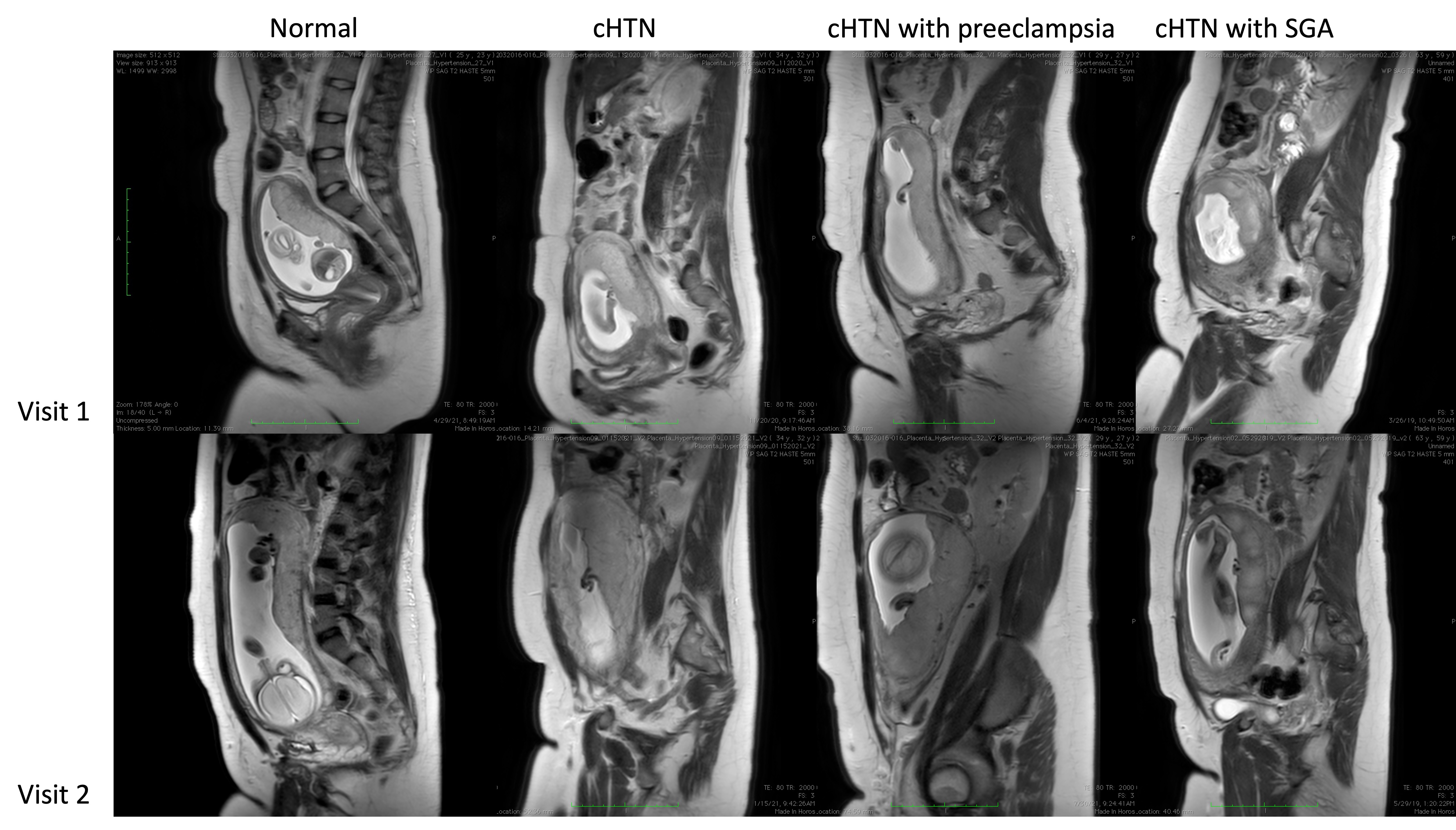
Of the 10 pregnancies in the healthy cohort, one pregnancy had SGA. Of the 15 pregnant patients with cHTN on antihypertensive therapy, 5 had superimposed preeclampsia with severe features and 4 SGA. Figure 1 demonstrates representative MR sagittal T2W images in normal pregnancy, cHTN, cHTN with preeclampsia with severe features, and cHTN with SGA.Figure 2 demonstrates no difference in radiomic features at visit 1 between normal(N=10) and cHTN(N=15) with one significant feature at visit 2. There were significant radiomic features differences at visit 1(7 features) and visit 2(2 features) comparing those with preeclampsia (N=5) versus without (20). There were also significant radiomic features differences at visit 1(1 feature), visit 2(13 features), and changes between visits(8 features) comparing those with SGA(N=4) versus those without(N=21).
Figure3 compares populations within chronic hypertension only. There were no radiomic features differences between those with and without preeclampsia with severe features. There were significant differences in texture features again demonstrated at visit 1(3 features), visit 2(11 features), and changes between visits(24 features).
Discussion
In a pilot study, we demonstrated that MR-T2W radiomic features as early as 16 weeks can identify those patients who develop preeclampsia with severe features and whose infants are born SGA. There are also significant radiomic features that develop between the early (visit1) and late second trimester (visit2). We believe significant differences may be attributed to differential spiral artery remodeling that begins in the late first trimester and continues throughout gestation. There was one feature that was different between normal and cHTN pregnancies in the late second trimester (visit2). This knowledge may be useful in the ability to predict placental-mediated disease prior to clinical manifestation given differences are apparent as early as 16 weeks (visit1). Future work will seek to incorporate these findings using regional analysis and automated segmentation for clinical predictive modeling.Conclusion
We investigated the difference in radiomics extracted from T2W-MR imaging of pregnancies under 4 conditions at two different visits. We demonstrated that MR-T2W radiomic features as early as 16 weeks can identify those patients who develop preeclampsia with severe features and whose infants are born SGA.Acknowledgements
No acknowledgement found.References
1. Say L, Chou D, Gemmill A, Tuncalp O, Moller AB, Daniels J, Gulmezoglu AM, Temmerman M, Alkema L. Global causes of maternal death: a WHO systematic analysis. Lancet Glob Health. 2014;2(6):e323-333.
2. Seely EW, Ecker J. Chronic hypertension in pregnancy. Circulation. 2014;129(11):1254-126.
3. Zwanenburg A, et al., The Image Biomarker Standardization Initiative: Standardized Quantitative Radiomics for High-Throughput Image-based Phenotyping. Radiology, 2020. 295(2): 328–338.
4. van Griethuysen JJM, et al., Computational Radiomics System to Decode the Radiographic Phenotype. Cancer Res, 2017. 77(21): e104–e107.
Figures