4305
Prognosticating Maternal and Neonatal Outcomes in Placenta Accreta Spectrum Disorders by Combining Placental Perfusion and MRI Features1Department of Radiology, The First Affiliated Hospital of Nanjing Medical University, Nanjing, China, 2MR Research Collaboration, Siemens Healthineers, Shanghai, China, Shanghai, China, 3Department of Obstetrics & Gynecology, The First Affiliated Hospital of Nanjing Medical University, Nanjing, China
Synopsis
Keywords: Placenta, Placenta, Diffusion-weighted imaging, intravoxel incoherent motion, perfusion, placenta accreta spectrum, postpartum hemorrhage, screening
Motivation: The factors to predict adverse maternal and neonatal outcomes in patients with placenta accreta spectrum disorders needed identification.
Goal(s): This study aimed to identify appropriate predictive indicators using magnetic resonance imaging (MRI) and intravoxel incoherent motion (IVIM) parameters.
Approach: The study examined the association between various influencing factors (clinical risk factors, MRI features, and IVIM parameters) and adverse outcomes using univariate and multivariate analyses.
Results: Independent predictors of a poor prognosis included previous cesarean sections, low-signal-intensity bands on T2WI, and D value.
Impact: The components of the prediction model in this study were more comprehensive and improved the prediction efficiency. The study analyzed the relationship between f value and postpartum hemorrhage and further examined the importance of IVIM in predicting adverse outcomes.
Introduction
In recent years, magnetic resonance imaging (MRI) has been applied to placenta research [1,2]. However, its application value in predicting the outcome of the placenta accreta spectrum (PAS) remains unclear. A known limitation of conventional MRI is its subjective nature, which can deter clinical application [3,4]. Recently, a novel technique has emerged for quantitatively analyzing placental perfusion. Specifically, the information on tissue diffusion and perfusion can be obtained by applying intravoxel incoherent motion (IVIM) analysis on diffusion-weighted imaging data acquired at multiple b values it is possible to gather [5]. This study aimed to (1) develop a model based on clinical features, MRI signs, and IVIM parameters for predicting the risk of maternal and neonatal clinical outcomes in patients with PAS, (2) compare the prognostic accuracy of models with and without IVIM parameters, and (3) investigate the relationship between IVIM parameters and postpartum hemorrhage (PPH).Methods
This study considered 56 singleton pregnancies at 33-39 weeks of gestation who underwent MRI, which included T2-weighted imaging (T2WI), T1-weighed imaging (T1WI), and IVIM with a spectrum of different b values (0, 50, 100, 150, 200, 500, and 800 s/mm2) before surgery using a 1.5T system (MAGNETOM Aera, Siemens Healthineers AG, Erlangen, Germany). T2W images were acquired using a conventional turbo spin echo sequence and T1W images using a gradient echo sequence. The IVIM parameters were as follows: TR/TE = 6450 /65.0 ms; FOV = 320 × 320 mm2; slice thickness = 5.5 mm; and acquisition time = 3 minutes 51 seconds. These patients all underwent MR examination in the third trimester of pregnancy and had surgical/pathological results available, and were not accompanied by hypertension, diabetes, or chronic kidney disease. Continuous variables were assessed via the Mann-Whitney U test or Student's t-test based on distribution normality, while categorical variables were evaluated using the χ2 test. All variables linked to poor outcome at P < 0.05 in univariate analysis were included in the multivariate logistic regression. Prediction models were built with and without IVIM parameters. The diagnostic performance of these models was evaluated by receiver-operating characteristic (ROC) analysis. Additionally, the correlations between f value and PPH and between f value and red blood cell (RBC) units transfused were studied using linear analysis.Results
Previous cesarean sections, gravidity, and transfused RBC units were remarkably different between the poor- and good-outcome groups (all P values <.05). The detailed results are illustrated in Table 1. Compared with patients with good outcomes, patients with poor outcomes were more likely to have low-signal-intensity bands on T2WI (68.75 vs 19.23%, P <.001) and focally interrupted myometrial border (90.62 vs 53.85%, P =.004), S2-invasion (68.75 vs 34.62%, P =.020), and abnormal vascularization of the placental bed (71.88 vs 42.31%, P =.044). Further, the patients exhibited higher D values (P =.047). In addition, no significant difference was observed in the f and D* values between the 2 groups (Table 2). Figure 1 provides an image example from 2 patients. A multivariable logistic regression analysis was conducted using the backward method. The results indicated that previous cesarean section, low-signal-intensity bands on T2WI, and a D value ≥1.67 × 10⁻³ mm2/s were independent negative predictors. The area under the ROC curve for the combination model with or without a D value was 0.903 and 0.862, respectively (Figure 2). A positive linear relationship was observed between the f value and postpartum hemorrhage (r = 0.305, P =.019) and between the f value and the number of RBC units transfused (r = 0.326, P =.012).Discussion
This was a preliminary feasibility study of MRI and IVIM for predicting adverse outcomes in patients with PAS. The findings suggested that previous cesarean section, low-signal-intensity bands on T2WI, and the D value were independent risk factors for predicting adverse outcomes. The combination model incorporating the D value outperformed the model without the D value in predicting poor outcomes, indicating that IVIM parameters could provide additional information and help improve the model. The f value positively correlated with PPH and the number of RBC units transfused. This correlation suggested that neovascularization and vasodilation of the vasculature became more pronounced with the progression of implantation depth. IVIM and placental descriptors from multiparametric placenta MRI may predict adverse outcomes of PAS.Conclusion
IVIM parameters and MRI features appear to be promising tools for predicting adverse outcomes in patients with PAS. The diagnostic accuracy of the combined model was higher than that of the model without IVIM.Acknowledgements
No acknowledgement found.References
[1] Q. Liu, W. Zhou, Z. Yan, D. Li, T. Lou, Y. Yuan, P. Rong, Z. Feng, Development and validation of MRI-based scoring models for predicting placental invasiveness in high-risk women for placenta accreta spectrum, Eur Radiol. (2023). https://doi.org/10.1007/s00330-023-10058-8IF: 5.9 Q1 .
[2] X. Wu, R. Zhou, M. Lin, Y. Li, W. Ying, L. Li, W. Ji, K. Zheng, The maximum length of T2-dark intraplacental bands may help predict intraoperative haemorrhage in pregnant women with placenta accreta spectrum (PAS), Abdominal Radiology. 47 (2022) 3594–3603. https://doi.org/10.1007/s00261-022-03619-wIF: 2.4 Q3 .
[3] F. Finazzo, F. D’antonio, G. Masselli, F. Forlani, J. Palacios‐jaraquemada, G. Minneci, S. Gambarini, I. Timor‐tritsch, F. Prefumo, D. Buca, M. Liberati, A. Khalil, G. Cali, Interobserver agreement in MRI assessment of severity of placenta accreta spectrum disorders, Ultrasound in Obstetrics & Gynecology. 55 (2020) 467–473. https://doi.org/10.1002/uog.20381IF: 7.1 Q1 .
[4] S. Thiravit, K. Ma, I. Goldman, P. Chanprapaph, P. Jha, D.S. Hippe, M. Dighe, Role of Ultrasound and MRI in Diagnosis of Severe Placenta Accreta Spectrum Disorder: An Intraindividual Assessment With Emphasis on Placental Bulge, American Journal of Roentgenology. 217 (2021) 1377–1388. https://doi.org/10.2214/AJR.21.25581IF: 5.0 Q1 .
[5] N. Siauve, P.H. Hayot, B. Deloison, G.E. Chalouhi, M. Alison, D. Balvay, L. Bussières, O. Clément, L.J. Salomon, Assessment of human placental perfusion by intravoxel incoherent motion MR imaging, The Journal of Maternal-Fetal & Neonatal Medicine. 32 (2019) 293–300. https://doi.org/10.1080/14767058.2017.1378334IF: 1.8 Q4 .
Figures
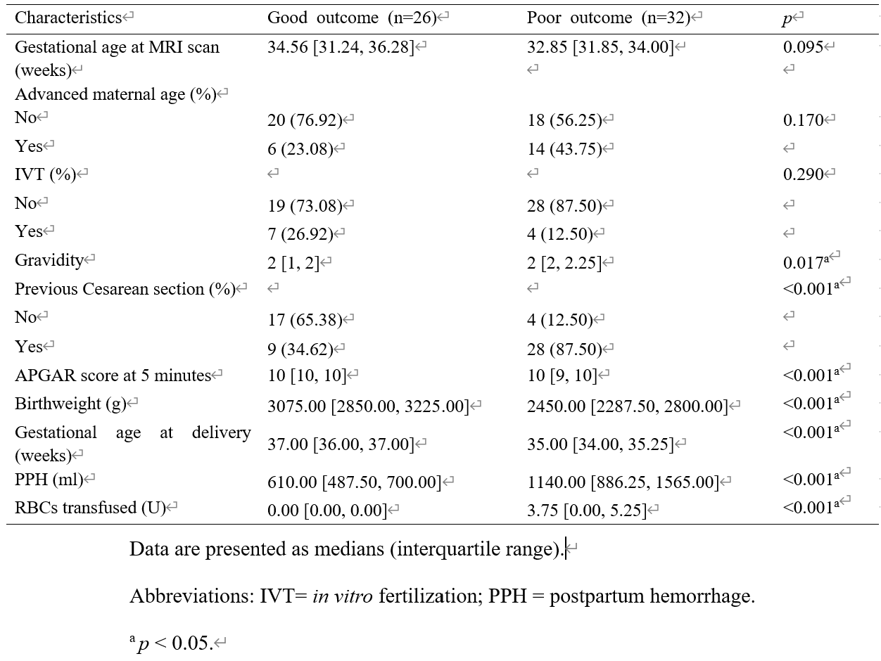
Table 1. Clinical and demographic patient data.
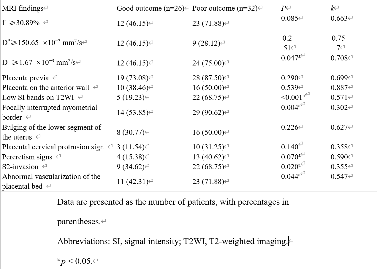
Table 2. Comparison of MRI findings and IVIM parameters between patients with and without good outcomes.
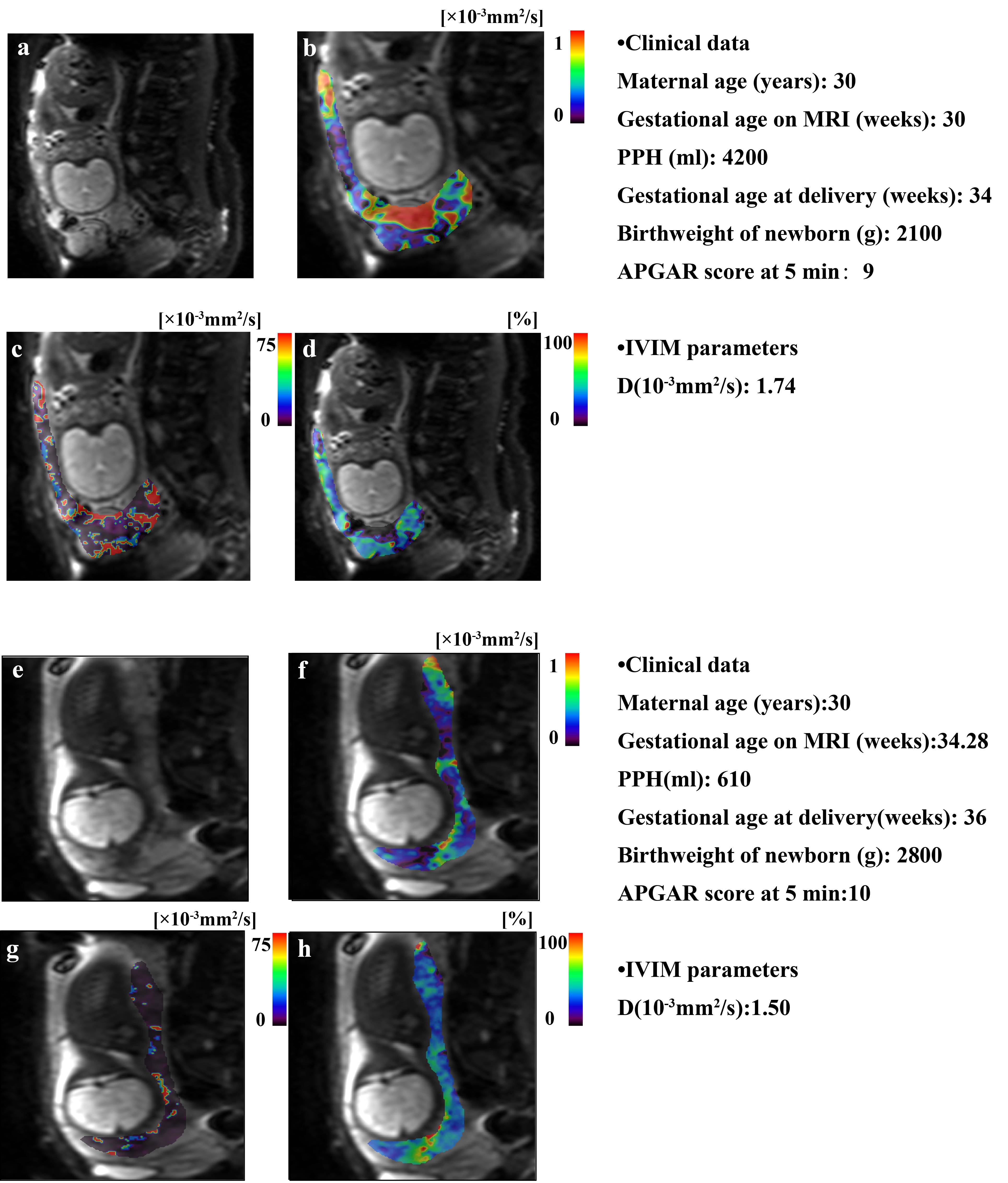
Figure 1. PPH of patient 1 was 4200 mL, neonatal birth weight was 2100 g, gestational age at delivery was 34 weeks, APGAR score at 5 minutes was 9, and the patient was from the poor–outcome group (a). MRI parameters: the D value was higher than the cutoff value (1.74 × 10 -3 mm2/s vs 1.67 × 10 -3 mm2/s) (b–d).
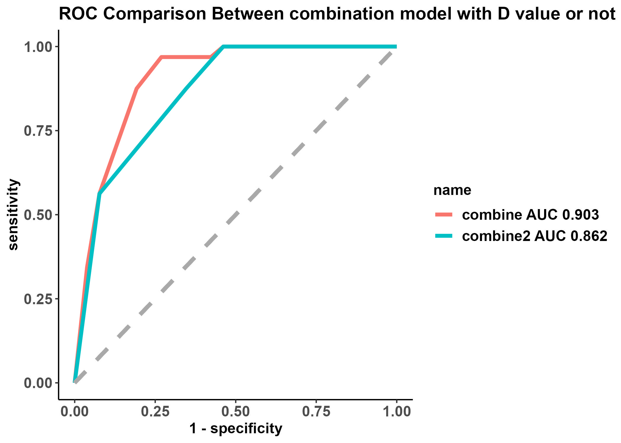
Figure 2. ROC curves of the combined model with and without a D value while differentiating between the poor- and good-outcome groups.