4304
Deep Learning-based Placental MRI Segmentation and Placental Boundary Vessel Recognition1Shanghai first maternity and infant hospital, Shanghai, China, Shanghai, China, 2Shanghai first maternity and infant hospital, Shanghai, China, 3MR Research, GE Healthcare, Beijing, China, 4University of Shanghai for Science and Technology, Shanghai, China
Synopsis
Keywords: Placenta, Placenta
Motivation: Accurate automatic segmentation of the placenta and identification of blood vessel distribution at the placental borders are crucial for diagnosing placental accrete spectrum (PAS) in MRI, yet effective methods are currently limited.
Goal(s): Introduce a refinement fusion method based on U-Net (RFU-Net) for accurately segmenting the placenta. And propose a boundary de-precision (BD) technique to identify the blood vessel distribution around the placental boundary.
Approach: MRI of 200 pregnant females were enrolled. RFU-Net and BD were conducted and evaluated.
Results: RFU-Net improved the accuracy of placenta segmentation (Dice = 0.9314). The BD resolved the blurring of the placenta boundary.
Impact: This study provided a novel method for the automatic identification of placental border vessel distribution.
Introduction
The placenta is an important organ for the exchange of material between the mother and the fetus, placental examination may yield information about serious fetal disorders, such as placental accrete spectrum (PAS) [1-3]. Doctors usually perform MR images on pregnant women who have abnormalities diagnosed with US [4]. However, quantitative assessment and further analysis of the human placenta in MRI requires precise segmentation, which is a challenge. Although existing models have achieved automatic segmentation of the placenta, most of them were targeted at US images or have limitations, such as relatively low performance. To solve the above problems, we proposed the Refinement positioning fusion U-Net(RFU-Net) for the segmentation of the placenta with variable position and shape in MRI images, with the boundary de-refinement (BD) to resolve placental border ambiguity and aid in exploring the distribution of peripheral blood vessels around the placenta.Materials and Methods
PatientsThe data set for this study consisted of 200 patients (103 normal and 97 with PAS) whose placental MRI images were collected, monitored, and recorded throughout their pregnancy. The dataset was divided into training, validation, and test sets at a ratio of 7:1:2.
MRI Acquisition
All patients were scanned for placental MRI by a 1.5T unit (OPTIMA MR360, GE Medical Systems, Milwaukee, WI) using an 8-channel phased-array coil. The FIESTA sequence was scanned in all three directions (axial, sagittal, and coronal). The acquisition parameters were as follows: repetition time/echo time [TR/TE], 3-5/minimum msec; slice thickness, 5-7 mm; matrix size, 224 ×224; flip angle, 55; bandwidth, 100 Hz/pixel. Each image volume consisted of 40 to 62 2D slices.
Model Construction
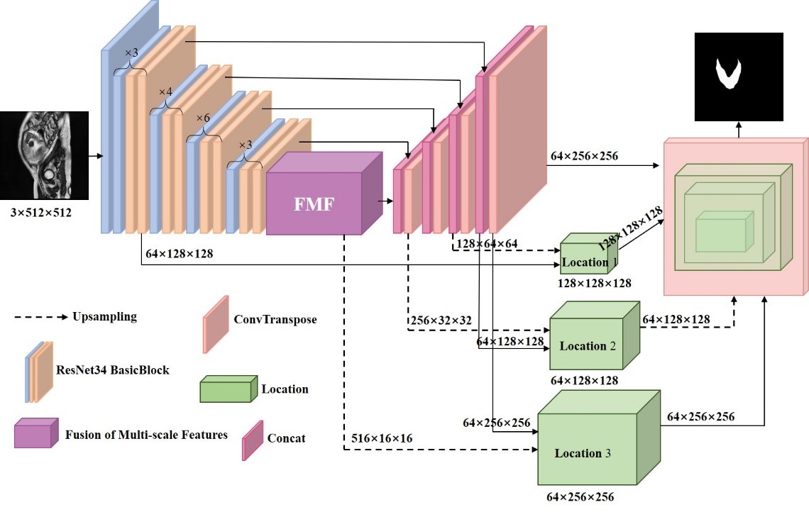
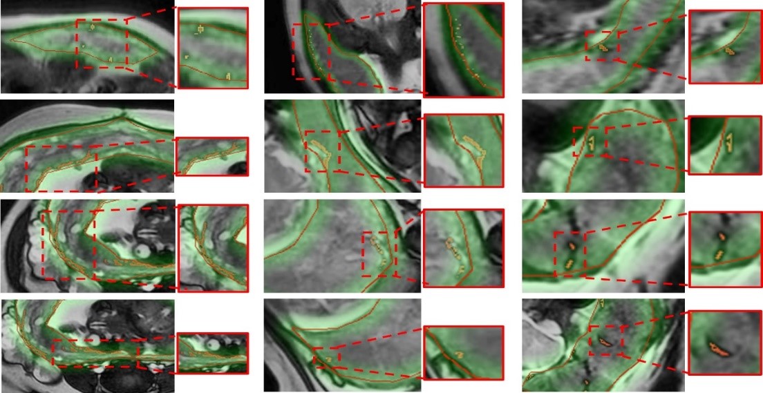
Figure 1 depicts the detailed structure of the segmentation algorithm: the RFU-Net network inherits the encoder-decoder idea from U-Net, to which the fusion multiscale feature and the refinement segmentation module are added. The predicted placental regions were classified using the modified spatial kernelized fuzzy C-mean clustering algorithm. The predicted placental boundary is used as the center, and the ROI expansion region of the placental boundary is formed according to the value of the adaptive expansion image, which completes the de-precision of the placental boundary.
Statistical Analysis
To accurately quantify the segmentation performance of the model, several metrics were introduced, including mean intersection over union (MIoU), dice coefficient (Dice), accuracy (Acc) and P value.
Results
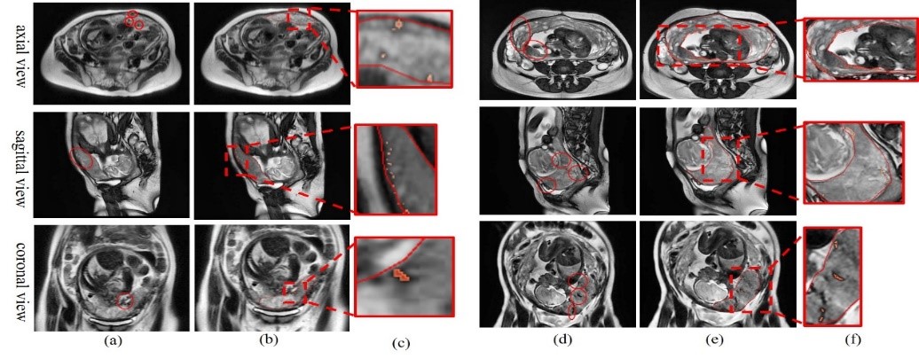
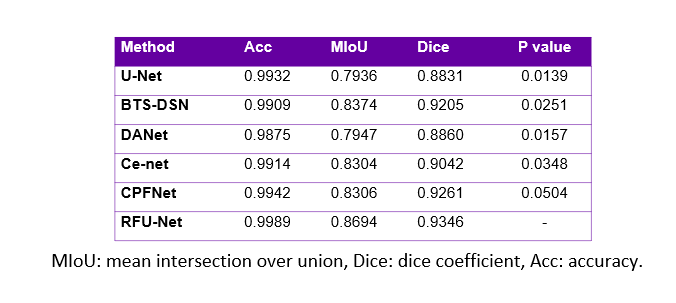
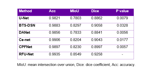
Tables 1 and 2 show the results for healthy and pregnant women with PAS in the test set, respectively. The proposed RFU-Net network achieved the best results in terms of Acc, MIoU, and Dice for the normal group with 0.9989, 0.8694 and 0.9346, respectively. The proposed RFU-Net network achieved the best results in terms of Acc, MIoU and Dice for the PAS group with 0.9935, 0.8549 and 0.9258, respectively. The RFU-Net network increased Acc, MIoU and Dice by 1.14%, 7.76% and 4.94%, respectively compared to the U-Net. The p-values for U-Net, BTS-DSN, DANet, and Ce-net are all less than 0.05, indicating significant differences between our method and these models. The p-values for CPFNet are around 0.05, indicating no significant statistical differences. This difference could be attributed to the relatively small sample size. Overall, it can be concluded that the improvements in the existing methods are significant. These results demonstrate that the RFU-Net performs well in placental segmentation. The segmentation results of the blood vessels at the placental border are shown in Figure 2. The boundary de-precision is designed to address the problem of reduced segmentation accuracy caused by ambiguous placental boundaries. The highlighted areas generated by the adaptively expanded pixels are shown in Figure 3.Discussion and Conclusion
In this study, we proposed a refinement fusion U-Net (RFU-Net) to address the challenging task of accurately segmenting the placenta in MRI images, arising from the varying shapes and positions of the placenta observed across different views and slices. Comparative experiments demonstrated that RFU-Net outperformed the alternative methods in placental segmentation. To tackle the issue of blurred placental boundaries, we also developed a boundary de-precision technique that effectively labeled the surrounding area of the placenta, which may contain blood vessels, thus providing valuable diagnostic assistance.Acknowledgements
No acknowledgement found.References
1. R. Vaughan, F. J. Rosario, T. L. Powell, et al. Regulation of placental amino acid transport and fetal growth. Prog Mol Biol Transl Sci 2017;145:217–51.
2. C. H. J. R. Jansen, A. W. Kastelein, C. E. Kleinrouweler, et al., Development of placental abnormalities in location and anatomy[J]. Acta Obstetricia et Gynecologica Scandinavica,2020, 99(8):983-993.
3. J. H. Dumolt, l T. L. Powel, T. Jansson, Placental function and the development of fetal overgrowth and fetal growth restriction[J]. Obstetrics and Gynecology Clinics, 2021, 48(2): 247-266.
4. A. Yang, X.H. Xiao, Z.L. Wang, et al., T2 -weighted balanced steady state free procession MRI evaluated for diagnosing placental adhesion disorder in late pregnancy [J]. Eur Radiol, 2018,28 ( 9 ): 3770-3778.
Figures