4303
Assessing Automated Vessel Segmentation Techniques of Feto-Placental Vasculature from MRI1School of Biomedical Engineering and Imaging Sciences (BMEIS), Kings College London, London, United Kingdom, 2Department of Medical Physics and Biomedical Engineering, University College London, London, United Kingdom, 3Elizabeth Garrett Anderson Institute for Women’s Health, University College London, London, United Kingdom, 4Auckland Bioengineering Institute, Auckland, New Zealand, 5University College London Hospital NHS Foundation Trust, London, United Kingdom
Synopsis
Keywords: Placenta, Placenta
Motivation: Placental insufficiency is a factor that contributes to multiple pregnancy complications such as Fetal Growth Restriction (FGR).
Goal(s): Providing better understanding to clinicians is important for future treatment planning and automatic detection of the feto-placental vasculature from imaging may provide a tool to guide clinical assessment.
Approach: This work compared the Frangi filter and an edge-based detection algorithm abilities to automatically identify feto-placental vasculature from MRI.
Results: The study found that both methods identified likely vascular structures, and both showed spatial trend similarities when compared with gold-standard/high resolution Micro-CT, as well as a showing differences between FGR and Control vessel segmentations.
Impact: Evaluating the most accurate method for automatically identifying feto-placental vasculature will go on to further aid quantifying placental insufficiency and improving understanding for predicting and clinically treating conditions such as fetal growth restriction.
Introduction
A well-functioning placenta is vital for healthy fetal growth and a large number of pregnancy complications are attributed to placental insufficiency. This includes conditions such as FGR, when the fetus is less than the 10th percentile, which leads to 40% of stillbirths in the UK.[1] Placental insufficiency can be caused by poor development of the highly vascularised placental circulatory system.The placental vasculature is difficult to observe in-vivo, and most existing literature focuses on quantifying its structure ex-vivo. Further developments in magnetic resonance imaging (MRI) in pregnancy are allowing for improved visualisation, due to fetal motion and resolution vessel segmentation used within other anatomy does not work in the same manner.This study aims to observe the effectiveness of different automated vessel segmentation for feto-placental vasculature.These are evaluated by vessel quality, loss functions and validation in comparison to ex-vivo Micro-CT.Methods
MRI data from 10 pregnant patients (6 with FGR(estimated fetal weight <10th centile) and 4 normally grown fetuses) at 24+2-33+6 weeks gestational age (GA) was acquired with a 1.5T Siemens Avanto under free-breathing.[2] For each case a manual mask of the chorionic plate was completed using ITK Snap.Then automated vessel segmentation methods were used: these were the Frangi filter[3] and the edge-based segmentation.[4] The Frangi Filter identifies aims to identify vessel-like structures. These filters are based on a Morpho-Hessian Filter which is used for the enhancement of curvilinear structures.[3,5]Edge Based Segmentation begins with an Adaptive Histogram Equalisation to highlight the edges and their intersections so that these are transformed into a voxel-based histogram.[6] Then from the adaptive histogram the edges are detected and filtered from the rest of the image using a thresholded gradient based edge detection.[7]Validation methods
Automated segmentations were compared using:
1.The vessel volume and range of radii of identified vessels.
2.Connectivity of the segmentations, quantified using skeletonisation and identification of branch points vessel lengths.From this the following metrics were quantified:
- Connectivity index, defined as total length of the vessel network divided by the total area of the segmentation. The higher the connectivity index the more interconnected the vessels are within the segmentation.[8]
- Looping of the segmentations was quantified by detecting the connected components in a closed or distinct loop.[9]
Validation compared with Micro CT
Micro-CT provides a high-resolution quantification but is ex-vivo. Vasculature measured from a gold standard ex vivo perfused placenta that had undergone micro-CT examination[2] was compared with vascular maps extracted from MRI.The placental vasculature was extracted and analysed using the same analysis as for the MRI.[4]The vessel trees were skeletonised and the endpoints identified.From these endpoints the vessel density was calculated and compared to the distance of each end point to the umbilical cord insertion.Discussion
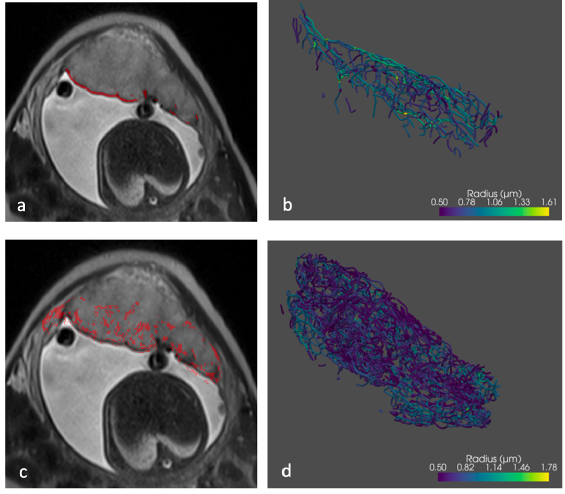
The Frangi Filter and edge-based detector were compared for their utility for automatically identifying feto-placental vasculature from MRI.The edge-based detection was found to have a larger volume of segmentation which can be seen visually in Figure 1, with a greater area segmented between a and c as well as a lot more vessel structures seen in b and c.The volume values are at least double in all cases, although the radii values are similar throughout, with minimum radius remaining the same due to the original image resolution.The volume of the of the segmented vasculature was double for the edge detection segmentations as seen in Table 1, and closer to the values from the ex-vivo Micro-CT data. Figure 2 shows again the far higher segmented objects from the edge detection method.The connectivity values for the segmentations were higher for the edge detection, most likely due to the higher volume segmented, dividing the connectivity value by the volume values showed the Frangi values of connectivity were higher per mm3.However, the looping by volume the Edge detection is lower, potentially due to this reduced connectivity.
Figure 3 showed that the segmentation methods and the Micro-CT data all showed similar spatial trends between vessel density and the distance from the umbilical cord. The coefficient for the segmentation methods showed that edge-based had a closer correlation to the Micro-CT, than the Frangi Filter.
Figure 4 showed that the data is showing a significance difference p=0.024(p<0.05) between the FGR and Control cases, although this is a small dataset.
In conclusion the edge-detection method segmenting the larger volume and found summary vasculature values closer to the Micro-CT, although had a lower connectivity than the Frangi Filter. Combining the two methods for the Frangi Filter to pick up the larger chorionic plate vessels and the Edge based method to identify the lower smaller vasculature could provide an improved visualisation in the future.
Acknowledgements
This research was funded by the Wellcome Trust (210182/Z/18/Z) and EPSRC (NS/A000027/1).References
[1] R. Aughwane, E. Ingram, E. D. Johnstone, L. J. Salomon, A. L. David, and A. Melbourne, “Placental MRI and its application to fetal intervention,” Prenat Diagn, vol. 40, no. 1, pp. 38–48, Jan. 2020, doi: 10.1002/pd.5526.
[2] R. Aughwane et al., “Magnetic resonance imaging measurement of placental perfusion and oxygen saturation in early-onset fetal growth restriction”, doi: 10.1111/1471-0528.16459.
[3] A. F. Frangi, W. J. Niessen, K. L. Vincken, and M. A. Viergever, “Multiscale Vessel Enhancement Filtering*.”
[4] J. Torrents-Barrena et al., “Fully Automatic 3D Reconstruction of the Placenta and its Peripheral Vasculature in Intrauterine Fetal MRI”.
[5] Olena Tankyevych, “(PDF) Filtering of thin objects : applications to vascular image analysis,” Filtering of thin objects : applications to vascular image analysis. Accessed: Jul. 14, 2022. [Online]. Available: https://www.researchgate.net/publication/281183711_Filtering_of_thin_objects_applications_to_vascular_image_analysis
[6] J. A. Stark, “Adaptive Image Contrast Enhancement Using Generalizations of Histogram Equalization,” IEEE TRANSACTIONS ON IMAGE PROCESSING, vol. 9, no. 5, 2000.
[7] J. A. M. Saif, M. H. Hammad, and I. A. A. Alqubati, “Gradient Based Image Edge Detection,” International Journal of Engineering and Technology, vol. 8, no. 3, pp. 153–156, Mar. 2016, doi: 10.7763/ijet.2016.v6.876.
[8] J. Zhang, F. Wu, W. Chang, and D. Kong, “Techniques and Algorithms for Hepatic Vessel Skeletonization in Medical Images: A Survey,” Entropy, vol. 24, no. 4. MDPI, Apr. 01, 2022. doi: 10.3390/e24040465.
[9] K. Li, X. Wu, D. Z. Chen, and M. Sonka, “Optimal surface segmentation in volumetric images - A graph-theoretic approach,” IEEE Trans Pattern Anal Mach Intell, vol. 28, no. 1, pp. 119–134, Jan. 2006, doi: 10.1109/TPAMI.2006.19.
[10] M. Byrne et al., “Structure-function relationships in the feto-placental circulation from in silico interpretation of micro-CT vascular structures,” J Theor Biol, vol. 517, May 2021, doi: 10.1016/j.jtbi.2021.110630.
Figures
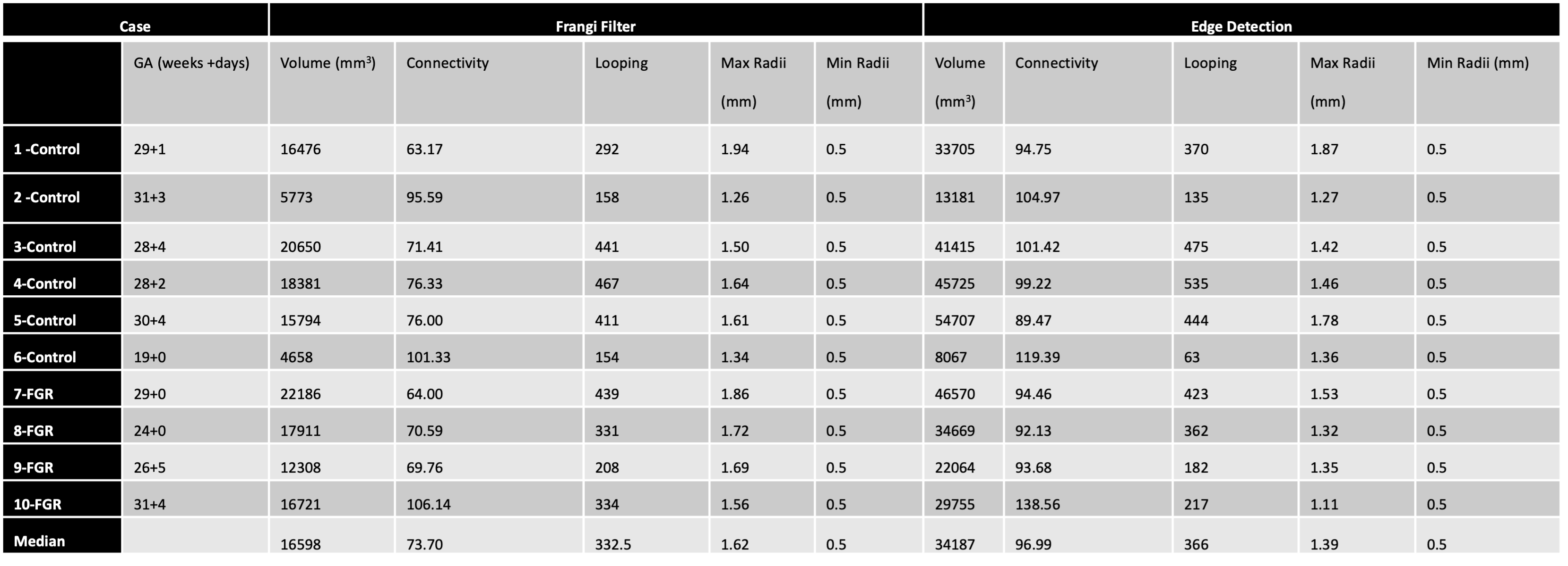
The comparison values from the Micro CT data are a volume of 89592mm3, connectivity of 21.06 and looping of 329.
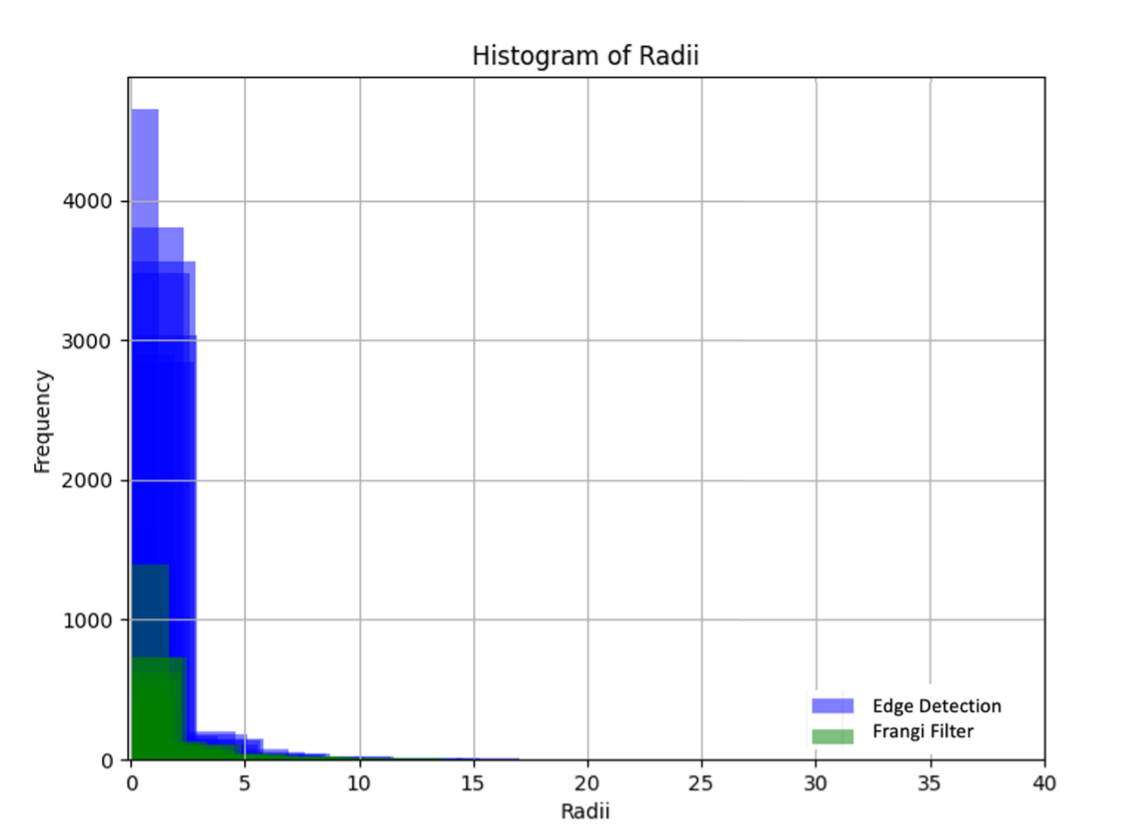
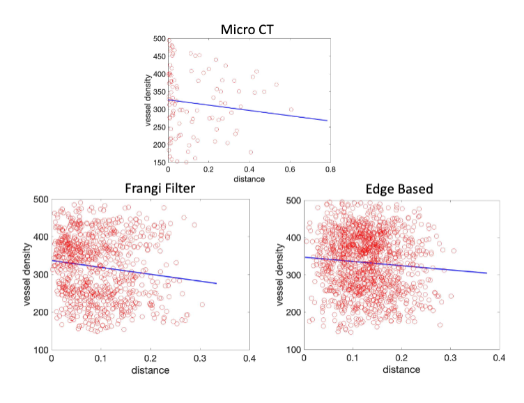
Figure 3 – The Micro CT vessel density plotted against the distance from the umbilical cord insertion at the top and on the left the Frangi Filter and the right Edge detection.
The Pearson Coefficient for the correlation between vessel density and distance from the umbilical cord was -0.1053, for the Frangi Filter cases it averaged at -0.1324 and for the Edge-based detection it averaged as -0.0810.
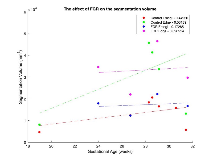
Figure 4 – The segmentation volume for the Frangi and Edge detector separating the control and FGR cases plotted against the GA with the Pearson correlation coefficient in the key for each group.
The effect of FGR on the segmentation volume, a statistical comparison of the correlation coefficients with a p-value of 0.024.