4295
Efficacy for asymmetric Fast 3D wheel technique on female pelvic MRI on contrast enhanced 3D T1-weighted image1Radiology, Fujita Health University School of Medicine, Toyoake, Japan, 2Diagnostic Radiology, Fujita Health University School of Medicine, Toyoake, Japan, 3Canon Medical Systems Corporation, Otawara, Japan, 4Radiology, Fujita Health University Bantane Hospital, Nagoya, Japan, 5Joint Research Laboratory of Advanced Medical Imaging, Fujita Health University School of Medicine, Toyoake, Japan
Synopsis
Keywords: Pelvis, Pelvis
Motivation: Fast 3D Wheel with Sequential filling and Asymmetric Fourier imaging (Fast 3Dwsa) which is one of the k-space based acceleration techniques can improve image quality and acquisition time.
Goal(s): The goal was to determine the utility of contrast-enhanced 3D T1-weighted image with Fast 3Dwsa on female pelvic MRI as compared with conventional parallel imaging (PI) and compressed sensing (CS).
Approach: Quantitative and qualitative indexes for image quality improvement and reducing acquisition time were statistically compared among all methods.
Results: Quantitative and qualitative indexes of Fast 3Dwsa was significantly better than that of PI and CS (<0.05).
Impact: Fast 3Dwsa is considered as useful for image quality improvement with reducing acquisition time on female pelvic MRI, when compared with conventional PI as well as CS.
Introduction
Contrast-enhanced 3D T1-weighted image, which can accurately delineate complex or small structures of the body is suggested as the fundamental sequence in clinical MR imaging and important components of gynecologic MR imaging1-3. However, one of the drawbacks of 3D MR imaging is relatively longer acquisition times, thereby limiting its clinical use4. Compressed sensing (CS) has been suggested as a new method for aiming to reduce the number of k-space samples by exploiting compressibility or sparsity in an appropriate transform domain in last several years, although o CS is suggested as having relatively lower signal-to-noise ratio (SNR) than parallel imaging (PI), which is frequently applied for various clinical purposes as well as sequences5. Under the above-mentioned situations, asymmetrically filled k-space with wheel encoding is introduced as Fast 3D wheel with sequential filling and Asymmetric Fourier Imaging (Fast 3Dwsa) technique by Canon Medical Systems, clinically applicable technique for k-space based acceleration technique and applied with PI for further reducing acquisition time. However, no major reports have been published about the utility of CS and Fast 3Dwsa on female pelvic MRI as compared with conventional PI. We hypothesized that Fast 3Dwsa can improve image quality and acquisition time on female pelvic MRI, when compared with conventional PI. The purpose of this study was to determine the utility of contrast enhanced 3D T1-weighted image with Fast 3Dwsa for improving image quality and acquisition time on female pelvic MRI as compared with that obtained by PI as well as CS.Materials and Methods
Twenty-two consecutive female patients with various pelvic diseases underwent contrasted-enhanced 3D T1-weighted imaging by Fast-Field Echo (FFE) pulse sequence (TR = 4.4ms, TE = 1.9ms, flip angle = 13 degree, slice thickness = 1.4 mm, NEX = 2, FOV = 300 × 300 mm, 30 slices, 256 × 256 matrix, 512 × 512 reconstruction matrix) with PI, CS and Fast 3Dwsa, randomly. Details of Fast 3Dwsa is shown in Figure 1. Then, all acquisition times were recorded in each patient. For quantitative assessment, SNR of muscle and CR between myometrium and muscle were determined on uterine corpus and cervical level by ROI measurements. For qualitative assessment, two board certified radiologists assessed overall image quality (OIQ), artifact and diagnostic confidence level (DCL) by 5-point scales. Then, each final score was determined as consensus of two readers. To compare the capability for acquisition time reduction, mean acquisition time was compared among all data sets by Tukey’s HSD test. To determine quantitative image quality improvement, SNRs and CRs were compared among all methods by Tukey’s HSD test. On qualitative image quality evaluations, inter-observer agreement on each data set was assessed by κ statistics followed by χ2 test. Finally, threeindexes among all methods by Wilcoxon signed-rank test.Results
Representative case is shown in Figure 2. On comparison of mean acquisition time, CS and Fast 3Dwsa (CS: 156.6 ± 4.8 sec, Fast 3Dwsa: 153.9 ± 2.7 sec) were significantly shorter than PI (PI: 313.0 ± 9.7 sec, p<0.001). Comparisons of SNR and CR among all methods are shown in Figure 3. SNR of Fast 3Dwsa on uterine corpus and cervical level (uterine corpus level: 17.5 ± 4.8, uterine cervical level: 18.2 ± 5.5) was significantly higher than that of PI and CS, although there was no significant difference of SNR between PI and CS on uterine corpus and cervical level (uterine corpus level; PI: 10.9 ± 3.7, CS: 12.9 ± 5.7, uterine cervical level; PI: 12.9 ± 5.7, CS: 13.9 ± 7.7). There was no significant difference of CR among three methods (p>0.05). Interobserver agreements for all qualitative index are shown in Figure 4. All inter-observer agreements were determined as ‘moderate’ or ‘excellent’ (0.53<κ<0.91, p<0.0001). Compared results of qualitative index among all methods are shown in Figure 5. OIQ and artifact of Fast 3Dwsa was significantly superior to those of PI and CS (p<0.05), although there was no significant difference of OIQ and artifact between PI and CS (p>0.05). There was no significant difference of DCL among three methods (p>0.05).Conclusion
Fast 3Dwsa is considered as more useful than PI and CS for image quality improvement with reducing acquisition time on female pelvic MRI.Acknowledgements
This work was technically and financially supported by Canon Medical Systems Corporation.References
- Cornfeld DM, Israel G, McCarthy SM, Weinreb JC. Pelvic imaging using a T1W fat-suppressed three-dimensional dual echo Dixon technique at 3T. J Magn Reson Imaging 2008;28(1):121-127.
- Rofsky NM, Lee VS, Laub G, Pollack MA, Krinsky GA, Thomasson D, Ambrosino MM, Weinreb JC. Abdominal MR imaging with a volumetric interpolated breath-hold examination. Radiology 1999;212(3):876-884.
- Israel GM. MRI of the kidney and urinary tract. J Magn Reson Imaging 2006;24(4):725-734.
- Mugler JP, 3rd. Optimized three-dimensional fast-spin-echo MRI. J Magn Reson Imaging 2014;39(4):745-767.
- Feng L, Benkert T, Block KT, Sodickson DK, Otazo R, Chandarana H. Compressed sensing for body MRI. J Magn Reson Imaging 2017;45(4):966-987.
Figures
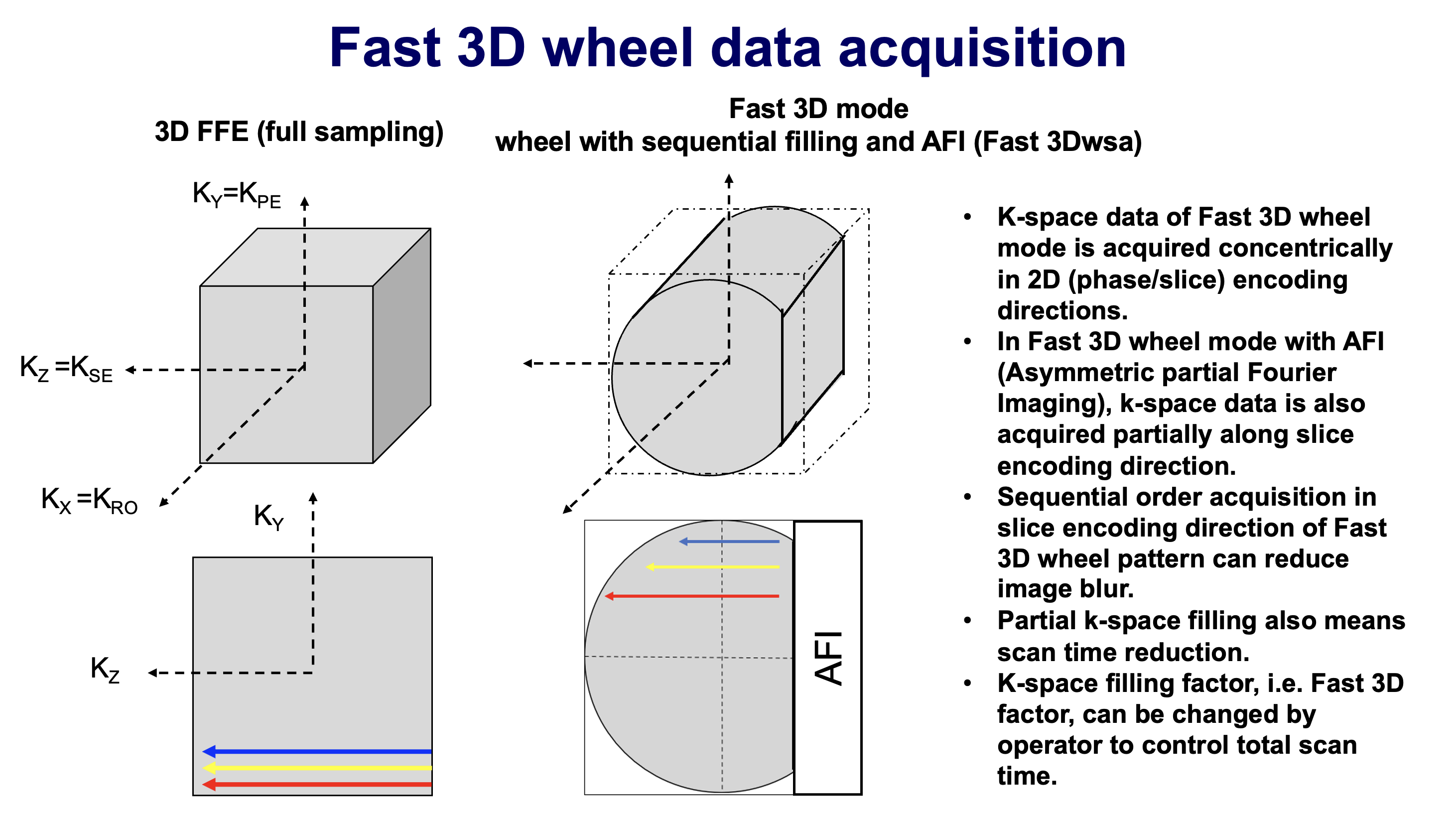
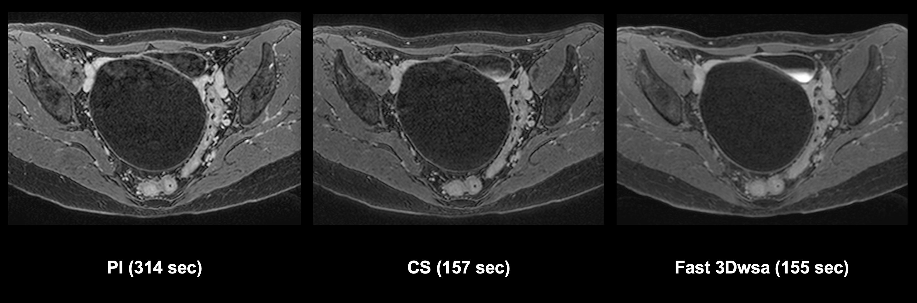
Figure 2. A 63-year-old female patient with ovarian tumor (L to R: Conventional PI, CS, Fast 3Dwsa)
Acquisition times for all methods are follows: PI, 314 sec; CS, 157 sec; Fast 3Dwsa, 155 sec. CS and Fast 3Dwsa could reduce examination times as compared with PI. All contrast-enhanced 3D T1 weighted-images obtained with PI, CS and Fast 3Dwsa clearly demonstrate ovarian tumor. Moreover, Fast 3Dwsa demonstrates ovarian tumor more clearly as compared with PI .
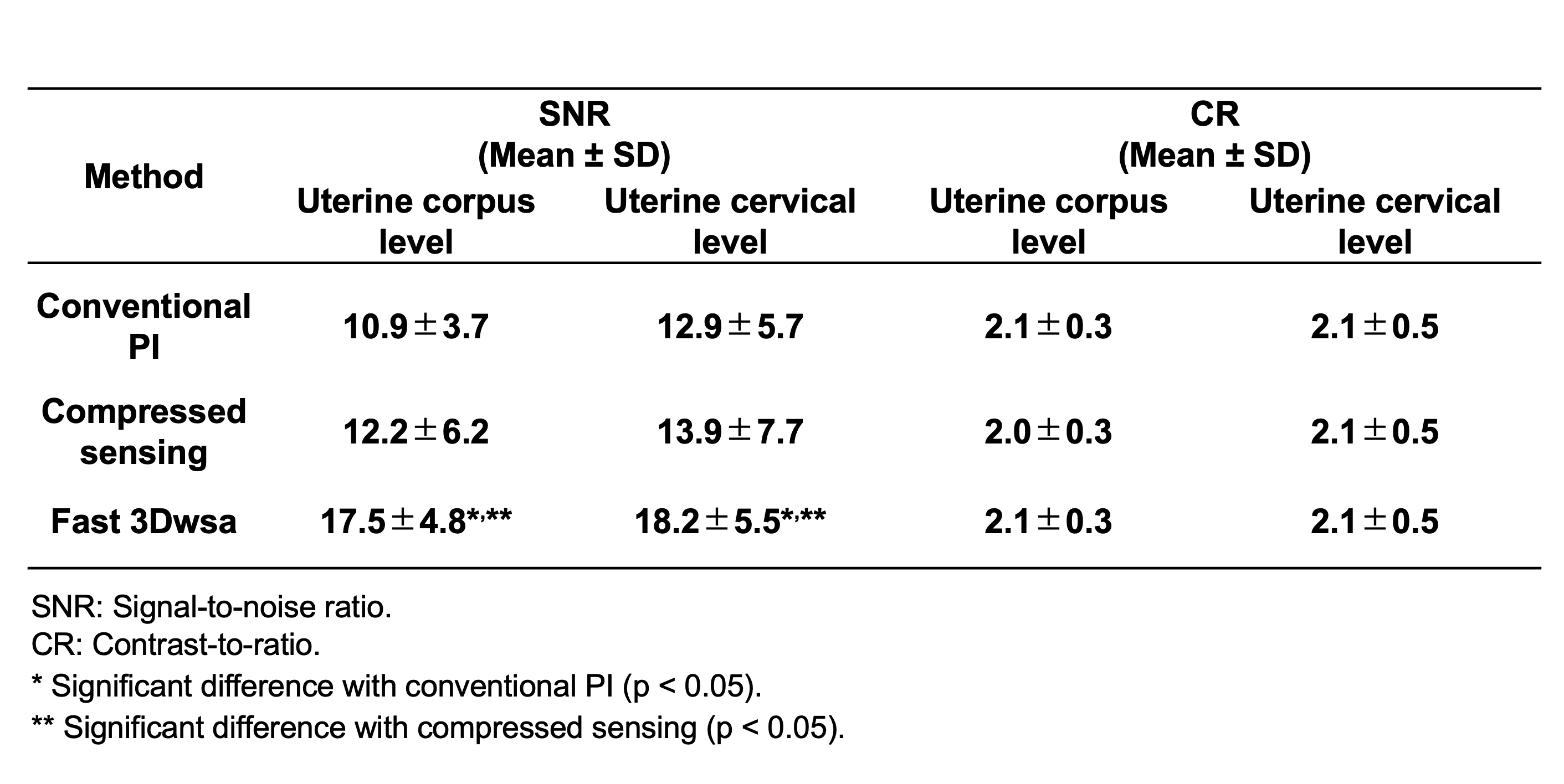
Figure 3. Compared results of SNR and CR on uterine corpus level and uterine cervical level among three methods
SNR of Fast 3Dwsa on uterine corpus and cervical level (uterine corpus level: 17.5 ± 4.8, uterine cervical level: 18.2 ± 5.5) was significantly higher than that of PI and CS, although there was no significant difference of SNR between PI and CS on uterine corpus and cervical level (uterine corpus level; PI: 10.9 ± 3.7, CS: 12.9 ± 5.7, uterine cervical level; PI: 12.9 ± 5.7, CS: 13.9 ± 7.7). There was no significant difference of CR among three methods (p>0.05).
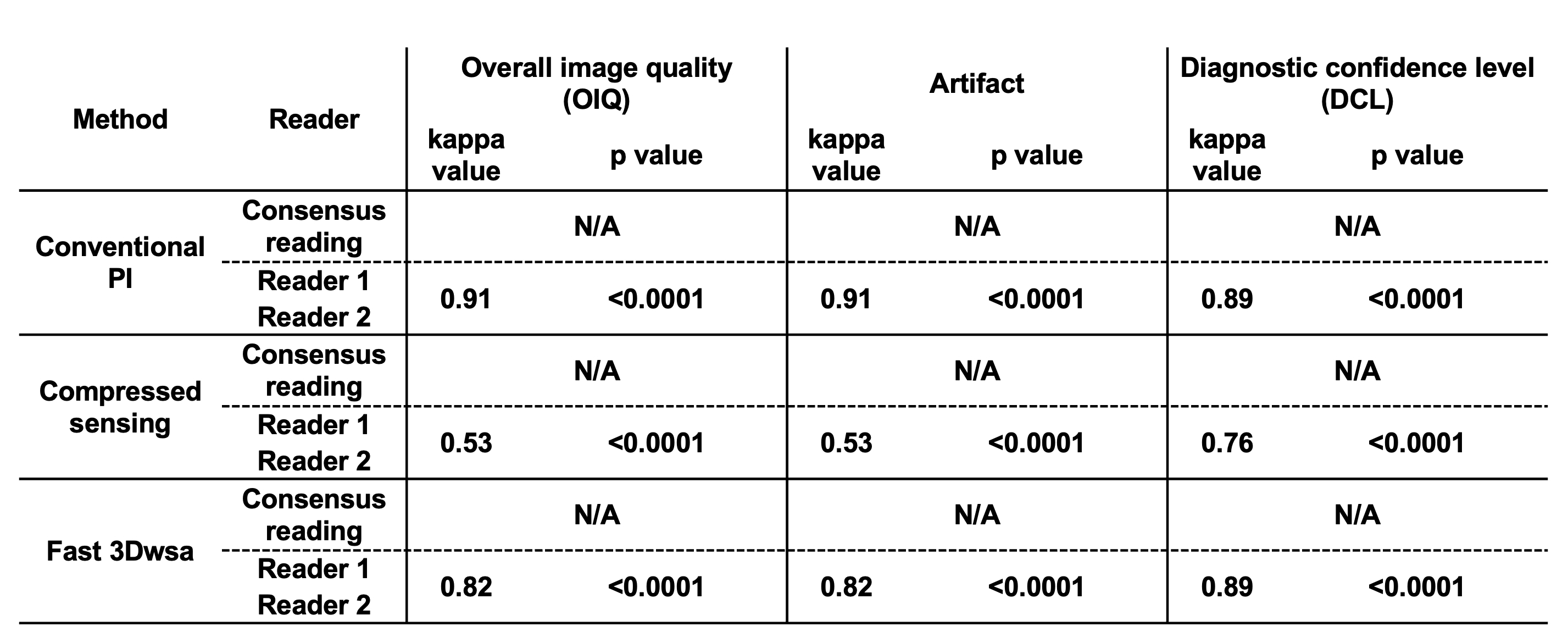
Figure 4. Compared results of inter-observer agreements among three methods
All inter-observer agreements were determined as ‘moderate’ or ‘excellent’ (0.53<κ<0.91, p<0.0001).
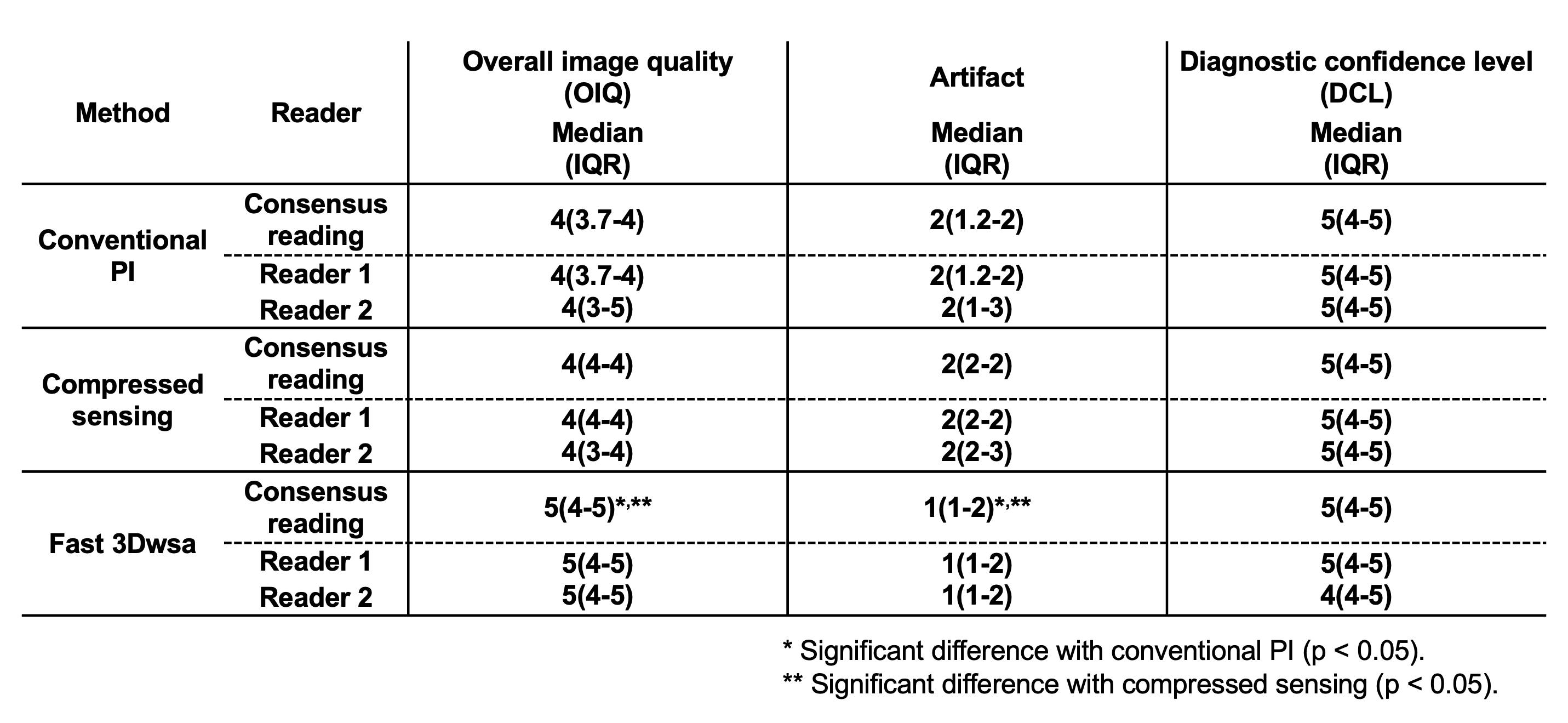
Figure 5. Compared results of overall image quality (OIQ), artifact and diagnostic confidence level (DCL) among three methods
OIQ and artifact of Fast 3Dwsa was significantly superior to those of PI and CS (p<0.05), although there was no significant difference of OIQ and artifact between PI and CS (p>0.05). There was no significant difference of DCL among three methods (p>0.05).