4294
Better diagnostic value and feasibility of Deep Learning DWI in uterine malignant neoplasms1The First Hospital of China Medical University, Shenyang, China, 2MR Research Collaboration, Siemens Healthineers, Bejing, China, 3MR Application Predevelopment, Siemens Healthineers AG, Erlangen, Germany
Synopsis
Keywords: Pelvis, Uterus
Motivation: Conventional diffusion-weighted imaging (c-DWI) of the uterus is time-consuming, and the lesion details are not well-defined.
Goal(s): To introduce a deep learning (DL) DWI sequence in uterine MRI and compare it with conventional DWI (c-DWI) to investigate its impact on examination time, image quality, lesion significance, diagnostic reliability, as well as contrast ratio (CN), signal-to-noise ratio (SNR), and contrast-to-noise ratio (CNR).
Approach: 10 patients with uterine malignancy disease were included in this study.
Results: There is no significant difference in objective assessment between the two techniques, while the overall image quality of DL-DWI is better than c-DWI (p < 0.01).
Impact: The research investigated the utilization of DL-DW in the uterus, which led to shorter examination times and significantly improved image quality. This analysis has the potential to examine other pelvic organs, such as the prostate, to assess pelvic lesions.
Introduction
Diffusion-weighted imaging (DWI) in pelvic MRI plays a vital role in providing valuable insights into tissue cellularity and detection and characterization of various lesions, particularly malignancies. However, implementation of DWI presents challenges, including inherent image blurring, extended acquisition time, geometric distortions affected by rectal air, susceptibility artifacts, and limited spatial resolution. These challenges often result in equivocal diagnosis and exclusion of benign lesion, thus impacting diagnostic confidence. Various approaches have been used to improve the image quality, such as increase averages, albeit at the cost of prolonged scanning time especially high b values. Deep learning-based reconstruction offer a promising solution to reduce examination time, mitigate blurring, and eliminate fold-over artifacts while requiring fewer averages and benefiting from high parallel imaging acceleration factors. These improvements have advantages in enhancing lesion conspicuity and image quality, augmenting diagnostic confidence and accuracy. In this research, we investigate the application of Deep learning DWI(DL-DWI) to provide more evidence in detecting and diagnosing endometrial cancer or cervical cancer, compared to conventional DWI (con-DWI).Methods
10 females with endometrial cancer or cervical cancer were prospectively enrolled. They underwent pelvic MRI on a 3T system (MAGNETOM Vida, Siemens Healthineers, Erlangen, Germany), including routine examinations, con-DWI and a DL-DWI research application. The detailed imaging parameters are listed in Table 1. For DL-DWI, the FOV and resolution were similar to con-DWI, while TR, TE, averages, and parallel imaging acceleration factor were adjusted to reduce acquisition time. For the qualitative analysis, the image quality was evaluated according to a 4-point rating scale, including the anatomic structure visualization (1: poorly visible anatomy and non-diagnostic; 2: Clear outline, blurred edges; 3: clean edges and nice curves.; and 4: Excellent sharpness), artifacts(1: grave and non-diagnostic; 2: moderate; 3: Slight; and 4: non-artifact), and overall image quality (1: poor, considered non-diagnostic; 2: fair, moderately poor diagnostics; 3: high, without sacrificing diagnostic accuracy; 4: excellent) of the DWI images. Diagnostic confidence1(1: DWI did not assist diagnose cancer; 2: Lesion characterization on DWI approximated conventional imaging diagnosis; 3: DWI verified conventional imaging's diagnosis; and 4: DWI detects invisible lesions in routine images), lesion conspicuity(1: Undetectable lesion; 2: merely recognizable lesion-to-background contrast; 3: middle lesion-to-background contrast or high contrast with unclear lesion margin; and 4: Sharp lesion-to-background contrast and margin) and lesion margin (1: undetectable; 2: obscure; 3: indistinct; and 4: distinct) were scored by 4-point rating scale based on the combination of DWI and conventional enhanced T1WI and T2WI. For the quantitative analysis, the signal-to-noise ratio (SNR) was calculated as the ratio between the mean signal intensity (SI) and the standard deviation of SI in the same ROI for myometrium and piriformis muscles at b value=50,800,1200. The contrast (CN) between malignancy and piriformis muscle was measured using the formula: CN = |SIA – SIB|/(SIA +SIB)2. Contrast-to-noise ratios (CNR) for different b-values was measured via|SIA – SIB|/√(SIA²+SIB²)3, where SIA and SIB are SI for tumors and piriformis muscles, and the SDA and SDB are corresponding standard deviation. Paired T-test was used for the comparison between groups with normal distribution of SNR, CNR and subject scores, and Wilcoxson signed-rank test was used for the comparison between groups with non-normal distribution of SNR, CNR and objective assessment score. All statistical analysis was performed with SPSS 26.0(SPSS Inc., Chicago, USA). P values below 0.05 were considered statistically significant.Results
For quantitative analysis, DL-DWI demonstrated significantly improved image quality compared to con-DWI with better anatomic structure visualization and overall image quality (anatomic structure visualization:3.55±0.51 vs 2.65±0.49; overall image quality:3.3±0.47 vs 2.90±0.31, both P < 0.05) as shown in Table 2. Furthermore, DL-DWI outperformed con-DWI in visualizing lesion conspicuity and lesion margin, with significantly increased objective scores (Lesion conspicuity, 3.85±0.37 vs 3.25±0.44; Lesion margin: 3.75±0.44 vs 3.0±0.79, both p<0.01, Table 3) . Additionally, there were no significant differences between con-DWI and DL-DWI in the CN, CNR, SNR of uterus malignant lesions (P > 0.05) as shown in Table 4. The acquisition time for DL-DWI was notably shorter, reducing scan time by over 20% while maintaining improved image quality and lesion visualization.Discussion
In this study, we investigated the utility of Deep Learning reconstruction in DWI in pelvic MRI for the detection of endometrial and cervical cancer, comparing it to con-DWI. DL-DWI offers superior image quality, shorter scan time without compromising SNR,and improved diagnostic capabilities, enhancing patient comfort and workflow efficiency. DL-DWI is a potential feasible option for the endometrial malignant neoplasms compared to c-DWI.Acknowledgements
Competing interests Yueluan Jiang is employed by MR Collaboration,Siemens Healthineers, Beijing, China. Thomas Benkert is employed by MR Application Predevelopment, Siemens Healthineers AG, Erlangen, Germany.The remaining authors declare that the research was conducted in the absence of any com-mercial or financial relationships that could be construed as a potential conflict of interest.
Ethical approval and consent to participate Our study complied with the Declaration of Helsinki. The study was approved by ethical committee of the First Hospital of China Medical University, written informed consent from the patients for use of data.
References
1.Liu J, Huang M, Ren Y, et al. Added value of zoomed-echo-planar imaging diffusion-weighted imaging for evaluation of periampullary carcinomas[J]. Abdominal Radiology,2023,48(10):3079-3090.
2.Tsuboyama T, Takei O, Okada A, et al. Comparison of HASTE with multiple signal averaging versus conventional turbo spin echo sequence: a new option for T2-weighted MRI of the female pelvis[J]. European Radiology,2020,30(6):3245-3253.
3.Ma Y, Chen X, Zhu W, et al. Speckle noise
reduction in optical coherence tomography images based on edge-sensitive
cGAN[J]. Biomedical Optics Express,2018,9(11): 5129-5146.
Figures
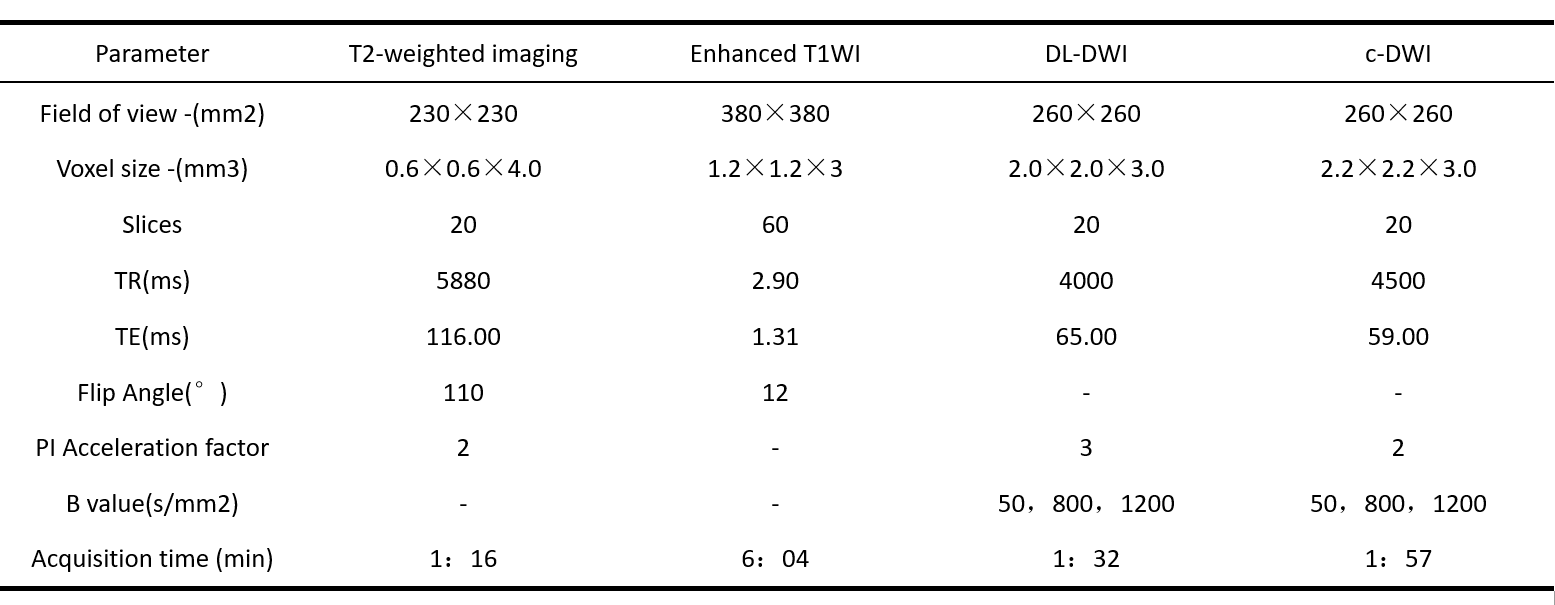
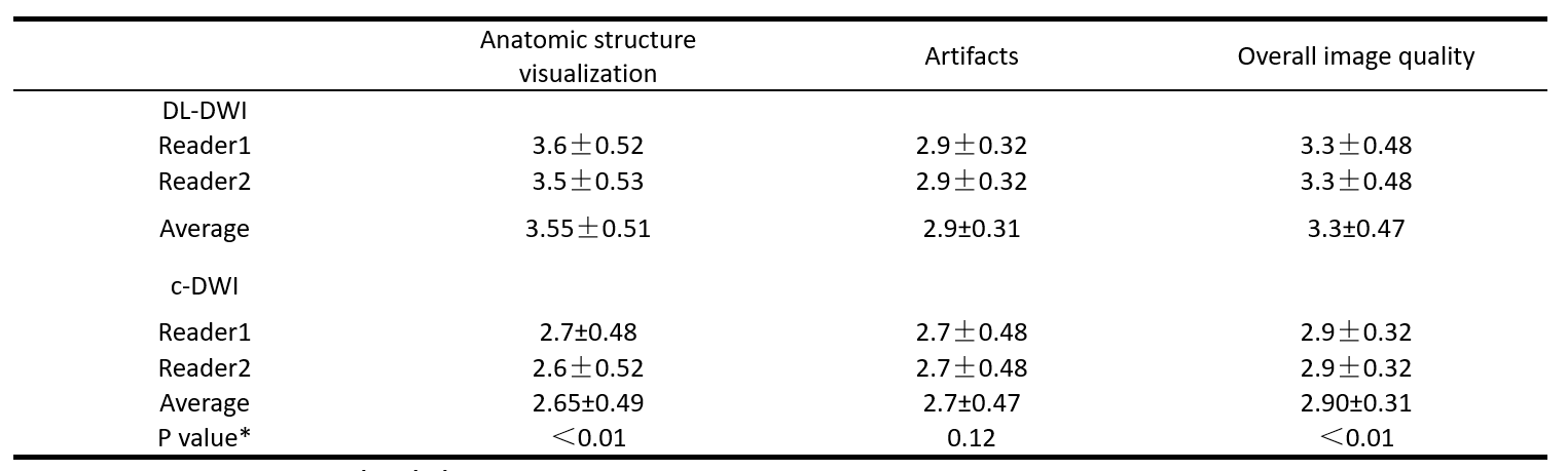
Table2. Comparison of image quality scores between DL-DWI and c-DWI diffusion-weighted imaging sequences (b = 800 sec/mm2)
Data are mean ± standard deviation.
*Wilcoxon signed-rank test was performed between DL-DWI and c-DWI sequences using averaged image quality scores of two readers
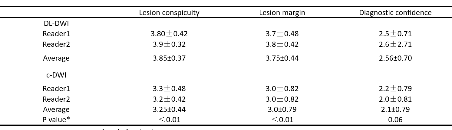
Table3.Comparison of lesion scores between DL-DWI and c-DWI sequences (b = 800 sec/mm2).
Data are mean ± standard deviation.
*Wilcoxon signed-rank test was performed between DL-DWI and c-DWI sequences using averaged lesion scores of two readers.
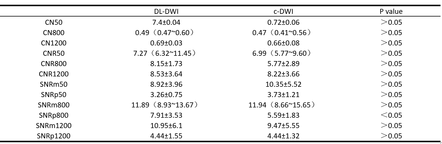
Table4. Comparison of CN, CNR and SNR(b=50,800,1200)
m, myometrium; p, piriformis muscle
Paired T-test was used for normally distributed data, and the Wilcoxson signed-rank test was used.