4293
The value of APT combined with mDIXON-Quant imaging in predicting lymph node metastasis of cervical cancer1the First Affiliated Hospital of Dalian Medical University, Dalian, China
Synopsis
Keywords: Pelvis, Tumor
Motivation: According to the literature, the 5year survival rate of early cervical cancer patients with LNM positive is only about 65%, which is lower than that of patients with LNM negative.
Goal(s): Evaluate the lymph node status before operation.
Approach: The APT, R2* and T2* values were measured by doctors with two years experience in imaging diagnosis. KS test was used to test the normality of the data, and independent sample T test or Mann-Whitney U test was used to compare the differences between the two groups.
Results: APT and mDixon-Quant multi-parameter imaging are helpful to predict the lymph node metastasis of cervical cancer.
Impact: APT and mDixon-Quant multi-parameter imaging are helpful to predict lymph node metastasis of cervical cancer, to make clinical decisions and to improve the prognosis of patients.
INTRODUCTION
Cervical cancer is the fourth most common malignant tumor among women in the world, and one of the important factors affecting the prognosis of patients with cervical cancer is lymph node metastasis (LNM). According to the literature, the 5-year survival rate of early cervical cancer patients with LNM positive is only about 65%, which is significantly lower than that of patients with LNM negative. Therefore, it is of great significance to accurately evaluate the lymph node status before operation. Amide proton transfer-weighted imaging (APTw), as a quantitative imaging technique, can detect the contents of protein and polypeptide in diseased tissues, which has certain value for differential diagnosis of diseases. Magic mirror imaging (mDixon-Quant) sequence is a quantitative technique for separating water and fat, which has been applied to the related research of quantitative evaluation of fat content, such as the quantitative study of liver fat content and the study of lumbar vertebral body fat content. As far as we know, there is no previous report on the use of quantitative parameters of mDixon-Quant and APT to evaluate the lymph node metastasis status of cervical cancer. Based on APT and mDixon-Quant multi-parameter imaging, this study aims to evaluate the lymph node metastasis status of cervical cancer.Materials and methods
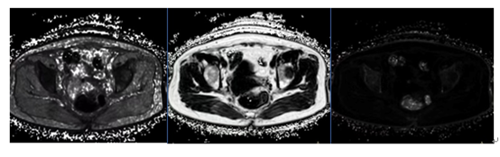
75 cases of cervical cancer confirmed by surgery and pathology from July 2019 to May 2023 were retrospectively analyzed, including 17 cases with lymph node metastasis(LM) and 58 cases with non-lymph node metastasis(NLM). MDIXON-Quant examination was performed before operation; The APT , R2* and T2* values were measured by doctors with two years experience in imaging diagnosis. Kolmogorov-Smirnov test was used to test the normality of the data, and independent sample T test or Mann-Whitney U test was used to compare the differences between the two groups. Logistic regression was used to calculate the predicted values of APTw and mDixon-Quant series joint parameter models. The diagnostic efficacy of different parameters alone or in combination was evaluated by receiver's operating characteristic (ROC) curve.Result
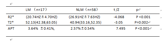
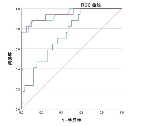
The APT value and T2* value in lymph node metastasis group (3.64% ±0.41%, 52.13(42.38,63.05)ms) were higher than those in non-lymph node metastasis group (2.57%±0.54%, 40.94(33.16,52.35)ms), and the difference was statistically significant (t=7.495, -3.05,P <0.001,P=0.002)The R2* value of lymph node metastasis group (20.74HZ±4.70HZ) was lower than that of non-lymph node metastasis group (26.91HZ±7.63HZ), and the difference was statistically significant (t/z=-4.068, P <0.001). There was no statistical difference in FF between lymph node metastasis group and non-lymph node metastasis group (z=-0.886, P=0.376). The AUC values of APT, R2* and T2* were 0.938, 0.258 and 0.744 respectively.Acknowledgements
No acknowledgement found.References
cite [1] Meng Xing, Liu Ailian. Progress in application of amide proton transfer imaging in MRI examination of uterus [J]. China Journal of Medical Imaging, 2023,31(04):413-417.
[2] Qin Li, Zhao Xiujuan, qi zhou. Progress in application of amide proton transfer imaging in gynecological malignant tumors [J]. China Journal of Medical Imaging, 2022,30(05):524-528.
Figures