4292
Predicting lymph node metastasis in early cervical cancer using spatial features at perfusion habitat Imaging based on DCE-MRI1China-Japan Union Hospital of Jilin university, Changchun, China, 2MR Research Collaboration, Siemens Healthineers,, Beijing, China
Synopsis
Keywords: Uterus, Cancer
Motivation: Cervical cancer has significant spatial heterogeneity, resulting in tumor recurrence and metastasis. The exploration of tumor spatial features may be valuable for predicting lymph node metastasis in cervical cancer.
Goal(s): Combined with landscape ecological analysis and DCE-MRI construction of blood perfusion landscape to predict lymph node metastasis of early cervical cancer.
Approach: Based on DCE-MRI pharmacokinetic parameter map, perfusion habitat imaging was constructed, and landscape ecological analysis was introduced to extract the spatial features of habitat imaging.
Results: The spatial heterogeneity features of blood perfusion obtained by landscape analysis can predict lymph node metastasis of early cervical cancer.
Impact: In this study, we innovatively introduced landscape analysis method to obtain the spatial heterogeneity features of blood perfusion, which demonstrated good performance for predicting lymph node metastasis of early cervical cancer.
Introduction
Surgical treatment is the primary choice for early cervical cancer[1-3]. Lymph node metastasis, one of the high-risk pathological factors for postoperative recurrence[4-5], pose a challenge in accurate preoperative diagnosis. In the International Federation of Gynecology and Obstetrics (FIGO) 2018 staging, once lymph node metastasis is found in patients with early cervical cancer in FIGO stageⅠ-Ⅱ, they are directly classified as FIGO Stage IIIC, leading to altered treatment plans as concurrent chemoradiotherapy. Therefore, accurate preoperative assessment of lymph node metastasis status is helpful to guide clinical treatment strategy and improve prognosis.As a solid tumor, cervical cancer has significant spatial heterogeneity, which is the main cause of tumor evolution, recurrence and metastasis[6-8]. The difference in spatial heterogeneity degree may be the reason for the different lymph node metastasis state of early cervical cancer with the same pathological grade, tumor size and stromal infiltration depth. However, there is a lack of methods to explore the spatial characteristics of tumors. In our study, we aimed to identify the spatial heterogeneity subregion of blood perfusion in early cervical cancer through DCE-MRI quantitative parameter maps. we introduced landscape ecological methods to extract the spatial heterogeneity characteristics, and study the relationship between spatial characteristics and lymph node metastasis to determine whether spatial features can be used as imaging biomarkers to predict lymph node metastasis in cervical cancer.
Method
A retrospective analysis based on a prospective study was conducted to collect the data of DCE-MRI in 88 patients with early-stage cervical cancer. DCE post-processing analysis were performed on Tissue 4D software (Siemens Healthineers) to calculate quantitative pharmacokinetic parameter maps (Ktrans, Kep, ve) based on standard Tofts’ model. Radiologist delineated ROI along the tumor margin at the largest tumor layer, avoiding cystic change and necrotic areas as much as possible. Based on the two-stage cluster analysis at the individual and population levels, each tumor was divided into several spatial subregions with the same hemodynamic phenotype, and the blood perfusion habitat image was constructed. Landscape ecological index was introduced to extract the spatial features of each subregion and the overall landscape of tumor. Student’s t-test or Mann-Whitney U test and ROC curve analysis were used to select the most representative spatial features of each subregion and the overall landscape of habitat image. Multivariate logistic regression analysis was used to further screen spatial features, and establish diagnosis models of lymph node metastasis for early-stage cervical cancer based on single habitat subregion, overall landscape and multi-scale combined habitat image.Result
After two-stage cluster analysis, blood perfusion habitat image of early-stage cervical cancer was constructed, and three spatial subregions (subregion1, subregion2, subregion3) with different characteristics of blood perfusion were found at the population level. Figure 1 showed the distribution of Ktrans, Kep and ve in each habitat subregion. The multi-scale combined diagnosis model based on spatial features from landscape and subregion2 of habitat images had the highest diagnostic efficiency in the identification of lymph node metastasis, with an AUC of 0.898(95%CI:0.788-0.963,P< 0.001). The detailed information of the multivariate logistic regression model of all models is shown in Table 1, the diagnostic efficiency is shown in Table 2, and the ROC curve is shown in Figure 2. Figure 3 showed two representative blood perfusion habitat images for early cervical cancer without and with lymph node metastasis.Discussion
In this study, a data-driven two-stage clustering algorithm was used to analyze the DCE-MRI data of early cervical cancer. We found three spatial subregions with different perfusion dynamic characteristics and phenotypes at the population level, realized the segmentation of different blood perfusion characteristic subregions within tumors, and constructed a habitat image to characterize the spatial heterogeneity of blood perfusion. Landscape ecological index was introduced to quantify the spatial characteristics of different subregions, and a diagnostic model for evaluating lymph node metastasis of early cervical cancer was further constructed. The results showed that the combined diagnosis model with multi-scale spatial features had the highest diagnostic efficiency, and the AUC value of predicting lymph node metastasis reached 0.898. Habitat imaging and landscape index quantifying spatial characteristics of subregions of the habitat can be useful methods to evaluate pathological risk factors of cervical cancer before treatment.Conclusion
Quantitative spatial features extracted from blood perfusion habitat imaging of early cervical cancer based on DCE-MRI are helpful for preoperative evaluation of lymph node metastasis.Acknowledgements
No acknowledgement found.References
[1] Abu-Rustum N R, Yashar C M, Bean S, et al. NCCN Guidelines Insights: Cervical Cancer, Version 1.2020 [J]. Journal of the National Comprehensive Cancer Network : JNCCN, 2020, 18(6): 660-6.
[2] Kim D Y, Shim S H, Kim S O, et al. Preoperative nomogram for the identification of lymph node metastasis in early cervical cancer [J]. British journal of cancer, 2014, 110(1): 34-41.
[3] Li Y, Ren J, Yang J J, et al. MRI-derived radiomics analysis improves the noninvasive pretreatment identification of multimodality therapy candidates with early-stage cervical cancer [J]. European radiology, 2022, 32(6): 3985-95.
[4] Sartori E, Tisi G, Chiudinelli F, et al. Early stage cervical cancer: adjuvant treatment in negative lymph node cases [J]. Gynecologic oncology, 2007, 107(1 Suppl 1): S170-4.
[5] Gemer O, Lavie O, Gdalevich M, et al. Evaluation of Clinical and Pathologic Risk Factors May Reduce the Rate of Multimodality Treatment of Early Cervical Cancer [J]. American journal of clinical oncology, 2016, 39(1): 37-42.
[6] Vitale I, Shema E, Loi S, et al. Intratumoral heterogeneity in cancer progression and response to immunotherapy [J]. Nature medicine, 2021, 27(2): 212-24.
[7] Dagogo-Jack I, Shaw A T. Tumour heterogeneity and resistance to cancer therapies [J]. Nature reviews Clinical oncology, 2018, 15(2): 81-94.
[8] Hunter M V, Moncada R, Weiss J M, et al. Spatially resolved transcriptomics reveals the architecture of the tumor-microenvironment interface [J]. Nature communications, 2021, 12(1): 6278.
Figures
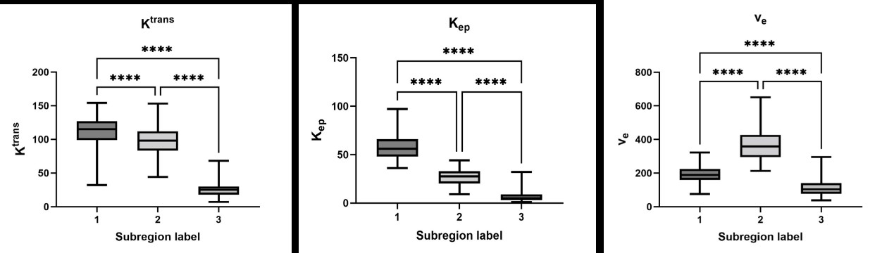
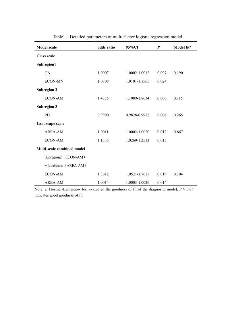
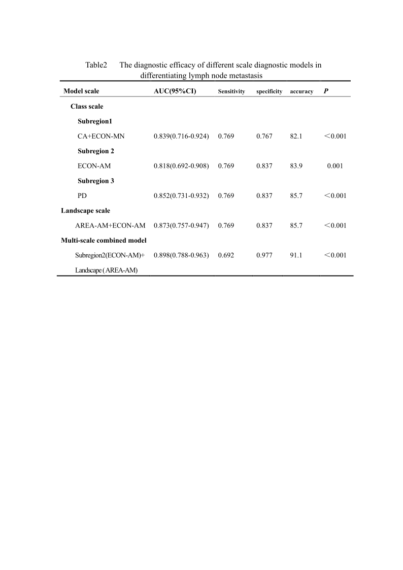
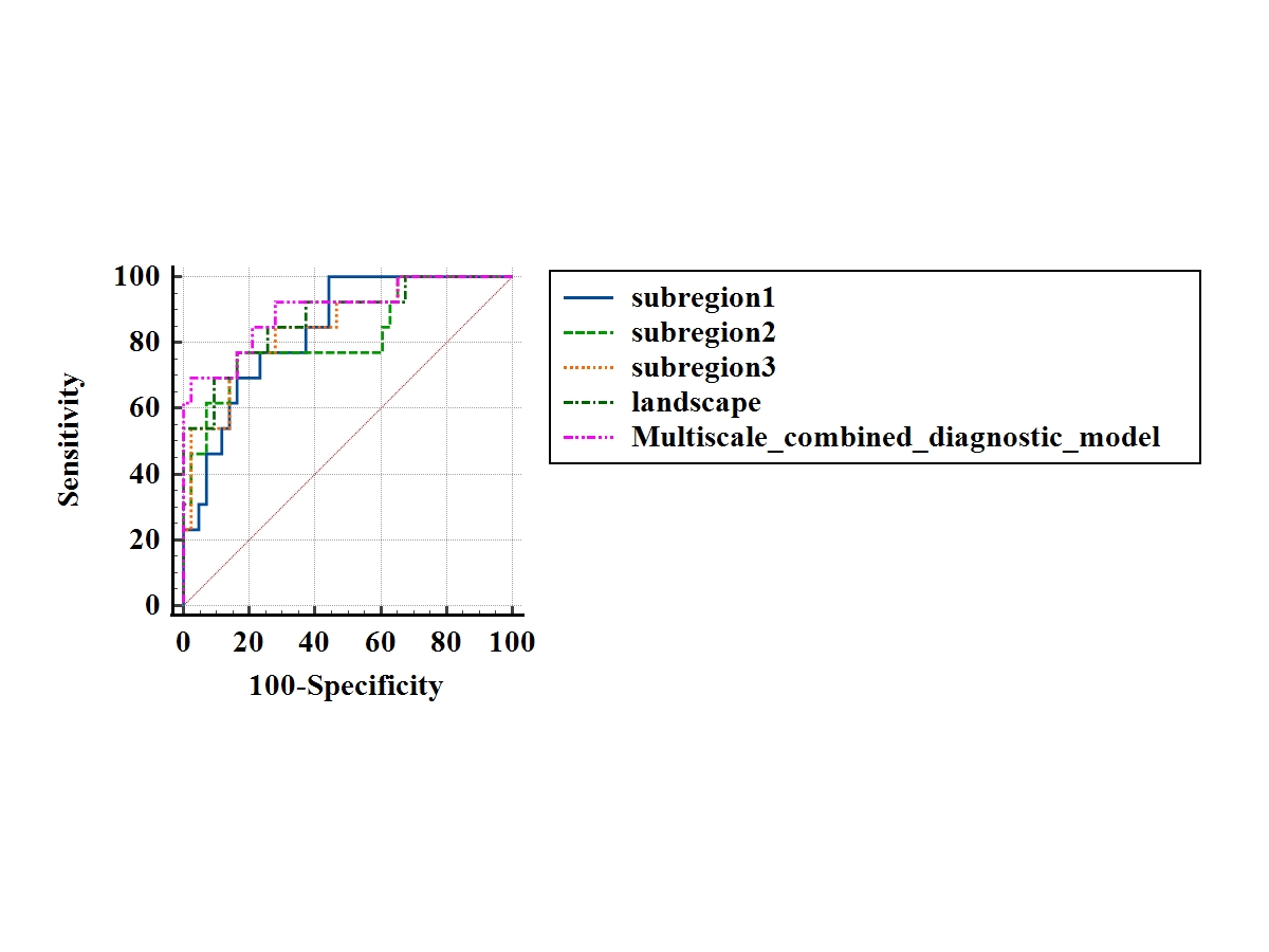
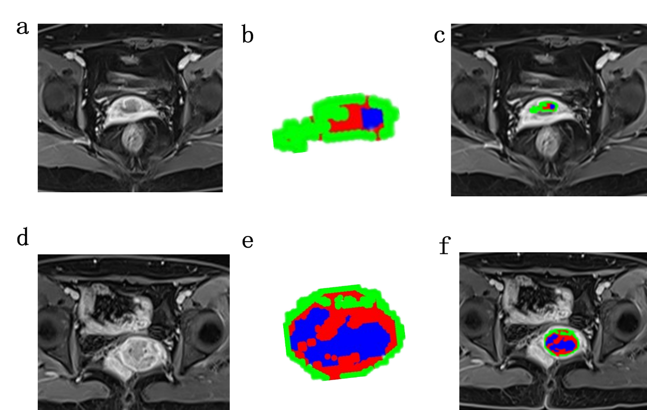
FIG.3 Images of blood perfusion habitats of two representative cases of early cervical cancer without and with lymph node metastasis.
Figure a-c: a 60-year-old female with FIGOⅠB2 stage without lymph node metastasis. Figure b: habitat imaging, subregion2 ECON-AM of 78.3632, Landscape AREA-AM of 0.2155.
Figure d-f, a 59-years-old female, FIGO ⅡA1 stage, with pelvic lymph node metastasis; Figure e, habitat imaging, subregion2 ECON-AM of 87.3343, Landscape AREA-AM of 0.5208.