4291
The Application of Time Dependent Diffusion Magnetic Resonance Imaging in Clinical Staging and Pathological Differentiation of Cervical Cancer1The First Affiliated Hospital of Xi'an Jiaotong University, Xi’an, China, 2Philips Healthcare, Xi’an, China, 3Philips Healthcare, Beijing, China, 4Philips Healthcare, Shenzhen, China
Synopsis
Keywords: Uterus, Cancer, Cervical Cancer;Time-dependent diffusion magnetic resonance imaging
Motivation: Time-dependent diffusion magnetic resonance imaging (TDD-MRI) remains underexplored in the clinical staging and pathological differentiation of cervical cancer.
Goal(s): To investigate the application value of parameters derived from TDD-MRI (d, vin, Dex) in the clinical staging and pathological differentiation of cervical cancer.
Approach: We collected traditional DWI and TDD-MRI data from cervical cancer patients, comparing the respective parameters' efficacy in clinical staging and pathological differentiation.
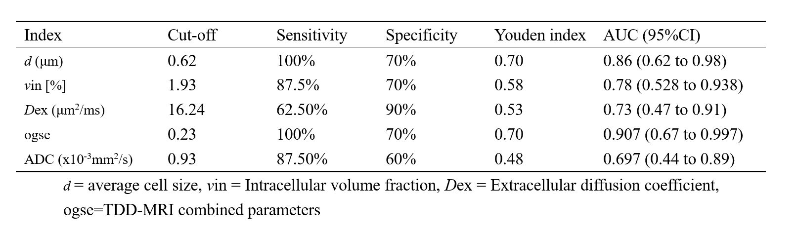
Results: TDD-MRI outperformed ADC in staging cervical cancer, with combined parameters yielding an AUC of 0.90, sensitivity of 100%, and specificity of 70% for early-stage detection.
Impact: This study confirms the advantages of TDD-MRI parameters in the clinical staging of cervical cancer, especially for discerning early from advanced stages, offering greater accuracy than traditional DWI-ADC.
Introduction
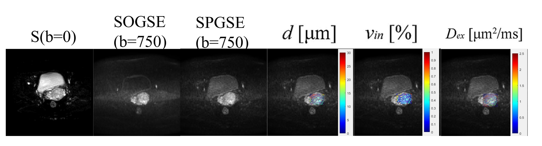
Cervical cancer is the fourth most prevalent malignancy among women worldwide[1]. Accurate staging and grading are crucial for tailored clinical management and prognosis[2]. Diffusion-weighted imaging (DWI) offers non-invasive quantitative analysis, with apparent diffusion coefficient (ADC) values varying significantly with tumor grades and FIGO stages[3-4]. Time-dependent diffusion MRI (TDD-MRI) can overcome DWI's limitations by detailing cellular-scale microstructural changes and assessing parameters such as cell diameter (d), intracellular volume fraction (vin), and extracellular diffusion coefficient (Dex). While TDD-MRI is increasingly applied to body tumors[5-6], its use in cervical cancer is limited. This study evaluates TDD-MRI's microstructural parameters against DWI's ADC values for assessing cervical cancer stages and histological differentiation, underscoring TDD-MRI's potential in clinical staging.Method
In this prospective study, 18 patients with biopsy-confirmed cervical carcinoma, aged 44-81 (mean 60.7±10.26), were enrolled between September and November 2023. Based on the 2018 edition of the International Federation of Gynecology and Obstetrics (FIGO) criteria stratified cases into early (<IIB) and mid-to-late (≥IIB) stages. All patients underwent pelvic MRI examinations using a 3.0T scanner (Ingenia CX, Philips, the Netherlands) with 16-channel torso phased-array coil. TDD-MRI utilized the IMPULSED bi-compartmental model for signal analysis, calculating microstructural parameters such as cell diameter, intracellular volume fraction, and extracellular diffusion coefficient. T-tests compared ADC and TDD-MRI parameters across cancer stages. Pearson correlation assessed their relationship with ADC, considering P<0.05 significant. The diagnostic efficacy of TDD-MRI microstructural parameters and ADC in cervical cancer was compared using the area under the curve (AUC) of the receiver operating characteristic (ROC) curves. A logistic regression model was employed to plot the combined parameter ROC curves and to analyze their diagnostic performance.Result
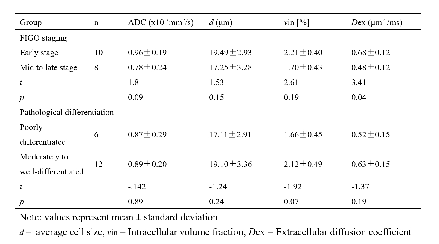
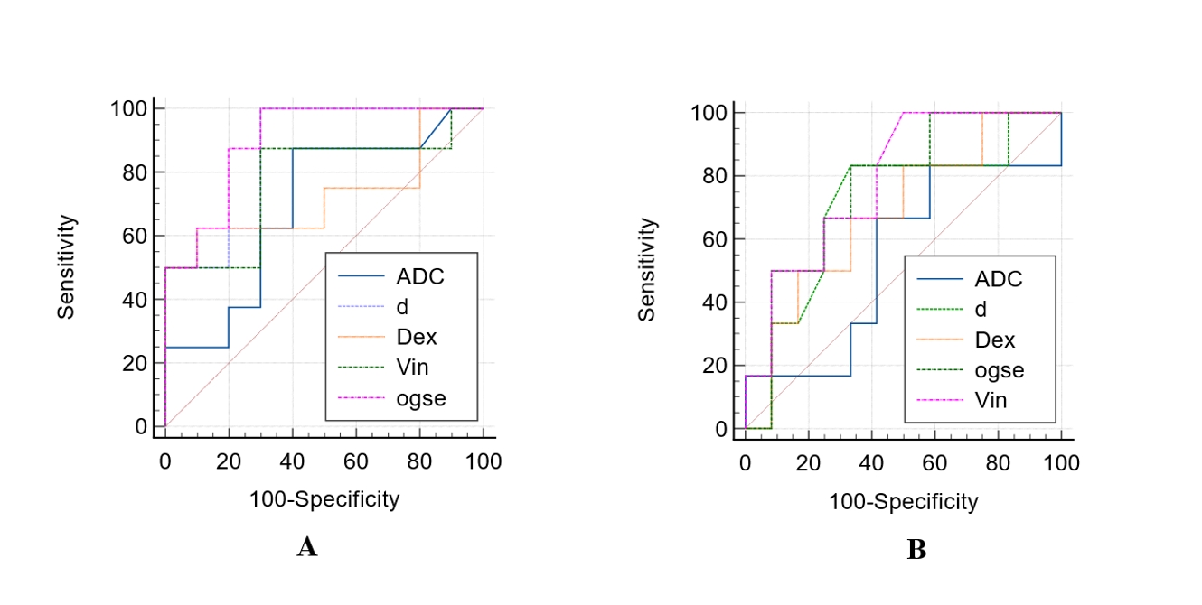
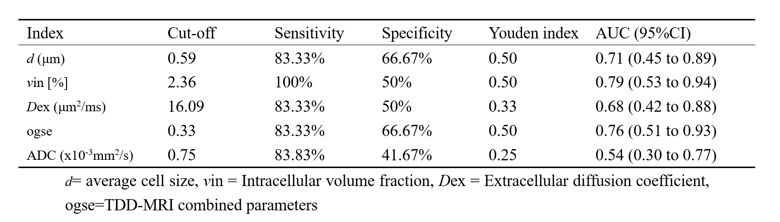
In the comparison of cervical carcinoma stages less than IIB (early stage) and stages IIB or greater (mid-to-late stage), all quantitative parameters, except for the extracellular diffusion coefficient (Dex) (P<0.05), did not show statistical significance between the groups; the TDD-MRI microstructural parameters and ADC values for early-stage cervical cancer tissues were higher than those for mid-to-late stage tissues; the TDD-MRI microstructural parameters and ADC values for poorly differentiated squamous cervical carcinoma tissues were lower than those for moderately to highly differentiated cervical cancer tissues (Table 1). The receiver operating characteristic (ROC) curves for differentiating early from mid-to-late stage cervical cancer based on TDD-MRI microstructural parameters and ADC values are presented in Figure 2, and the diagnostic efficacy is shown in Tables 2 and 3. In the differentiation of early versus mid-to-late stage cervical cancer and low versus moderate-to-high differentiation of cervical cancer, the efficacy of the TDD-MRI microstructural parameter values was superior to that of ADC values, though the difference in efficacy was not statistically significant. Pearson correlation analysis indicated a very low correlation between d, Dex, and ADC values (r=0.25 and 0.20, respectively, both P>0.05). A moderate correlation was found between vin and ADC (r= 0.56, P=0.02<0.05). The clinical staging of cervical cancer corresponds with divergent treatment regimens[2], and accurate preoperative staging aids physicians in devising superior surgical and therapeutic strategies.Discussion
Our study highlights the significant role of non-invasive TDD-MRI microstructural parameters in diagnosing cervical cancer and determining its clinical stage to guide treatment and prognosticate clinical outcomes. Conventional diffusion MRI (ADC) values are sensitive to pathological changes in cervical cancer but are limited in characterizing the microstructural cellular heterogeneity of tumors. In our research, the utilization of a combined diagnostic approach with TDD-MRI microstructural parameters yielded higher accuracy in distinguishing between early and mid-to-late-stage cervical cancer, with an area under the receiver operating characteristic curve of 0.90, surpassing that of traditional ADC measurements. Our findings indicate that TDD-MRI microstructural parameters and ADC values for early-stage cervical cancer tissues exceed those of mid-to-late-stage tissues, suggesting significant microstructural changes may occur during disease progression. This presents specific means to detect the status of cancer tissues, particularly for diagnosis and prognosis, and is poised to have potential value in preclinical and clinical applications.Conclusion
In summary, we have demonstrated the diagnostic potential of TDD-MRI-based microstructural mapping to non-invasively probe the pathological features of cervical cancer within a clinical context. However, our findings require further validation in a larger cohort.Acknowledgements
We are grateful to all the participants for their cooperation and patienceReferences
[1] Razlog R, Kruger CA, Abrahamse H. Enhancement of Conventional and Photodynamic Therapy for Treatment of Cervical Cancer with Cannabidiol. Integr Cancer Ther. 2022; 21
[2] LAI BJ, LI JX, YAN ZH, et al. RESOLVE-ADC value correlates with clinical staging and pathological differentiation of cervical cancer[J]. International Journal of Medical Radiology, 2020, 43(5):525
[3] Downey K, Riches SF, Morgan VA et al (2013) Relationship between imaging biomarkers of stage I cervical cancer and poor-prognosis histologic features: quantitative histogram analysis of diffusion-weighted MR images. AJR Am J Roentgenol 200:314–320
[4] SALEH M, VIRARKAR M, JAVADI S,et al . Cervical canc-er:2018 revised international federation of gynecology and obstetrics staging system and the role of imaging[J]. AJR Am J Roentgenol, 2020, 214(5)
[5] Wu D, Jiang K, Li H, et al. Time-Dependent Diffusion MRI for Quantitative Microstructural Mapping of Prostate Cancer. Radiology. 2022, 303 (3):578-587.
[6] Xu J, Jiang X, Li H, et al. Magnetic resonance imaging of mean cell size in human breast tumors. Magn Reson Med., 2020, 83(6)
Figures