4290
Quantitative Assessment of Perineural Invasion in Cervical Carcinoma using IVIM Combined with mDixon-Quant Multiparameter Imaging1Department of Radiology, the First Affiliated Hospital of Dalian Medical University, Dalian, China
Synopsis
Keywords: Uterus, Uterus
Motivation: Perineural invasion (PNI) is a significant prognostic factor in cervical carcinoma(CC). The utilization of quantitative parameters derived from IVIM and mDixon-Quant MRI offers a new approach for evaluating PNI in CC.
Goal(s): This study aimed to investigate the potential value of IVIM and mDixon-Quant multiparameter imaging in quantitatively assessing PNI in CC.
Approach: MRI scans were conducted on 36 patients with CC to explore the correlation and diagnostic efficacy between MRI quantitative parameters and PNI in CC.
Results: The combined assessment of IVIM and mDixon-Quant quantitative parameters demonstrated a correlation with PNI in CC ,could determine the status of CC PNI.
Impact: The quantitative parameters derived from IVIM and mDixon-Quant MRI are significantly associated with PNI in cervical carcinoma. These parameters hold promising clinical application prospects for evaluating neural invasion in cervical carcinoma.
Abstract
Introduction: Perineural invasion (PNI) is a crucial factor that impacts the prognosis of cervical carcinoma. The utilization of Incoherent Motion Imaging (IVIM) and mDixon-Quant MRI quantitative parameters provides a new approach for evaluating PNI in cervical carcinoma. This study aims to explore the application value of IVIM combined with mDixon-Quant multiparameter imaging in the quantitative assessment of PNI in cervical carcinoma.Methods: Retrospective analysis was conducted on imaging data from 36 cervical carcinoma patients who were pathologically confirmed at our hospital from July 2019 to May 2023, including 12 patients with PNI(PNI(+)) and 24 patients without PNI(PNI(-)). All patients underwent magnetic resonance imaging (MRI) examination before surgery, which included scanning sequences such as T1WI, T2WI, IVIM, mDixon-Quant, etc. Two independent and blind observers measured the apparent diffusion coefficient (ADC), pure apparent diffusion coefficient (D), pseudo-apparent diffusion coefficient (D*), and perfusion fraction (f) from the IVIM sequence, as well as the fat fraction (FF), R2* value, and T2* value from the mDixon-Quant sequence. The intra-class correlation coefficient (ICC) was used to evaluate interobserver consistency, and the independent samples t-test or Mann-Whitney U test was used to compare parameter differences. The efficacy of parameters to predict PNI status in cervical carcinoma was assessed using receiver operating characteristic (ROC) curves. The Delong test was employed to compare differences among the area under the curves (AUCs).
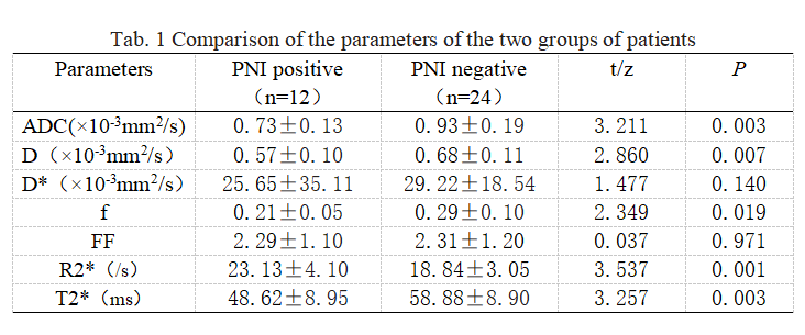
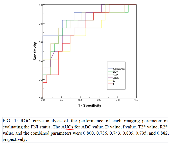
Results: There was good agreement between the measurements of the two observers (ICC > 0.75). The PNI-positive group exhibited lower values of ADC (0.73±0.13 (×10-³mm²/s)), D (0.57±0.10 (×10-³mm²/s)), f (0.21±0.05), and T2* (48.62±8.95 ms) compared to the PNI-negative group (ADC: 0.93±0.19 (×10-³mm²/s), D: 0.68±0.11 (×10-³mm²/s), f: 0.29±0.10, T2*: 58.88±8.90 ms). The R2* value (23.13±4.10 /s) in the PNI-positive group was higher than in the PNI-negative group (18.84±3.05 /s). These differences were statistically significant (P < 0.05). There were no statistically significant differences in D2* value and FF value between the two groups (P > 0.05).The AUCs of ADC value, D value, f value, T2*value, R2*value and the combined parameters for evaluating the PNI status of cervical carcinoma were 0.800,0.736,0.743,0.809,0.795and0.882,respectively.All differences among the AUCs were statistically significant.
Conclusions: Both IVIM and mDixon-Quant multiparameter imaging can quantitatively evaluate PNI in cervical carcinoma. The combination of the two significantly improves prediction performance and holds strong clinical application prospects.
Acknowledgements
We thank all the healthcare workers who fight against cervical carcinoma. We would like to thank my teacher and my team.References
1.Ozan H, Ozuysal S, Ediz B. Perineural invasion in early-stage cervical carcinoma. Eur J Gynaecol Oncol. 2009;30(4):379-83. PMID: 19761126.
2.Martínez-Flores R, Gómez-Soto B, Lozano-Burgos C, Niklander SE, Lopes MA, González-Arriagada WA. Perineural invasion predicts poor survival and cervical lymph node metastasis in oral squamous cell carcinoma. Med Oral Patol Oral Cir Bucal. 2023 Sep 1;28(5):e496-e503. doi: 10.4317/medoral.25916. PMID: 37330960; PMCID: PMC10499348.
3.Gadducci A, Pistolesi S, Cosio S, Naccarato AG. Is Perineural Invasion a Novel Prognostic Factor Useful to Tailor Adjuvant Treatment in Patients Treated With Primary Surgery for Cervical and Vulvar Carcinoma? Anticancer Res. 2020 Jun;40(6):3031-3037. doi: 10.21873/anticanres.14283. PMID: 32487596.
4.Hurník P, Chyra Z, Ševčíková T, Štembírek J, Trtková KS, Gaykalova DA, Buchtová M, Hrubá E. Epigenetic Regulations of Perineural Invasion in Head and Neck Squamous Cell Carcinoma. Front Genet. 2022 Apr 27;13:848557. doi: 10.3389/fgene.2022.848557. PMID: 35571032; PMCID: PMC9091179.
5.Schmitd LB, Perez-Pacheco C, Bellile EL, Wu W, Casper K, Mierzwa M, Rozek LS, Wolf GT, Taylor JMG, D'Silva NJ. Spatial and Transcriptomic Analysis of Perineural Invasion in Oral Cancer. Clin Cancer Res. 2022 Aug 15;28(16):3557-3572. doi: 10.1158/1078-0432.CCR-21-4543. PMID: 35819260; PMCID: PMC9560986.
6.Wan T, Cai G, Gao S, Feng Y, Huang H, Liu L, Liu J. Preoperative Evaluation of Perineural Invasion in Cervical Cancer: Development and Independent Validation of a Novel Predictive Nomogram. Front Oncol. 2021 Dec 23;11:774459. doi: 10.3389/fonc.2021.774459. PMID: 35004296; PMCID: PMC8733474.
Figures